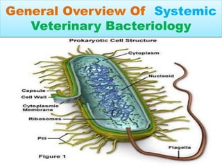
General Overview Of Systemic
Veterinary Bacteriology
INTRODUCTION TO BACTERIA
• Prokaryotic(primitive neucleus, without nuclear
memberane)
• They are most abundant microorganisms.
• Cell wall made up of peptidoglycan(abscent in
Mycoplasma)
• Grouped under 4 categories acc to shape:
• Spherical (coccus), rod shaped (bacillus)
• Comma shaped (vibrium) & spiral shaped
(spirillum).
STAPHYLOCOCCUS
• Gram positive ,flagella abscent, non spore forming.
• Coagulase +ve, catalase +ve, Oxidase –ve.
• Selective media : MANNITOL SALT AGAR & BEARD-
PARKER AGAR.
• Bunch of grapes like colony.
• Shows Double hemolysis / hot-cold hemolysis (S.hycus
is non hemolyic).
• S.aureus: shows TSS (Toxic Shock Syndrome)
• Protein A present which prevents opsonisation.
• Biofilm formation occurs in Staphylococcus.
• Diseases: Mastitis in cattle,Botryomycosis in horse & sows,
Bumble-foot disease in poultry(S.aureus).
STREPTOCOCCUS
• Gram +ve, non motile, chain of cocci.
• TRANSLUSCENT colonies.
• Catalase –ve, coagulase +ve & oxidase –ve.
• Hemolysis (Beta- complete , Alpha –partial).
• Peptidoglycan layer of Strep. Contains C substance.
• S.pneumoniae shows QUELLING reaction.
• Selective media : EDWARD’S MEDIA & E.faecalis can
grow on MacConkey agar.
• Diagnosis: CAMP test for S.agalactiae.
• Diseases: Scarlet fever in humans (S.pyogenes) (DICK
test is done ),Chronic Mastitis(S.agalactiae),acute
mastitis(S.dysagalactiae), STRANGLES in horses
(S.equi)
CORYNEBACTERIUM
• Gram+ve, Pleomorphic,non motile, INTRACELLULAR bacteria.
• Cell wall contains DAP, Arabinogalactin,Mycolic acid.
• Shows PALLISADE or CRISS CROSS ARRANGEMENT,
also called as” CHINESE LETTER APPEARANCE”.
• C.renale :found in urine & CAMP +ve.
• “BABES –ERNST” Granules are found in this bacteria.
• C.pseudotuberculosis shows ONION RING app in lymph
nodes of Sheep.
• Mostly non capsulated except Rhodococcus equi.
• R.equi shows MZN.
• Diseases: Balanoposthitis &”Pizzle rot” in
rams(C.renale),”Piegonfever”(C.pseudotuberculosis),”SUM
MER MASTITIS” (Arachnobacter.pyogens).
LISTERIA
• Gram +ve ,motile ( Peritrichous flagella).
• L.monocytogens &L.ivanovii are HEMOLYTIC.
• PALCAM & OXFORD AGAR are selective media.
• Shows “TUMBLING” motility.
• Shows UMBRELLA like pattern in semisolid media.
• Diagnosis: CAMP test & “ANTONS EYE TEST” for
L.monocytogens.
• Diseases: “CIRCLING DISEASE/ SILAGE DISEASE”
(L.monocytogens),also cause Abortion in 3rd trimester.
• L.monocytogens show “COLD ENRICHMENT
PHENOMENON” & “ASCULIN hydrolysis”.
CLOSTRIDIUM
• Gram +ve , Motile(Peritichous flagella) except
C.perfringens.
• Strictly anaerobic (5-10% CO2) except
C.perfringens.& non capsulated except C.perfringens.
• “ROBERTSON COOKED MEAT MEDIA” is special
media for Clostridium.
• C.tetani shows “SWARMING GROWTH”& ”DRUM
STICK” appearance.
• C.chauvoei is CIGAR shaped & cause “BLACK QUARTER”
disease.
• C.botulinum type C causes “SHAKER ‘s FOAL
SYNDROME”or “FLOPPY BABY SYNDROME”
C.tetani & C.botulinum
C. tetani
• Shows activity at Synaptic
junctions.,inhibits inhibitory
Synaptic receptors.
• Neurotoxin is
TETANOSPASMIN
• Cause “SPASTIK PARALYSIS” or
“SAW HORSE/LOCK JAW”
appearance
C. botulionum
• Shows activity at
Neuromuscular
junctions,Acetyl choline is
unable to bind.
• Have 7 antigenic variants.
• Cause “FLACID PARALYSIS”.
• “Wasp waste appearance”
C.perfringens
• Shows CAMP test & “NAGLAR
REACTION”(alpha toxin present)
• C.perfringens & C.novyi shows LECITHINASE
ACTIVITY.
• Shows “STORMY CLOT”.
• Show DOUBLE ZONE OF HEMOLYSIS.
• Type D causes “Pulpy kidney disease”
MYCOBACTERIUM
• Gram positive,Aerobic,Non spore forming ,Non motile,
Rod shaped acid fast bacilli.
• They are Intracellular pathogens.
• STONE BRICK’S & Lowenstein-Jensen medium
media is used for growth.
• Cell wall of Mycobacterium contains Mycolic acid.
• Cell wall is stained by Ziel Nelson staining.
M.tuberculosis :Shows EUGONIC GROWTH,
M.Bovis: Shows DYSGONIC GROWTH.
M.paratuberculosis: grows best on HAROLD’S EGG YOLK
media.
• M.tuberculosis have rough colonies and
do not break easily whereas M.Bovis have
cream coloured colonies & easily break.
• M.Bovis shows Delayed type
Hypersensitivity(Type 4).
• Diseases:M.bovis cause bovine tuberculosis.
• M.paratuberculosis: causes Johne's disease
in cattle.
SALMONELLA
• Gram –ve , motile.
• Selective media : XLD AGAR (Xylene Lysine
Deoxycholate agar) , H2S Production takes place.
• S.pullorum cause “Bacillary white diarrhoea “in
chicks, S.gallinarum causes “Fowl typhoid” in adult
birds, S.dublin causes “joint ill “ in calves.
• Diagnosis :XLD agar
• BRILLIANT GREEN AGAR : RED coloured colonies.
• Sub culture on TSI (Triple Sugar Iron).
• Diagnosis : IMVIC test:
I(negative),M(positive),VI(negative),C(positive).
• Phage typing in S.typhimurium
Escherichia coli
• Gram +ve ,Motile, peritrichous flagella, Show alpha
Hemolysis on blood agar, Coliform bacteria.
• They are ZOONOTIC.
• Grow on MacConkey agar, blood agar, EMB agar.
• Colonies on MacConkey agar are bright pink.
• On EMB (Eosin Methylene Blue ) agar , colonies show
“green metallic sheen”.
• COLIGRANULOMA or HJARRE’S disease in poultry.
• “Fading puppy Syndrome” (Colisepticemia) in dogs.
• “Mushy Yolk disease”(omphalitis) in poultry.
• Diagnosis : IMVIC test:
I(positive),M(positive),VI(negative),C(negative).
BRUCELLA
• Gram –ve , non motile , cocobacillary MZN +ve.
• Organism is Capnophilic, catalase +ve ( except
B.ovis & B.neotomae), Urease +ve ( except B.ovis).
• Selective media : SERUM DEXTROSE AGAR,
COLUMBIAAGAR , CRYPTOSE SOYAAGAR.
• B.abortus , B.malitensis & B.suis are smooth
& shows small, glistening, bluish&
transluscent colonies
• B.ovis & B.canis are rough form & shows
dull,yellowish, opaque & friable colonies.
• B.abortus is also called as “BANGS
BACILLUS”& Causes ABORTION .
• B.malitensis causes undulant fever in humans
called as “MALTA FEVER”.
• Brucella are NON HEMOLYTIC.
• B.abortus Infects mature animals with
prediliction for reproductive tract of males and
females because of presence of
“ERYTHRITOL” which stimulates its growth.
PASTEURELLA
• Gram –ve, rod or coccobacillus, non motile.
• Catalase +ve , Oxidase +ve.
• Growth occurs in BLOOD AGAR .
• Shows “BIPOLAR STAINING”.
• P.multocida do not grow on MacConkey agar.
• P.multocida type A causes Bovine pneumonic
pasteurellosis or “SHIPPING FEVER “in cattle
• Type B causes H.S , Type D causes “Atropic Rhinitis”
in pigs.
• Tissue samples stained with Giemsa stain shows
BIPOLAR STAINING.
BACILLUS ANTHRACIS
• Gram +ve, Resistant endospore formation, non motile.
• Catalase +ve, Oxidase-ve.
• On Nutrient agar colonies show “ MEDUSA HEAD”
appearance & when observed microscopically shows
“BAMBOO STICK “ appearance.
• B.anthracis have greyish colonies & shows
“GROUND GLASS APPEARANCE”.
• On gelatin slab shows “INVERTED FIR
APPEARANCE.”
• Causes ANTHRAX (ZOONOTIC DISEASE).
• Diagnosis : MacFydean reaction , ASCOLI’S test.
LEPTOSPIRA
• Leptospira are aerobic, gram-negative spirochetes.
• They have characteristic corkscrew-like motility.
• the infection can be transmitted venereally or
transplacentally.
• L interrogans pomona causes “MOON
BLINDNESS” in horses also called as EQUINE
RECURRENT UVEITIS/ PERIODIC
OPTHALMIA.
• EMJH media is used.
• Diagnosis: The microscopic agglutination test
(MAT) is most frequently used.
ACTINOBACILLUS
• Gram –ve, Pleomorphic, non motile.
• A.equilli is HEMOLYTIC.
• A.suis & A.equilli show pink colonies on MacConkey agar except
A.pleuropneumoniae & A.seminis does not grow on MacConkey agar.
• A lignieresii ,A suis & A equuli has SLIME formation on media.
• Catalase +ve except A.semenis.
• A.ligneresi cause “WOODEN TONGUE” disease in cattle,
A.equilli cause “SLEEPY FOAL “ disease .
• Selective media for A.pleuropneumoniae is CHOCOLATE
AGAR.
• A.pleuropneumoniae have capsule, abscent in others & it
shows CAMP test.
ACTINOMYCES
• Gram +ve ,non spore forming, non motile.
• A.suis gives Coryneform type colonies.
• All species affect oral mucosa except A.
hordeovulnaris.
• A.bovis causes “LUMPY JAW” disesase in
cattle due to rarifying Osteomyelitis.
CAMPYLOBACTER
• Gram –ve rods in “Gull winged shapes” & spiral
forms.
• Motile, non fermentive , Oxidase +ve.
• Many species grow on MacConkey Agar.
• Most of the species are +nt in intenstinal tract but
C.fetus subsp veneralis is +nt in bovine
reproductive tract(bovine preputial mucosa).
• C.fetus subsp fetus causes Still birth & Abortion
in Sheep & goats.
• C.jejuni cause Enteritis in dogs.
CHLAMYDIAE
• Spherical intracellular bacteria with unique
developmental cycle.
• “ELIMENTARY BODIES “are present
• Cell wall lacks Peptidoglycan but contains genus
specific Lipopolysaccharides.
• Staining by MZN.
• C.abortus causes “Enzootic abortion” in ewes.
• C.felis causes “Conjuctivitis “ in Cats.
MYCOPLASMA
• “CELL WALL IS ABSENT”.
• Smallest prokaryote, faccultative anaerob.
• Also called as PPLO & it is also used as a media in
mycoplasma.
• Colonies observed in light it shows “fried egg
appearance” .
• Knob like colonies or “umbonate” colonies.
• Diseases : “ CBPP” by M.mycoites(on P.M tissues
show “Marble like appearance”, “CCPP “in
caprines by M.capripneumonae , “CRD” in poultry
by M.gallisepticum.
THANK YOU
SUBMITTED BY: SUBMITTED TO:
Vivek Kumar (2015V157B-IIVER) Dr. Mohd.YOUNIS
BVSc. & A.H 3rd year Assistant Professor, IIVER
Veterinary Microbiology

bacteria (2).pptx

  • 1.
    General Overview OfSystemic Veterinary Bacteriology
  • 2.
    INTRODUCTION TO BACTERIA •Prokaryotic(primitive neucleus, without nuclear memberane) • They are most abundant microorganisms. • Cell wall made up of peptidoglycan(abscent in Mycoplasma) • Grouped under 4 categories acc to shape: • Spherical (coccus), rod shaped (bacillus) • Comma shaped (vibrium) & spiral shaped (spirillum).
  • 3.
    STAPHYLOCOCCUS • Gram positive,flagella abscent, non spore forming. • Coagulase +ve, catalase +ve, Oxidase –ve. • Selective media : MANNITOL SALT AGAR & BEARD- PARKER AGAR. • Bunch of grapes like colony. • Shows Double hemolysis / hot-cold hemolysis (S.hycus is non hemolyic). • S.aureus: shows TSS (Toxic Shock Syndrome) • Protein A present which prevents opsonisation. • Biofilm formation occurs in Staphylococcus. • Diseases: Mastitis in cattle,Botryomycosis in horse & sows, Bumble-foot disease in poultry(S.aureus).
  • 4.
    STREPTOCOCCUS • Gram +ve,non motile, chain of cocci. • TRANSLUSCENT colonies. • Catalase –ve, coagulase +ve & oxidase –ve. • Hemolysis (Beta- complete , Alpha –partial). • Peptidoglycan layer of Strep. Contains C substance. • S.pneumoniae shows QUELLING reaction. • Selective media : EDWARD’S MEDIA & E.faecalis can grow on MacConkey agar. • Diagnosis: CAMP test for S.agalactiae. • Diseases: Scarlet fever in humans (S.pyogenes) (DICK test is done ),Chronic Mastitis(S.agalactiae),acute mastitis(S.dysagalactiae), STRANGLES in horses (S.equi)
  • 5.
    CORYNEBACTERIUM • Gram+ve, Pleomorphic,nonmotile, INTRACELLULAR bacteria. • Cell wall contains DAP, Arabinogalactin,Mycolic acid. • Shows PALLISADE or CRISS CROSS ARRANGEMENT, also called as” CHINESE LETTER APPEARANCE”. • C.renale :found in urine & CAMP +ve. • “BABES –ERNST” Granules are found in this bacteria. • C.pseudotuberculosis shows ONION RING app in lymph nodes of Sheep. • Mostly non capsulated except Rhodococcus equi. • R.equi shows MZN. • Diseases: Balanoposthitis &”Pizzle rot” in rams(C.renale),”Piegonfever”(C.pseudotuberculosis),”SUM MER MASTITIS” (Arachnobacter.pyogens).
  • 6.
    LISTERIA • Gram +ve,motile ( Peritrichous flagella). • L.monocytogens &L.ivanovii are HEMOLYTIC. • PALCAM & OXFORD AGAR are selective media. • Shows “TUMBLING” motility. • Shows UMBRELLA like pattern in semisolid media. • Diagnosis: CAMP test & “ANTONS EYE TEST” for L.monocytogens. • Diseases: “CIRCLING DISEASE/ SILAGE DISEASE” (L.monocytogens),also cause Abortion in 3rd trimester. • L.monocytogens show “COLD ENRICHMENT PHENOMENON” & “ASCULIN hydrolysis”.
  • 7.
    CLOSTRIDIUM • Gram +ve, Motile(Peritichous flagella) except C.perfringens. • Strictly anaerobic (5-10% CO2) except C.perfringens.& non capsulated except C.perfringens. • “ROBERTSON COOKED MEAT MEDIA” is special media for Clostridium. • C.tetani shows “SWARMING GROWTH”& ”DRUM STICK” appearance. • C.chauvoei is CIGAR shaped & cause “BLACK QUARTER” disease. • C.botulinum type C causes “SHAKER ‘s FOAL SYNDROME”or “FLOPPY BABY SYNDROME”
  • 8.
    C.tetani & C.botulinum C.tetani • Shows activity at Synaptic junctions.,inhibits inhibitory Synaptic receptors. • Neurotoxin is TETANOSPASMIN • Cause “SPASTIK PARALYSIS” or “SAW HORSE/LOCK JAW” appearance C. botulionum • Shows activity at Neuromuscular junctions,Acetyl choline is unable to bind. • Have 7 antigenic variants. • Cause “FLACID PARALYSIS”. • “Wasp waste appearance”
  • 9.
    C.perfringens • Shows CAMPtest & “NAGLAR REACTION”(alpha toxin present) • C.perfringens & C.novyi shows LECITHINASE ACTIVITY. • Shows “STORMY CLOT”. • Show DOUBLE ZONE OF HEMOLYSIS. • Type D causes “Pulpy kidney disease”
  • 10.
    MYCOBACTERIUM • Gram positive,Aerobic,Nonspore forming ,Non motile, Rod shaped acid fast bacilli. • They are Intracellular pathogens. • STONE BRICK’S & Lowenstein-Jensen medium media is used for growth. • Cell wall of Mycobacterium contains Mycolic acid. • Cell wall is stained by Ziel Nelson staining. M.tuberculosis :Shows EUGONIC GROWTH, M.Bovis: Shows DYSGONIC GROWTH. M.paratuberculosis: grows best on HAROLD’S EGG YOLK media.
  • 11.
    • M.tuberculosis haverough colonies and do not break easily whereas M.Bovis have cream coloured colonies & easily break. • M.Bovis shows Delayed type Hypersensitivity(Type 4). • Diseases:M.bovis cause bovine tuberculosis. • M.paratuberculosis: causes Johne's disease in cattle.
  • 12.
    SALMONELLA • Gram –ve, motile. • Selective media : XLD AGAR (Xylene Lysine Deoxycholate agar) , H2S Production takes place. • S.pullorum cause “Bacillary white diarrhoea “in chicks, S.gallinarum causes “Fowl typhoid” in adult birds, S.dublin causes “joint ill “ in calves. • Diagnosis :XLD agar • BRILLIANT GREEN AGAR : RED coloured colonies. • Sub culture on TSI (Triple Sugar Iron). • Diagnosis : IMVIC test: I(negative),M(positive),VI(negative),C(positive). • Phage typing in S.typhimurium
  • 13.
    Escherichia coli • Gram+ve ,Motile, peritrichous flagella, Show alpha Hemolysis on blood agar, Coliform bacteria. • They are ZOONOTIC. • Grow on MacConkey agar, blood agar, EMB agar. • Colonies on MacConkey agar are bright pink. • On EMB (Eosin Methylene Blue ) agar , colonies show “green metallic sheen”. • COLIGRANULOMA or HJARRE’S disease in poultry. • “Fading puppy Syndrome” (Colisepticemia) in dogs. • “Mushy Yolk disease”(omphalitis) in poultry. • Diagnosis : IMVIC test: I(positive),M(positive),VI(negative),C(negative).
  • 14.
    BRUCELLA • Gram –ve, non motile , cocobacillary MZN +ve. • Organism is Capnophilic, catalase +ve ( except B.ovis & B.neotomae), Urease +ve ( except B.ovis). • Selective media : SERUM DEXTROSE AGAR, COLUMBIAAGAR , CRYPTOSE SOYAAGAR. • B.abortus , B.malitensis & B.suis are smooth & shows small, glistening, bluish& transluscent colonies • B.ovis & B.canis are rough form & shows dull,yellowish, opaque & friable colonies.
  • 15.
    • B.abortus isalso called as “BANGS BACILLUS”& Causes ABORTION . • B.malitensis causes undulant fever in humans called as “MALTA FEVER”. • Brucella are NON HEMOLYTIC. • B.abortus Infects mature animals with prediliction for reproductive tract of males and females because of presence of “ERYTHRITOL” which stimulates its growth.
  • 16.
    PASTEURELLA • Gram –ve,rod or coccobacillus, non motile. • Catalase +ve , Oxidase +ve. • Growth occurs in BLOOD AGAR . • Shows “BIPOLAR STAINING”. • P.multocida do not grow on MacConkey agar. • P.multocida type A causes Bovine pneumonic pasteurellosis or “SHIPPING FEVER “in cattle • Type B causes H.S , Type D causes “Atropic Rhinitis” in pigs. • Tissue samples stained with Giemsa stain shows BIPOLAR STAINING.
  • 17.
    BACILLUS ANTHRACIS • Gram+ve, Resistant endospore formation, non motile. • Catalase +ve, Oxidase-ve. • On Nutrient agar colonies show “ MEDUSA HEAD” appearance & when observed microscopically shows “BAMBOO STICK “ appearance. • B.anthracis have greyish colonies & shows “GROUND GLASS APPEARANCE”. • On gelatin slab shows “INVERTED FIR APPEARANCE.” • Causes ANTHRAX (ZOONOTIC DISEASE). • Diagnosis : MacFydean reaction , ASCOLI’S test.
  • 18.
    LEPTOSPIRA • Leptospira areaerobic, gram-negative spirochetes. • They have characteristic corkscrew-like motility. • the infection can be transmitted venereally or transplacentally. • L interrogans pomona causes “MOON BLINDNESS” in horses also called as EQUINE RECURRENT UVEITIS/ PERIODIC OPTHALMIA. • EMJH media is used. • Diagnosis: The microscopic agglutination test (MAT) is most frequently used.
  • 19.
    ACTINOBACILLUS • Gram –ve,Pleomorphic, non motile. • A.equilli is HEMOLYTIC. • A.suis & A.equilli show pink colonies on MacConkey agar except A.pleuropneumoniae & A.seminis does not grow on MacConkey agar. • A lignieresii ,A suis & A equuli has SLIME formation on media. • Catalase +ve except A.semenis. • A.ligneresi cause “WOODEN TONGUE” disease in cattle, A.equilli cause “SLEEPY FOAL “ disease . • Selective media for A.pleuropneumoniae is CHOCOLATE AGAR. • A.pleuropneumoniae have capsule, abscent in others & it shows CAMP test.
  • 20.
    ACTINOMYCES • Gram +ve,non spore forming, non motile. • A.suis gives Coryneform type colonies. • All species affect oral mucosa except A. hordeovulnaris. • A.bovis causes “LUMPY JAW” disesase in cattle due to rarifying Osteomyelitis.
  • 21.
    CAMPYLOBACTER • Gram –verods in “Gull winged shapes” & spiral forms. • Motile, non fermentive , Oxidase +ve. • Many species grow on MacConkey Agar. • Most of the species are +nt in intenstinal tract but C.fetus subsp veneralis is +nt in bovine reproductive tract(bovine preputial mucosa). • C.fetus subsp fetus causes Still birth & Abortion in Sheep & goats. • C.jejuni cause Enteritis in dogs.
  • 22.
    CHLAMYDIAE • Spherical intracellularbacteria with unique developmental cycle. • “ELIMENTARY BODIES “are present • Cell wall lacks Peptidoglycan but contains genus specific Lipopolysaccharides. • Staining by MZN. • C.abortus causes “Enzootic abortion” in ewes. • C.felis causes “Conjuctivitis “ in Cats.
  • 23.
    MYCOPLASMA • “CELL WALLIS ABSENT”. • Smallest prokaryote, faccultative anaerob. • Also called as PPLO & it is also used as a media in mycoplasma. • Colonies observed in light it shows “fried egg appearance” . • Knob like colonies or “umbonate” colonies. • Diseases : “ CBPP” by M.mycoites(on P.M tissues show “Marble like appearance”, “CCPP “in caprines by M.capripneumonae , “CRD” in poultry by M.gallisepticum.
  • 24.
    THANK YOU SUBMITTED BY:SUBMITTED TO: Vivek Kumar (2015V157B-IIVER) Dr. Mohd.YOUNIS BVSc. & A.H 3rd year Assistant Professor, IIVER Veterinary Microbiology