This document provides an overview of cerebellar development in mice. It discusses how signaling centers establish the cerebellar territory and define its boundaries. Two primary progenitor zones give rise to cerebellar cells - the ventricular zone and rhombic lip. The rhombic lip generates glutamatergic neurons, including granule neuron progenitors that form the external granule layer and drive cerebellar growth. The ventricular zone produces GABAergic neurons and interneurons. Finally, it notes that while mouse studies provide insights into human cerebellar development and disease, direct study of human fetal cerebella remains important due to species differences.
2. mouse models to dissect the basic biology of cerebellar
development and directly model human cerebellar disease.
In this chapter, we will discuss three major overlap-
ping stages of cerebellar development with a focus on
the mouse literature. First, we will review how the cere-
bellar territory of the neural tube is defined and patterned
by the expression of a series of transcription factors and
signaling molecules. We will then describe how three
germinal zones arise within this territory and undergo
extensive proliferation to give rise to cell types of the
adult cerebellum, followed by an overview of how these
cells then undergo migration and acquire their appropri-
ate positions within the cerebellum as they initiate their
differentiation programs. The establishment of cerebellar
efferents and afferents and the cerebellar circuit itself is
beyond the scope of this review and is covered in detail
elsewhere in this book.
Our current mouse-centric knowledge base of cere-
bellar development has enabled a multitude of insights
into the pathogenesis of human cerebellar disease. How-
ever, mice are not human. We therefore have chosen to
end our review by highlighting the limited literature
available specifically describing human fetal cerebellar
development. We will emphasize species similarities
and differences that may be relevant to disease and
underline the importance of ongoing human fetal
research.
OVERVIEW OF CEREBELLAR
DEVELOPMENT
The spatial complexities of cerebellar morphogenesis
and the interrelatedness of cellular populations can be
difficult to conceptualize. Therefore, before diving into
the details of cerebellar developmental biology,
Figure 2.1 provides an overview of the major epochs
of development.
The cerebellum is a derivative of the anteriormost dor-
sal hindbrain and development starts as soon as the mid/
hindbrain boundary is established at neural plate stages.
The isthmic organizer (IsO), a transient embryonic sig-
naling center that is a derivative of this boundary,
secretes several molecules to define the cerebellar terri-
tory along the anterior–posterior axis of the developing
neural tube (Sato and Joyner, 2009; Harada et al.,
2016). The roof of the hindbrain over the fourth ventricle
roof plate is another signaling center required to establish
the dorsal–ventral extent of the cerebellum within the
anterior hindbrain (Chizhikov et al., 2006). In overlap-
ping waves of neurogenesis, from e10.5 in the mouse
until late mouse embryogenesis, progenitors in the cere-
bellar ventricular zone give rise to GABAergic neurons
of the cerebellar nuclei, and Purkinje cells in addition to
cerebellar interneuron progenitors, which migrate into
the developing cerebellar anlage (Butts et al., 2014b;
Green and Wingate, 2014).
By e10.5 a secondary germinal zone, the cerebellar
rhombic lip, is established at the junction of the cerebel-
lar ventricular zone and dorsal roof plate. The glutama-
tergic neurons of the cerebellar nuclei emerge from
this zone and migrate over the top of the anlage to form
the nuclear transitory zone, which is a staging zone for
cerebellar nuclei assembly. By e11.5, large numbers of
granule neuron progenitors (GNPs) emerge from the
rhombic lip to migrate over the anlage to form the exter-
nal granule layer (EGL), which resides on the pial surface
of the developing anlage, but under the developing
meninges. Within the EGL, GNPs divide extensively.
In mice, peak EGL proliferation occurs around postnatal
day (P) 7 in mice and is complete by P15. Exponential
GNP proliferation in the EGL drives cerebellar growth
and foliation. GNP differentiation occurs continually
from P0 to P14. As granule neurons exit the cell cycle,
they migrate tangentially within the inner EGL and then
exit the EGL migrating radially inward to settle below the
developing Purkinje cell layer to form the internal gran-
ule layer (IGL), resulting in the final laminar arrange-
ment of the mature cerebellum (Millen and Gleeson,
2008; Butts et al., 2014a; Marzban et al., 2014; Leto
et al., 2016).
INITIAL DEFINITION OF THE
CEREBELLAR TERRITORY BY
TRANSIENT EMBRYONIC SIGNALING
CENTERS
The cerebellar territory emerges from the anteriormost
segment (rhombomere 1) of the hindbrain.
The mid/hindbrain boundary is the first segmental
division of the developing neural plate and forms due
to activation of a gene cascade at neural plate stages cul-
minating in juxtaposed expression of two key transcrip-
tion factors: orthodenticle homeobox 2 (Otx2) and
gastrulation brain homeobox 2 (Gbx2). Otx2 is expressed
in the forebrain and midbrain, with its posterior limit at
the presumptive mid/hindbrain boundary. Concurrently,
Gbx2 is expressed in the posterior central nervous sys-
tem, with an anterior boundary at the presumptive mid/
hindbrain boundary. Loss of Otx2 shifts the mid/hind-
brain boundary anteriorly, enlarging the cerebellar
anlage at the expense of posterior midbrain tissue and
loss of Gbx2 shifts the mid/hindbrain boundary posteri-
orly, causing an expansion of the midbrain at the expense
of cerebellar tissue. The establishment of juxtaposed
Otx2 and Gbx2 results in formation of a transient signal-
ing center called the IsO straddling the mid/hindbrain
boundary. The IsO secretes fibroblast growth family 8
and WNT1, which are required for cell survival and
30 P. HALDIPUR ET AL.
3. pattern the adjacent tissue from e8 to 11.5 in mice (Sato
and Joyner, 2009; Harada et al., 2016).
The posterior limit of the cerebellum is defined by
Hoxa2, which is expressed in the caudal central nervous
system with its anterior boundary at the rhombomere 1/2
boundary. Loss of Hoxa2 results in a caudal enlargement
of the cerebellum at the expense of more posterior hind-
brain structures (Gavalas et al., 1997). Ectopic Hoxa2
expression in rhombomere 1 suppresses the specification
of cerebellar neurons (Eddison et al., 2004). Hoxa2
expression is normally excluded from rhombomere 1
via repression by fibroblast growth family 8 from the
IsO (Irving and Mason, 2000; Mason et al., 2000).
Much less is known regarding the mechanisms which
define the dorsal nature of the cerebellum; however, the
dorsal roof plate, another transient signaling center,
clearly plays a role. The roof plate forms on the dorsal
midline of the early neural tube and expresses bone mor-
phogenetic protein (BMP) and WNT-secreted factors. In
rhombomere 1, roof plate-derived Wnt expression is
required to drive early cerebellar anlage ventricular zone
proliferation, while secreted BMP gene expression is
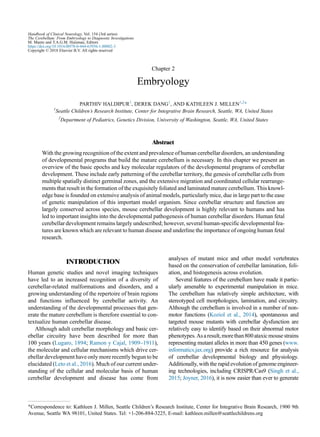
Fig. 2.1. (A) Schematic representation of an e12.5 mouse cerebellum (CB) sectioned along the sagittal plane. The cerebellum is
derived from the dorsal region rhombomere 1 under the influence of signaling factors from the isthmic organizer (IsO) and roof
plate (RP). 4th V, fourth ventricle; MB, midbrain. (B) The developing cerebellum has two zones of neurogenesis, the ventricular
zone (VZ) and the rhombic lip (RL). The cerebellar ventricular zone that consists of a lining radial glia (RG) gives rise to all cer-
ebellar GABAergic neurons and interneurons. GABAergic cerebellar nuclei neurons are produced first, followed by Purkinje cells
and PAX2-expressing cerebellar interneuron progenitors. Bergmann glia are also derived from the cerebellar ventricular zone. The
rhombic lip on the other hand gives rise to the three major glutamatergic neuronal subtypes that populate the cerebellum. Firstly,
cerebellar nuclei projection neurons migrate from the rhombic lip into the nuclear transitory zone (NTZ) over the anlage as the
rostral migratory stream. As embryonic development proceeds, granule neuron progenitors (GNPs) next migrate out of the rhombic
lip between embryonic days 12.5 and 16. These cell progenitors migrate tangentially under the pial surface to establish the external
granule layer (EGL) of the developing cerebellum in an anterior-to-posterior manner. The rhombic lip also gives rise to unipolar
brush cells (UBC) that migrate into the cerebellar anlage. (C) The EGL is a secondary germinal zone, or transit amplifying center.
The EGL is composed of two sublayers – a proliferating external zone and an inner differentiating zone. Proliferation of GNPs
takes place during postnatal days P0–P14. This proliferation is largely driven by the mitogen sonic hedgehog (SHH) secreted from
Purkinje cells which have formed the Purkinje layer (PL) under the EGL. (D) Proliferation of GNPs in the EGL is responsible for
the dramatic size increase of the postnatal mouse cerebellum. As granule neurons exit the cell cycle, they migrate tangentially
within the inner EGL and then exit the EGL, migrating radially inward to settle below the developing Purkinje cell layer to form
the internal granule layer (IGL), resulting in the final laminar arrangement of the mature cerebellum. (E) Schematic representation
of the multiple cell types that arise in the cerebellar ventricular zone and rhombic lip. CN, cranial nerve; CPe, choroid plexus
epithelium; ML, medial/lateral; WM, white matter.
EMBRYOLOGY 31
4. required to induce the cerebellar rhombic lip and cor-
rectly pattern expression of pancreatic transcription fac-
tor (Ptf1a) in the ventricular zone of the nascent
cerebellar anlage (Mishima et al., 2009; Millen et al.,
2014; Yamada et al., 2014). Loss of Ptf1a leads to trans-
formation of cerebellar ventricular zone fates into more
ventral brainstem fates (Millen et al., 2014). To date,
Ptf1a is the sole known gene defining the ventral bound-
ary of the cerebellar territory of rhombomere 1, although
the molecular cascades that precisely regulate this ventral
limit of Ptf1a cerebellar expression remain unknown.
THE CEREBELLAR RHOMBIC LIP GIVES
RISE TO CEREBELLAR GLUTAMATERIC
NEURONS
The cerebellar rhombic lip, which forms adjacent to the
fourth ventricle roof plate in rhombomere 1, is one of two
primary progenitor zones in the developing cerebellum.
Fate-mapping studies have demonstrated that the cere-
bellar rhombic lip gives rise to cells that populate extra-
cerebellar regions of the hindbrain, including the pontine
nuclei (Machold and Fishell, 2005; Wang et al., 2005;
Rose et al., 2009). It also gives rise to the three major glu-
tamatergic neuronal subtypes that populate the cerebel-
lum. These lineages are derived in overlapping waves
of neurogenesis. Beginning at e10.5 and terminating
at e12.5, cerebellar nuclei projection neurons migrate
from the rhombic lip into the aforementioned nuclear
transitory zone over the anlage as the rostral migratory
stream. As embryonic development proceeds, GNPs
next migrate out of the rhombic lip between e12.5 and
e16. These cell progenitors migrate tangentially under
the pial surface to establish the EGL of the developing
cerebellum in an anterior to posterior manner. Addition-
ally, between e13.5 and the early neonatal period, the
rhombic lip gives rise to unipolar brush cells (UBCs),
which migrate through the developing white matter in
the nascent cerebellum and eventually settle in the IGL
(Englund et al., 2006).
BMP signaling from the roof plate and choroid plexus
is essential to induce the rhombic lip and the expression
of the bHLH transcription factor gene atonal homologue
1 (Atoh1) (Alder et al., 1996; Chizhikov et al.,
2006,2010). Atoh1 is expressed in all rhombic lip deriv-
atives as they exit the rhombic lip and Atoh1 is currently
the only gene known to be required to generate all deriv-
atives of the rhombic lip (Wang et al., 2005; Rose
et al., 2009). In early Atoh1-expressing cells have many
downstream gene targets, and a molecular cascade in-
volving paired box protein 6 (PAX6), NEUROD,
T-box brain 1 (TBR1), and TBR2 proteins is variably
expressed within each rhombic lip lineage contributing
tocelltype-specificmorphologiesandmigratorypathways
(Englund et al., 2006). For example, PAX6 is expressed in
all rhombic lip derivatives. It is not required to generate
rhombic lip-derived cerebellar nuclei neurons, but rather
is needed for their survival as they transit to the nuclear
transitory zone over the pial surface (Yeung et al., 2016).
PAX6 is also not required to generate GNPs that form
the EGL but regulates their migration and the migration
of granule neurons as they differentiate from GNPs
(Swanson and Goldowitz, 2011). In contrast, PAX6 is
required to generate UBCs (Yeung et al., 2016). Although
ATOH1 is required for all rhombic lip lineages, there is no
evidence that ATOH1 actually regulates rhombic lip-
derivative identity. There is, however, emerging evidence
that cell fate identity is imparted within the rhombic lip
itself, prior to ATOH1 expression. Although the mecha-
nisms directing rhombic lip neurogenesis remain largely
unknown,Lmx1aandWlshavebeenshown tobeinvolved
in rhombic lip maintenance and cell fate determination
(Chizhikov et al., 2010; Yeung et al., 2014, 2016; Yeung
and Goldowitz, 2017).
External granule layer development
and cerebellar foliation
Given their sheer number, cerebellar granule neurons
represent perhaps the most important rhombic lip deriv-
ative. Indeed, of all cerebellar cell types, the develop-
ment of this population is the most studied. GNPs
continue to proliferate as they exit the rhombic lip,
migrating over the cerebellar anlage to form the EGL.
Rhombic lip exit is at least partially directed by SDF1a –
a chemotaxic signal secreted from the overlying pia and
posterior fossa mesenchyme and received by the CXCR4
receptor expressed by GNPs (Hartmann et al., 1998a;
Zou et al., 1998; Reiss et al., 2002; Zhu et al., 2002;
Vilz et al., 2005; Tiveron and Cremer, 2008; Yu et al.,
2010; Haldipur et al., 2017). The anterior limit of migra-
tion at the mid/hindbrain boundary is at least partially
controlled by UNC5H3 (Ackerman et al., 1997;
Leonardo et al., 1997). Temporal fate mapping of rhom-
bic lip progenitors using inducible cre recombinase
under the control of an Atoh1 promoter shows that
GNPs generated the earliest tend to populate the ante-
rior lobes of the adult cerebellum, whilst the distribution
of the cells exiting the rhombic lip progressively becomes
more caudal as development progresses (Machold and
Fishell, 2005).
The EGL is a secondary germinal zone, or transit amp-
lifying center. The EGL is composed of two sublayers – a
proliferating external zone and an inner differentiating
zone. Proliferating GNPs undergo a dramatic clonal
expansion, which in mice takes place during postna-
tal days P0–P14. This proliferation is largely driven
by the mitogen sonic hedgehog (SHH) secreted from
32 P. HALDIPUR ET AL.
5. Purkinje cells which form the Purkinje cell plate under
the EGL (Wallace, 1999; De Luca et al., 2016), although
other factors, including Wnt 5a (Subashini et al., 2017),
insulin-like growth factor (IGF1) (Fernandez et al.,
2010), NOTCH2 (Solecki et al., 2001), and SDF1a
(Klein et al., 2001), have also been implicated as GNP
mitogens. GNPs are maintained in their proliferative
niche by the signaling protein SDF1a which is secreted
from the overlying developing meninges, which acti-
vates the CXCR4 receptor in GNPs and regulates cell
adhesion (Hartmann et al., 1998a, b; Zou et al., 1998;
Reiss et al., 2002; Zhu et al., 2002; Vilz et al., 2005;
Tiveron and Cremer, 2008; Yu et al., 2010; Huang
et al., 2014; Haldipur et al., 2017).
Proliferation of GNPs in the EGL is responsible
for the dramatic size increase of the postnatal mouse cer-
ebellum. Since the size of the posterior fossa does not
dramatically increase, increased cerebellar size is accom-
modated within the posterior fossa by folding along the
anterior/posterior axis. In mice, the circumference of the
cerebellar medial anterior/posterior axis increases 17.6-
fold between E17.5 and P14 compared with only a
2.2-fold increase in the mediolateral axis (Legue et al.,
2015). Intriguingly, the basic pattern of cerebellar folia-
tion is conserved across evolution. Foliation is initiated
by the formation of multiple “anchoring centers” at ste-
reotypic places across the anlage which will form the
base of each fissure separating two folia (Sudarov and
Joyner, 2007). The patterning mechanisms that position
these anchoring centers are incompletely defined, but
are related to underlying Purkinje cell patterning and
clustering and at least partially involve function of the
homeobox engrailed genes (En1/2) (Sillitoe and Joyner,
2007; Sudarov and Joyner, 2007). In the mouse cerebellar
vermis, four anchoring centers giverise to the fivecardinal
lobules. Anchoring centers are first characterized by local-
ized increase in proliferation and inward thickening of
the granule cell precursors and subsequent coordinated
Bergmann glial fiber rearrangement. Anterior/posterior-
oriented cell division with the EGL drives the outward
and preferential anterior/posterior expansion of cerebellar
folia, while differential timing of granule neuron produc-
tion in each lobule is responsible for the overall shaping of
the mature cerebellar form (Sillitoe and Joyner, 2007;
Legue et al., 2016).
The mechanisms responsible for cell cycle exit and
subsequent differentiation of GNPs into granule neurons
are poorly understood. As GNPs exit the cell cycle, they
move to the inner layer of the EGL where they undergo a
brief period of tangential migration (Komuro et al.,
2001), which transitions into glial-guided radial migra-
tion along the radial glial fibers of the Bergmann glia
(Rakic, 1971; Edmondson and Hatten, 1987; Hatten,
1999). These migratory phases are associated with
dramatic morphologic transformations. Outer EGL pro-
liferative GNPs are round with multiple short processes.
In the inner EGL, newly differentiating granule neurons
reorganize their cytoskeleton reorganized to form two
short horizontal processes that elongate during their jour-
ney to the molecular layer. After a brief latency at the
molecular layer, they acquire an elongated spindle
shape and a vertical process (Komuro et al., 2001). It
is currently understood that upregulated activity of
the Par cell polarity complex is coupled to cell cycle
exit, as well as changes in cellular adhesion and the
initiation of migration. Zinc finger and homeobox tran-
scription factor-1 (Zeb1), a master regulator of epithe-
lial polarity, is highly expressed in unpolarized GNPs
and controls neuronal differentiation by transcriptionally
repressing polarity genes in GNPs. ZEB1 expression
itself is maintained by SHH expression. Posttransla-
tional ubiquitination of Par complex protein PARD3A
via Siah, a Par complex-interacting E3 ubiquitin ligase
expressed in the EGL, also regulates Par complex activ-
ity in the EGL (Famulski and Solecki, 2013; Ramahi
and Solecki, 2014; Singh and Solecki, 2015; Singh
et al., 2016).
Since an imbalance between GNP proliferation and
differentiation can lead to either cerebellar hypoplasia
or tumor formation, it is unsurprising that redundant sys-
tems regulate GNP differentiation. Several pathways are
also thought to provide negative regulation of GNP
proliferation, including BMP4, WNT3, and the anaphase-
promoting complex/cyclosome (APC/CCDH1) ubiquitin
ligase (Leto et al., 2016). WNT3 suppresses GNP growth
through a noncanonic WNTsignaling pathway, activating
prototypic mitogen-activated protein kinases (MAPKs),
the RAS-dependent extracellular signal-regulated kinases
1/2 (ERK1/2) and ERK5, instead of the classical
b-catenin pathway (Anne et al., 2013). WNT3 also
inhibits GNP proliferation by downregulating prolifera-
tive target genes of the mitogen Shh and the bHLH tran-
scription factor ATOH1 (Anne et al., 2013). CK1d is
another novel regulator of GNP expansion as a loss of
casein kinase (CK) 1d or treatment of GNPs with a
highly selective small-molecule CK1d inhibitor inhibits
GNP expansion. CK1d is targeted for proteolysis via
APC/CCdh1 ubiquitin ligase, and conditional deletion
of the APC/CCdh1 activator, Cdh1 in cerebellar GNPs
results in higher levels of CK1d, suggesting an impor-
tant role for the APC/CCdh1 complex in GNP cell cycle
exit (Penas et al., 2015).
Finally, epigenetic modification of SHH down-
stream effectors, the Gli genes, are contributors to
GNP cell cycle exit (Zanin et al., 2016). For example,
activation of the p75 neurotrophin receptor p75NTR
prevents deacetylation of acetylated gene-regulatory
regions surrounding Gli1 and Gli2 by inhibiting histone
EMBRYOLOGY 33
6. deacetylases such as HDAC1, thereby ensuring that they
remain transcriptionally inactive (Canettieri et al., 2010).
Indeed, dramatic chromatin reorganization is also required
to execute the entire granule neuron differentiation pro-
gram as these neurons migrate through the molecular layer
and establish their connectivity in the IGL (Zhu et al.,
2016). A thorough discussion of this extensive literature
however is beyond the scope of this review.
Glutamatergic cerebellar nuclei and UBCs
Unlike GNPs, both glutamatergic cerebellar nuclei neu-
rons and UBCs exit the rhombic lip as postmitotic newly
differentiating neurons. Cerebellar nuclei neurons exit
the rhombic lip early to migrate over the anlage to form
the nuclear transitory zone. Recent work has determined
that these rhombic lip-derived cerebellar nuclei neurons
are defined by a temporal transcriptional program (Leto
et al., 2016). LHX9+ cells are born first and are destined
to form the lateral nucleus (projecting to midbrain and
thalamus), followed by a TBR1+ medial (fastigial)
group, which sends axons to the hindbrain via the fascic-
ulus uncinatus, or hook bundle (Green and Wingate,
2014). The progressive deposition of cells in more dorsal
(ultimately medial) positions reflects a decreasing sensi-
tivity to netrin signaling from ventral midline in migrat-
ing cells (Alcantara et al., 2000; Gilthorpe et al., 2002).
Netrin receptors are also responsible for determining the
laterality of the projections of cerebellar nuclei axons
(Tamada et al., 2008),which extend seamlessly from
the leading processes of migrating cells (Gilthorpe
et al., 2002). Target selection (rostral or caudal central
nervous system) appears to be a property of LHX9 versus
TBR1 expression (Green and Wingate, 2014).
UBCs are late derivatives exiting the rhombic lip
directly into the cerebellar anlage and remained unrecog-
nized until 1994 (Mugnaini and Floris, 1994; Englund
et al., 2006). Other than the requirement of PAX6 in
the rhombic lip for their generation (Yeung et al.,
2016), little is known of their molecular drivers of spec-
ification, migration, or differentiation (Leto et al., 2016).
THE CEREBELLAR VENTRICULAR ZONE
GIVES RISE TO ALL CEREBELLAR
GABAERGIC NEURONS
The other major primary progenitor zone of the develop-
ing cerebellum is the cerebellar ventricular zone which
gives rise to all cerebellar GABAergic neurons and
interneurons, including in overlapping periods of neuro-
genesis from e11.5 to e14.5 (Sudarov et al., 2011).
GABAergic cerebellar nuclei neurons are produced first,
followed by Purkinje cells, then PAX2-expressing
cerebellar inhibitory interneuron progenitors (PIPs),
which will continue to divide within the presumptive
cerebellar white matter, to eventually produce the inter-
neurons of the cerebellar cortex, including basket,
stellate, Golgi, Lugaro, globular, and candelabrum
neurons. Bergmann glia are also derived from the cere-
bellar ventricular zone.
The mechanisms that specify each derivative of
the ventricular zone are only just beginning to be eluci-
dated. Fate-mapping studies have clearly demonstrated
that Ptf1a expression defines the cerebellar ventricular
zone and Ptf1a itself is required to generate all GABAer-
gic derivatives (Hoshino et al., 2005; Yamada et al.,
2014). Further, a complex interaction between PTF1a
and ATOH1 maintains ventricular zone versus rhombic
lip identity at the interface of these two progenitor zones
(Pascual et al., 2007; Yamada et al., 2014). Additional
recent fate-mapping studies have identified two spatially
and temporally distinct progenitor zones within the
PTF1a+ ventricular zone. Expression of oligodendro-
cyte-specific bHLH transcription factor (Olig2) defines
Purkinje cell progenitors, while PIPs are derived from a
PTF1a+ subzone-expressing homeodomain-containing
transcription factor, GSX1. At e12.5 in the developing
mouse embryo, OLIG2+ Purkinje cell progenitors com-
prise a predominant portion of the PTF1a+ ventricular
zone domain. By e14.5, GSX1 has swept across the ven-
tricular zone and PIPs become the output of the ventric-
ular zone. GSX1 inhibits Olig2 expression and acts as
a brake for temporal identity transition and there is
some evidence that Olig gene expression is required
for the Purkinje cell progenitors identity (Seto et al.,
2014). However, the molecular regulatory details of
these dramatic changes in gene expression and subse-
quent changes in ventricular zone output remain to be
determined.
Purkinje cell development
As detailed above, Purkinje cells are specified within the
PTF1a+ OLIG2+ ventricular zone. As they exit the ven-
tricular zone starting at e10.5 in mice, they cease prolif-
eration and initiate migration towards the nascent EGL to
form a multilayered structure called the Purkinje cell
plate. Early-born Purkinje cells, predominantly born in
the posterior region of the ventricular zone, are the first
cells to migrate to the nascent Purkinje cell plate by
e14.5. These cells utilize a tangential migratory pathway
at first, similar to that used by cells derived from the
rhombic lip. However, at e13.5, these cells abruptly
change orientation upon sending their trailing processes
into a cortical region where protein reelin is abundant,
secreted from newly arriving GNPs in the forming
EGL. This quick, reelin-dependent transition from tan-
gential to radial migration guides the nascent Purkinje
cells to the Purkinje cell plate (Miyata et al., 2010;
34 P. HALDIPUR ET AL.
7. Leto et al., 2016). Later-born Purkinje cells, as well as
cells produced during the early phase but in an anterior
region, must migrate further than their earlier-born coun-
terparts, utilizing a radial glial scaffold away from the
ventricular zone. The role of reelin in the migration of
these later-born cells to the Purkinje cell plate has yet
to be elucidated.
Early- and late-born Purkinje cells have distinct
molecular identities essential to the formation of the sag-
ittally striped topographic map of the mature cerebellum
(Brochu et al., 1990; Armstrong et al., 2000; Sarna et al.,
2006; White and Sillitoe, 2013). Indeed, the functional
topographic map of the mature cerebellum is organized
in medial/lateral stripes of afferents and efferents that
directly relate to clusters of Purkinje cells with shared
expression patterns of genes, arrayed in medial/lateral
stripes. This mediolateral Purkinje cell code is estab-
lished during development and is at least partially related
to Purkinje cell birth date (Fujita et al., 2012). One par-
ticularly well-known marker of Purkinje cell molecular
heterogeneity is zebrinII (ZII) – named for the inter-
spersed zebra-like sagittal arrays of ZII+ and ZII– seen
in the mature cerebellum. Early-born Purkinje cells
become ZII+ and late-born Purkinje cells remain
ZII– (Larouche et al., 2006; Namba et al., 2011).
Beginning around e14.5, the cerebellar plate begins to
reorganize itself, forming more than 50 distinct clusters
by e18.5. These clusters are composed of either ZII+ or
ZII– Purkinje cells. The mechanisms driving cluster
formation are not understood; however, differential
expression of cell-cell adhesion molecules, especially
cadherins, has been promoted as possible organizing
molecules (Vibulyaseck et al., 2017). Clusters transform
to sagittal stripes as the cerebellum expands along the
anterior/posterior axis due to GNP proliferation and
inward migration of granule neurons, changing the mul-
ticellular Purkinje cell layer into a Purkinje cell mono-
layer residing on top of the IGL. The relationship
between embryonic Purkinje cell clusters and mature
Purkinje cell stripes is highly complicated. One cluster
may give rise to a singular stripe or multiple stripes, or
combine with other clusters to form a singular stripe.
Although we have molecular markers for these Purkinje
cell clusters from early to mature stages, the mechanisms
that pattern and define these clusters in early develop-
ment remain incompletely understood, although the
Engrailed genes (En1 and En2) are involved (Sillitoe
and Joyner, 2007; Sillitoe et al., 2010; Fujita et al., 2012).
As the Purkinje cell layer transforms into a mono-
layer, each Purkinje cell initiates neurite formation
around P4–P5 in mice, resulting in the iconic and elabo-
rate flat dendritic arbor and extended single axon of the
mature Purkinje cell. Neurite-patterning mechanisms
also remain largely unknown. There is evidence that
intrinsic programs may drive initiation and some aspects
of Purkinje cell dendrite patterning (Sotelo and Dusart,
2009; Tanaka, 2009, 2015; Shih et al., 2015). However,
Purkinje cell development is clearly also influenced by
GNPs and granule neurons. They provide essential Pur-
kinje cell trophic factors. In addition, granule neuron
defects nonautonomously cause delays in Purkinje cell
dendrite formation and disruptions of the planarity of
Purkinje cell dendritic trees (Baptista et al., 1994; Hirai
and Launey, 2000; Ohashi et al., 2014).
INHIBITORY INTERNEURON
DEVELOPMENT
In contrast to Purkinje cells which are born directly from
the ventricular zone, cerebellar inhibitory interneurons
are derived from proliferative precursors that delaminate
into the developing anlage, otherwise called the prospec-
tive white matter (Leto et al., 2016). These precursors
remain mitotically active, with peak proliferation during
the first postnatal week in mice (Leto et al., 2006, 2012).
Heterotopic/heterochronic transplantation experiments
indicate that these interneuron progenitors maintain full
developmental potential up to the end of cerebellar devel-
opment and acquire mature phenotypes under the influ-
ence of environmental cues present in the prospective
white matter. Furthermore, the final fate choice occurs
in postmitotic cells, rather than dividing progenitors
(Leto et al., 2009). Fates are acquired in an inside-out
manner, with cerebellar nuclei-resident interneurons
born first and molecular layer-resident basket and stellate
cells born last.
The molecular details of the cell autonomous and non-
autonomous effectors of PIP differentiation remain
largely obscure. SHH disseminated by Purkinje cell
axons in the prospective white matter is an important
mitotic driver for PIPs in the prospective white matter
(De Luca et al., 2015; Fleming and Chiang, 2015). Pur-
kinje cells are also important for the terminal differenti-
ation and morphogenesis of cerebellar interneurons. For
example, the complexity of basket/stellate cell axonal
arborizations and their positioning on Purkinje cells is
critically dependent on neurofascin and also Semaphorin
(SEMA3A)/neuropilin-1-mediated signaling between
Purkinje cells and differentiating medial/lateral interneu-
rons (Ango et al., 2004; Buttermore et al., 2012; Cioni
et al., 2013; Leto et al., 2016). Additionally, granule
neuron-dependent signals are known to direct the sur-
vival and growth of early postmigratory basket/stellate
interneurons (Mertz et al., 2000; Konno et al., 2014).
Gliogenesis
The ventricular zone is the origin of all cerebellar
astrocytes, a subpopulation of oligodendrocytes, and
EMBRYOLOGY 35
8. Bergmann glia. Notch signaling, which begins at e10.5
in the developing mouse cerebellar anlage, is essential
to promote the astrocytic cell fate. Ablation of Notch
activity in the nascent cerebellum leads to precocious
neural differentiation with reduced astrocyte formation.
Constitutive Notch activation leads to the overpro-
duction of astrocytes along with a concomitant decrease
in neurons. Astrocyte production is also regulated by
Achaete-scute homolog 1 (ASCL1), another protein tran-
siently expressed in the ventricular zone between e10.5
and e13.5. Ablation of Ascl1 results in increased numbers
of astrocytes, while overexpression of Ascl1 suppresses
astrocytic cell fate and promotes GABAergic inter-
neuronal cell fates. Although a small number of oligo-
dendrocytes are derived from the cerebellar ventricular
zone, targeted in utero electroporation experiments have
pointed to an extracerebellar source for most of these
cells (Grimaldi et al., 2009). Mouse fate-mapping studies
have determined that most originate from the Olig2-
expressing neuroepithelial domain in the ventral rhom-
bomere 1 (Hashimoto et al., 2016). Chick quail chimeras
and in ovo transplants implicate a region of the mesence-
phalic ventricular zone as the origin of the majority of
avian cerebellar oligodendrocytes (Mecklenburg et al.,
2011), indicating potential species differences.
Bergmann glia are an essential cell type in the devel-
oping cerebellum. They are generated directly from the
radial glia that line the cerebellar ventricular zone from
e13.5, after Purkinje cells are born (Sudarov and
Joyner, 2007). Initially, the ventricular zone radial glia
form the scaffold upon which Purkinje cells migrate to
the Purkinje cell plate. However, after Purkinje cells
are generated, radial glia lose their apical attachment
and their cell bodies migrate through the cerebellar
anlage radially upwards, terminating their journey within
the Purkinje cell layer. While the protein PTPN11 has
been shown to aid in the transformation of radial glia into
Bergmann glia (Li et al., 2014; Heng et al., 2017), their
arrangement within the Purkinje cell layer is influenced
by the interaction of Bergmann glial-expressed Notch
with delta/Notch-like EGF-related receptor (DNER) on
Purkinje cells (Eiraku et al., 2005).
Bergmann glia possess multiple ascending processes
that traverse the molecular layer and EGL. As already
discussed, granule cells migrate inward from the EGL
to the IGL along these processes. Bergmann glia have
also been shown to direct migration of molecular layer
interneurons (Ango et al., 2008). The Bergmann glial
end feet hook on to the basement membrane of the pos-
terior fossa mesenchyme. Aberrations in end feet attach-
ment can drastically affect Bergmann glial function
which in turn can lead to abnormalities in cerebellar lam-
ination and foliation (Moore et al., 2002; Qu and Smith,
2005; Satz et al., 2008). In rodents, the number of
Bergmann glial fibers increases significantly during
the first 2 postnatal weeks, particularly during peak pro-
liferation and migration of granule cell progenitors, sug-
gesting that GNPs play a role in Bergmann glial fiber
formation. The molecular identity of signals from granule
cells to Bergmann glia however remains largely unex-
plored. However, it is known that the differentiation
and maintenance of Bergmann glia are heavily dependent
on Purkinje cell secretion of SHH. Blocking SHH func-
tion results in reduced Bergmann glial survival. Con-
versely, Purkinje cell survival is also dependent on
Bergmann glia (Wang et al., 2011), yet the molecular reg-
ulators of this interaction remain unknown.
COMPLEX INTERRELATIONSHIPS
BETWEEN MULTIPLE CELLULAR
POPULATIONS ARE REQUIRED FOR
NORMAL DEVELOPMENT
In the sections above, we have generally discussed the
genesis and initial differentiation of each cerebellar cell
type in isolation. This is an artificial reflection of the true
nature of cerebellar development. Indeed, we have briefly
outlined several complex interactions between multiple
developing cells that are essential for normal cerebellar
development. These include interdependencies between
developing Purkinje cells, interneurons, granule neurons,
and Bergmann glial cells for generation, survival, and his-
togenesis. There remain however two notable complex
interrelationships that are worthy of separate discussion
as they are active areas of ongoing research.
Signaling from the choroid plexus and
posterior fossa mesenchyme
The cerebellar territory is established under the influence
of the IsO and roof plate, both extracerebellar structures.
The central nervous system-derived roof plate differenti-
ates into the choroid plexus epithelium and is juxtaposed
by vascular elements derived from head mesenchyme.
Genetic loss-of-function studies have demonstrated that
the choroid vascular elements are a source of SHH oper-
ating through a transventricular route to maintain the last
phase of proliferative cerebellar ventricular zone from
e13.5 (Huang et al., 2010).
The vascular elements of the choroid plexus are not
the only posterior fossa mesenchymal derivatives
influencing cerebellar development. More recently, it
has become evident that nearly all aspects of cerebellar
development are influenced by extracerebellar signaling
from the developing meninges adjacent to the cerebellar
anlage which is derived from head mesenchyme. Early
experimental removal of the meninges during peak post-
natal EGL proliferation causes fragmentation of the EGL
and disruption of the radial fibers of the radial glial cells
36 P. HALDIPUR ET AL.
9. in hamster studies (Sievers et al., 1994). We have more
recently shown that the developing meninges has a more
extensive and much earlier influence on cerebellar
development. Loss of FOXC1 – a transcription factor
expressed exclusively in the mesenchyme adjacent to
the early cerebellar anlage – causes dramatic cerebellar
hypoplasia in mice and contributes to Dandy–Walker
malformation in humans (Aldinger et al., 2009). Loss
of this transcription factor nonautonomously disrupts
cerebellar development because Foxc1 regulates the
expression of a number of genes encoding secreted pro-
teins that are required for normal cerebellar develop-
ment. Most notable is SDF1a (Cxcl12). This ligand
activates the CXCR4 receptor which is expressed by
all cerebellar progenitor cells in the ventricular zone,
rhombic lip, and EGL. Mutant analysis demonstrates
that SDF1a-CXCR4 signaling is required for ventricu-
lar zone radial glial proliferation and survival in addi-
tion to maintenance and organization of radial glial
fibers used by Purkinje cell to migrate to the Purkinje
cell plate. Further, meningeal SDF1a is required to gen-
erate Bergmann glia and maintain Bergmann glial fiber
organization (Haldipur et al., 2014). It is also required to
maintain proliferation in the rhombic lip, attract rhom-
bic lip-derived cerebellar neurons and GNPs away from
the rhombic lip, and even direct the normal internal
migratory route of UBCs (Haldipur et al., 2017). Given
the vast influence of the meninges on multiple cerebel-
lar developmental programs, it is clearly not valid to
conceive of the cerebellum as an autonomously devel-
oping brain region.
Nucleogenesis of the cerebellar nuclei
Most of the focus of the cerebellar developmental field
has been on development of the cerebellar cortex. The
cerebellar nuclei of the cerebellum are clusters of gray
matter lying within the white matter at the core of the cer-
ebellum. They are, with the minor exception of the nearby
vestibular nuclei, the sole sources of output from the cer-
ebellum. From lateral to medial, the four deep cerebellar
nuclei are the dentate, emboliform, globose, and fastigial
nuclei. Some animals, including humans, do not have dis-
tinct emboliform and globose nuclei, instead having a sin-
gle, fused interposed nucleus. Few, if any, mouse mutants
have been described that specifically or preferentially
affect the cerebellar nuclei. In combination with a lack
of specific molecular markers and even a full description
of the glutamatergic and GABAergic constituent neurons
of the mature cerebellar nuclei, there is an extraordinary
gap in our understanding of how mature cerebellar nuclei
form. As discussed above, genetic lineage-mapping stud-
ies revealed that the neurons of the cerebellar nuclei are
composed of two populations of distinct ventricular zone
and rhombic lip lineal origin and that the glutamatergic
rhombic lip-derived cerebellar nuclei neurons have
some temporal encoding of their final positional fate
within the nuclear transitory zone (Leto et al., 2016).
However, it remains completely unknown how these
two populations intercalate to form the mature cerebellar
nuclei distribution and structure. Cerebellar nucleogen-
esis remains perhaps the biggest unexplored area of cer-
ebellar development
HUMAN CEREBELLAR DEVELOPMENT
Cerebellar foliation, lamination, and neuronal morphol-
ogy and circuitry are conserved from mice to humans,
although there are some species-specific differences.
For example, the cerebellar hemispheres are greatly
expanded in humans, with a concomitant increase in
the size of the dentate nucleus. Although foliation com-
plexity is also increased in humans, the cardinal fissures
present in mice underlie the folial pattern in humans.
Additionally, cellular ratios are different across species.
For example in mice there are 200 GNPs per Purkinje
cell. In humans there are 3000 GNPs per Purkinje cell
(Lange, 1975). Human cerebellar development however
is highly protracted compared to mice (Fig. 2.2). In mice,
the cerebellum develops over a period of 30–35 days,
with peak EGL expansion, foliation, and IGL formation
and Purkinje cell maturation occurring during the first 2
postnatal weeks (Marzban et al., 2014). In striking con-
trast, human cerebellar development extends from the
early first trimester to final circuit maturity, which is
achieved by the end of the second postnatal year. Despite
this extended postnatal maturation period, a significant
portion of human cerebellar development occurs in utero,
including peak proliferation of GNPs and folia formation
during the last trimester (Raaf, 1944; Rakic and Sidman,
1970). Regardless of these differences, available data
indicate that principal developmental programs are con-
served between humans and mice (Fig. 2.2). Here we
overview published human cerebellar developmental
data to familiarize the reader with the timing of human
cerebellar development and some aspects of human cer-
ebellar development that render it particularly sensitive
to developmental insult.
Descriptions of human fetal cerebellar development
are unsurprisingly rare with few molecular details. The
earliest available data are from 8 gestational weeks
(Larsell, 1947; Rakic and Sidman, 1970; Zecevic and
Rakic, 1976; Haldipur et al., 2011, 2012). At this
early stage, both zones of neurogenesis, the rhombic
lip, and ventricular zone are visible. By 10–11 gestational
weeks, a stream of cells can be seen present along the pial
surface connecting to the rhombic lip, which presumably
is the EGL, but cannot be confirmed due to lack of
EMBRYOLOGY 37
10. immunohistochemical data (Rakic and Sidman, 1970).
The ventricular zone is thinner than that of the mouse at
e11.5, indicating that Purkinje cells and PIPs were likely
born prior to this stage. This is supported by the fact that
a broad multilayered Purkinje cell layer extending from
the ventricular zone to the nascent molecular layer is evi-
dent between 10 and 13 gestational weeks. Calbindin-
expressing Purkinje cells remain as immature neurons
until 16 gestational weeks, which constitutes their first
stage of maturation. During this stage, Purkinje cells lack
their characteristic dendritic morphology and remain
mostly spindle-shaped and arranged as a multilayer. The
second stage of Purkinje cell maturation occurs between
the 16th and 28th gestational weeks, during which cere-
bellar volume increases as a result of the expansion of
the EGL. At this stage, Purkinje cells begin to spread,
resulting in decreased Purkinje cell plate thickness with
a Purkinje cell monolayer achieved by 20–24 gestational
weeks, although Purkinje cells remain structurally multi-
polar and stellate. Inthe third trimester, Purkinje cellsenter
the final stage of maturation initiating development of
their characteristic extensive and flattened dendritic arbor
and long axon (Rakic and Sidman, 1970; Zecevic and
Rakic, 1976; Haldipur et al., 2011, 2012). This final mat-
uration period is sixfold longer in humans versus mice
(Zecevic and Rakic, 1976). The maturation of the human
cerebellar Purkinje cell monolayer coincides with the
phase during which a high number of neuron glia interac-
tions take place as well as peak EGL proliferation, as
evidenced by increased EGL thickness.
Fig. 2.2. Summary of neurogenesis in the developing (A) mouse and (B) human cerebellum. Human cerebellar development is
highly protracted compared to mice. In mice, the cerebellum develops over a period of 30–35 days with the formation of gluta-
matergic deep cerebellar nuclei (DCN), peak external granule layer (EGL) expansion, foliation, and internal granule layer forma-
tion and Purkinje cell (PC) maturation occurring during the first 2 postnatal weeks. In striking contrast, human cerebellar
development extends from the early first trimester to final circuit maturity, which is achieved by the end of the second postnatal
year. Also, a significant portion of human cerebellar development occurs in utero, including peak proliferation of granule neuron
progenitors and folia formation during the last trimester.
38 P. HALDIPUR ET AL.
11. The human fetal EGL achieves its maximum thick-
ness between the 20th and 32nd gestational week
(Rakic and Sidman, 1970), indicating peak proliferation
of GNPs. Indeed, the human cerebellum increases in size
fivefold from gestational weeks 24 to 40 (Limperopoulos
et al., 2005b; Volpe, 2009) due to extensive EGL prolif-
eration, which also correlates with increased folial com-
plexity. The human cerebellum is conspicuous by the
lack of foliation prior to 11 gestational weeks. The pri-
mary fissure first appears around the 11th gestational
week while the secondary fissure, seen around the 16th
gestational week, becomes more prominent with age.
Foliation between the 20th and 32nd gestational week
increases dramatically as the cerebellum continues to
increase in size and volume. Peak EGL thickness coin-
cides with the secondary and tertiary phases of Purkinje
cell maturation, during which the Purkinje cells coalesce
beneath the EGL (Rakic and Sidman, 1970; Zecevic and
Rakic, 1976). It is assumed that the increase in EGL
thickness is a result of increased proliferation which is
itself a result of SHH secretion by the Purkinje cell layer.
However, studies indicate that, much like in the mouse,
the nascent EGL can be a source of SHH (Haldipur
et al., 2012). EGL thickness remains constant between
32 gestational weeks and birth.
The developing human cerebellar cortex has a unique
transient layer – the lamina dissecans. The lamina disse-
cans is a cell-sparse region that is present between the
maturing Purkinje cells and the nascent IGL between
the 20th and 32nd gestational week. The lamina disse-
cans disappears after the 32nd gestational week. No func-
tion has been ascribed to this layer; however, electron
microscopic studies indicate that the lamina dissecans
shows the presence of many cellular processes which
may indicate a site of incipient synapse formation
(Rakic and Sidman, 1970).
At birth, the human cerebellar cortex has four layers:
the EGL, the molecular layer, the Purkinje cell layer, and
the IGL. The number of layers reduces from four to three
in a matter of 12–24 months as the EGL gradually
decreases in thickness as a result of decreased prolifera-
tion and migration of granule neurons into the IGL. By
the end of the second postnatal year the EGL ceases to
exist while the thickness of the molecular layer and
length of the Purkinje layer increase with increased cer-
ebellar volume (Raaf, 1944; Rakic and Sidman, 1970;
Haldipur et al., 2011, 2012).
The well-understood developmental interdependence
between cerebellar cell types together with the protracted
nature of human cerebellar development make the human
cerebellum particularly vulnerable to developmental dis-
ruption. A multitude of human cerebellar developmental
disorders are recognized. A full discussion of these disor-
ders and their presumed developmental pathogenesis is
well beyond the scope of this chapter. The genetic land-
scape of these disorders has begun to be elucidated, which
has led to key insights into their developmental patho-
genesis (Aldinger and Doherty, 2016). However, it is
important to note that not all cerebellar developmental
malformations are genetic in nature. The protracted nature
of human cerebellar development makes the human cere-
bellum particularly vulnerable to nongenetic developmen-
tal disruption and injury. During the third trimester, many
critical events occur, including foliation, peak prolifera-
tion, and migration of GNPs; interneuron differentiation,
late stages of Purkinje cell differentiation, and synapse
formation all take place in utero (Rakic and Sidman,
1970; Zecevic and Rakic, 1976). Therefore it is per-
haps not surprising that premature birth is a major
risk factor for cerebellar injury and is associated with
deficits in motor coordination and cognition and is
also associated with reduced cerebellar volume
(Limperopoulos et al., 2005b, 2007; Peralta-Carcelen
et al., 2017). Preterm infants are prone to cerebellar
hemorrhage, which often leads to cerebellar atrophy
(Limperopoulos et al., 2005a). Additionally, prenatal or
neonatal exposure to glucocorticoids (Heine et al.,
2011), hypoxia (Darnall et al., 2017), and hyperoxia
(Scheuer et al., 2017) can dramatically alter GNP prolif-
eration and migration, contributing to cerebellar injury.
Cerebellar regions that interconnect with sensorimo-
tor cortices are associated with motor impairments when
damaged; disruption to posterolateral cerebellar regions
that form circuits with association cortices impact
long-term cognitive outcomes; and midline posterior
vermal damage is associated with behavioral dysregu-
lation and an autism-like phenotype (Stoodley and
Limperopoulos, 2016). In addition to white-matter and
circuit damage evidenced by diffusion tensor imaging
studies (Brossard-Racine et al., 2017), postmortem ana-
lyses of preterm cerebella indicate a reduction in EGL
thickness, increased packing density of GNPs, and
increased Bergmann glial fiber density. SHH signaling
is also reduced in the preterm cerebella (Haldipur
et al., 2011). Together, these recent studies emphasize
the need to fully define the mechanisms of genetic and
environmental influence on perinatal cerebellar develop-
ment and devise treatment paradigms that protect cere-
bellar developmental programs in preterm infants and
other children at risk for significant cerebellar-related
neurodevelopmental disorders.
CONCLUSIONS
Thereisagrowingrecognitionoftheextentandprevalence
of human cerebellar developmental disorders/disruptions.
To define disease pathogenesis, an understanding of
basic mechanisms that build the cerebellum is required.
EMBRYOLOGY 39
12. Here, we have outlined the key developmental programs
that generate the elegant and stereotypic morphologies
of cerebellar neurons and the exquisitely laminated and
foliated mature cerebellum. We have also emphasized
several key areas of active research. While there has been
extensive progress over the last two decades, much
remains to be discovered. Most relevant to this audience,
we note that most of our knowledge base is derived from
the study of animal models, particularly mice. Very little
is specifically known regarding normal or pathologic
human cerebellum development. Although there is exten-
sive conservation of cerebellar form and function, we do
know there are important species-specific differences.
A better appreciation of these phenomena will facilitate
improved diagnosis and the development of treatment par-
adigmsfor theseimportant neurodevelopmentaldisorders.
ACKNOWLEDGMENT
This work was supported by the following US National
Institutes of Health grants to KJM: R01NS080390,
R01NS050375, and R01NS095733.
REFERENCES
Ackerman SL, Kozak LP, Przyborski SA et al. (1997). The
mouse rostral cerebellar malformation gene encodes an
UNC-5-like protein. Nature 386: 838–842.
Alcantara S, Ruiz M, De Castro F et al. (2000). Netrin 1 acts as
an attractive or as a repulsive cue for distinct migrating neu-
rons during the development of the cerebellar system.
Development 127: 1359–1372.
Alder J, Cho NK, Hatten ME (1996). Embryonic precursor
cells from the rhombic lip are specified to a cerebellar gran-
ule neuron identity. Neuron 17: 389–399.
Aldinger KA, Doherty D (2016). The genetics of cerebellar
malformations. Semin Fetal Neonatal Med 21: 321–332.
Aldinger KA, Lehmann OJ, Hudgins L et al. (2009). FOXC1 is
required for normal cerebellar development and is a major
contributor to chromosome 6p25.3 Dandy-Walker malfor-
mation. Nat Genet 41: 1037–1042.
Ango F, di Cristo G, Higashiyama H et al. (2004). Ankyrin-
based subcellular gradient of neurofascin, an immunoglob-
ulin family protein, directs GABAergic innervation at
purkinje axon initial segment. Cell 119: 257–272.
Ango F, Wu C, Van der Want JJ et al. (2008). Bergmann glia
and the recognition molecule CHL1 organize GABAergic
axons and direct innervation of Purkinje cell dendrites.
PLoS Biol 6: e103.
Anne SL, Govek EE, Ayrault O et al. (2013). WNT3 inhibits
cerebellar granule neuron progenitor proliferation and
medulloblastoma formation via MAPK activation. PLoS
One 8: e81769.
Armstrong CL, Krueger-Naug AM et al. (2000). Constitutive
expression of the 25-kDa heat shock protein Hsp25 reveals
novel parasagittal bands of Purkinje cells in the adult
mouse cerebellar cortex. J Comp Neurol 416: 383–397.
Baptista CA, Hatten ME, Blazeski R et al. (1994). Cell-cell
interactions influence survival and differentiation of puri-
fied Purkinje cells in vitro. Neuron 12: 243–260.
Brochu G, Maler L, Hawkes R (1990). Zebrin II: a polypeptide
antigen expressed selectively by Purkinje cells reveals
compartments in rat and fish cerebellum. J Comp Neurol
291: 538–552.
Brossard-Racine M, Poretti A, Murnick J et al. (2017).
Cerebellar microstructural organization is altered by com-
plications of premature birth: a case-control study. J Pediatr
182 (28–33): e1.
Buttermore ED, Piochon C, Wallace ML et al. (2012). Pinceau
organization in the cerebellum requires distinct functions
of neurofascin in Purkinje and basket neurons during post-
natal development. J Neurosci 32: 4724–4742.
Butts T, Green MJ, Wingate RJ (2014a). Development of the
cerebellum: simple steps to make a ‘little brain.
Development 141: 4031–4041.
Butts T, Hanzel M, Wingate RJ (2014b). Transit amplification
in the amniote cerebellum evolved via a heterochronic shift
in NeuroD1 expression. Development 141: 2791–2795.
Canettieri G, Di Marcotullio L, Greco A et al. (2010). Histone
deacetylase and Cullin3-REN(KCTD11) ubiquitin ligase
interplay regulates Hedgehog signalling through Gli acet-
ylation. Nat Cell Biol 12: 132–142.
Chizhikov VV, Lindgren AG, Currle DS et al. (2006). The roof
plate regulates cerebellar cell-type specification and prolif-
eration. Development 133: 2793–2804.
Chizhikov VV, Lindgren AG, Mishima Y et al. (2010). Lmx1a
regulates fates and location of cells originating from the
cerebellar rhombic lip and telencephalic cortical hem.
Proc Natl Acad Sci U S A 107: 10725–10730.
Cioni JM, Telley L, Saywell V et al. (2013). SEMA3A signal-
ing controls layer-specific interneuron branching in the cer-
ebellum. Curr Biol 23: 850–861.
Darnall RA, Chen X, Nemani KV et al. (2017). Early postnatal
exposure to intermittent hypoxia in rodents is proinflam-
matory, impairs white matter integrity, and alters brain
metabolism. Pediatr Res 82: 164–172.
De Luca A, Parmigiani E, Tosatto G et al. (2015). Exogenous
Sonic hedgehog modulates the pool of GABAergic inter-
neurons during cerebellar development. Cerebellum 14:
72–85.
De Luca A, Cerrato V, Fuca E et al. (2016). Sonic hedgehog
patterning during cerebellar development. Cell Mol Life
Sci 73: 291–303.
Eddison M, Toole L, Bell E et al. (2004). Segmental identity
and cerebellar granule cell induction in rhombomere 1.
BMC Biol 2: 14.
Edmondson JC, Hatten ME (1987). Glial-guided granule neu-
ron migration in vitro: a high-resolution time-lapse video
microscopic study. J Neurosci 7: 1928–1934.
Eiraku M, Tohgo A, Ono K et al. (2005). DNER acts as a
neuron-specific Notch ligand during Bergmann glial devel-
opment. Nat Neurosci 8: 873–880.
Englund C, Kowalczyk T, Daza RA et al. (2006). Unipolar
brush cells of the cerebellum are produced in the rhombic
lip and migrate through developing white matter.
J Neurosci 26: 9184–9195.
40 P. HALDIPUR ET AL.
13. Famulski JK, Solecki DJ (2013). New spin on an old transition:
epithelial parallels in neuronal adhesion control. Trends
Neurosci 36: 163–173.
Fernandez C, Tatard VM, Bertrand N et al. (2010). Differential
modulation of Sonic-hedgehog-induced cerebellar granule
cell precursor proliferation by the IGF signaling network.
Dev Neurosci 32: 59–70.
Fleming J, Chiang C (2015). The Purkinje neuron: a central
orchestrator of cerebellar neurogenesis. Neurogenesis
(Austin) 2: e1025940.
Fujita H, Morita N, Furuichi T et al. (2012). Clustered fine
compartmentalization of the mouse embryonic cerebellar
cortex and its rearrangement into the postnatal striped con-
figuration. J Neurosci 32: 15688–15703.
Gavalas A, Davenne M, Lumsden A et al. (1997). Role of
Hoxa-2 in axon pathfinding and rostral hindbrain pattern-
ing. Development 124: 3693–3702.
Gilthorpe JD, Papantoniou EK, Chedotal A et al. (2002). The
migration of cerebellar rhombic lip derivatives.
Development 129: 4719–4728.
Green MJ, Wingate RJ (2014). Developmental origins of
diversity in cerebellar output nuclei. Neural Dev 9: 1.
Grimaldi P, Parras C, Guillemot F et al. (2009). Origins and
control of the differentiation of inhibitory interneurons
and glia in the cerebellum. Dev Biol 328: 422–433.
Haldipur P, Bharti U, Alberti C et al. (2011). Preterm delivery
disrupts the developmental program of the cerebellum.
PLoS One 6: e23449.
Haldipur P, Bharti U, Govindan S et al. (2012). Expression of
Sonic hedgehog during cell proliferation in the human cer-
ebellum. Stem Cells Dev 21: 1059–1068.
Haldipur P, Gillies GS, Janson OK et al. (2014). Foxc1 depen-
dent mesenchymal signalling drives embryonic cerebellar
growth. elife 3.
Haldipur P, Dang D, Aldinger KA et al. (2017). Phenotypic
outcomes in mouse and human Foxc1 dependent Dandy-
Walker cerebellar malformation suggest shared mecha-
nisms. elife 6.
Harada H, Sato T, Nakamura H (2016). Fgf8 signaling for
development of the midbrain and hindbrain. Develop
Growth Differ 58: 437–445.
Hartmann D, Schulze M, Sievers J (1998a). Meningeal cells
stimulate and direct the migration of cerebellar external
granule cells in vitro. J Neurocytol 27: 395–409.
Hartmann D, Ziegenhagen MW, Sievers J (1998b). Meningeal
cells stimulate neuronal migration and the formation of
radial glial fascicles from the cerebellar external granular
layer. Neurosci Lett 244: 129–132.
Hashimoto R, Hori K, Owa T et al. (2016). Origins of oligoden-
drocytes in the cerebellum, whose development is controlled
by the transcription factor, Sox9. Mech Dev 140: 25–40.
Hatten ME (1999). Central nervous system neuronal migra-
tion. Annu Rev Neurosci 22: 511–539.
Heine VM, Griveau A,ChapinC etal.(2011).A small-molecule
smoothened agonistpreventsglucocorticoid-induced neona-
tal cerebellar injury. Sci Transl Med 3: 105ra104.
Heng X, Guo Q, Leung AW et al. (2017). Analogous mecha-
nism regulating formation of neocortical basal radial glia
and cerebellar Bergmann glia. elife 6.
Hirai H, Launey T (2000). The regulatory connection between
the activity of granule cell NMDA receptors and dendritic
differentiation of cerebellar Purkinje cells. J Neurosci 20:
5217–5224.
Hoshino M, Nakamura S, Mori K et al. (2005). Ptf1a, a bHLH
transcriptional gene, defines GABAergic neuronal fates in
cerebellum. Neuron 47: 201–213.
HuangX,LiuJ,KetovaTetal.(2010).Transventriculardelivery
of Sonic hedgehog is essential to cerebellar ventricular zone
development. Proc Natl Acad Sci U S A 107: 8422–8427.
Huang GJ, Edwards A, Tsai CY et al. (2014). Ectopic cerebel-
lar cell migration causes maldevelopment of Purkinje cells
and abnormal motor behaviour in Cxcr4 null mice. PLoS
One 9: e86471.
Irving C, Mason I (2000). Signalling by FGF8 from the isth-
mus patterns anterior hindbrain and establishes the anterior
limit of Hox gene expression. Development 127: 177–186.
Joyner AL (2016). From cloning neural development genes to
functional studies in mice, 30 years of advancements. Curr
Top Dev Biol 116: 501–515.
Klein RS, Rubin JB, Gibson HD et al. (2001). SDF-1 alpha
induces chemotaxis and enhances Sonic hedgehog-induced
proliferation of cerebellar granule cells. Development 128:
1971–1981.
Komuro H, Yacubova E, Rakic P (2001). Mode and tempo of
tangential cell migration in the cerebellar external granular
layer. J Neurosci 21: 527–540.
Konno K, Matsuda K, Nakamoto C et al. (2014). Enriched
expression of GluD1 in higher brain regions and its
involvement in parallel fiber-interneuron synapse forma-
tion in the cerebellum. J Neurosci 34: 7412–7424.
Koziol LF, Budding D, Andreasen N et al. (2014). Consensus
paper: the cerebellum’s role in movement and cognition.
Cerebellum 13: 151–177.
Lange W (1975). Cell number and cell density in the cerebellar
cortex of man and some other mammals. Cell Tissue Res
157: 115–124.
Larouche M, Che PM, Hawkes R (2006). Neurogranin expres-
sion identifies a novel array of Purkinje cell parasagittal
stripes during mouse cerebellar development. J Comp
Neurol 494: 215–227.
Larsell O (1947). The development of the cerebellum in man in
relation to its comparative anatomy. J Comp Neurol 87:
85–129.
Legue E, Riedel E, Joyner AL (2015). Clonal analysis reveals
granule cell behaviors and compartmentalization that
determine the folded morphology of the cerebellum.
Development 142: 1661–1671.
Legue E, Gottshall JL, Jaumouille E et al. (2016). Differential
timing of granule cell production during cerebellum devel-
opment underlies generation of the foliation pattern. Neural
Dev 11: 17.
Leonardo ED, Hinck L, Masu M et al. (1997). Vertebrate
homologues of C. elegans UNC-5 are candidate netrin
receptors. Nature 386: 833–838.
Leto K, Carletti B, Williams IM et al. (2006). Different types of
cerebellar GABAergic interneurons originate from a com-
mon pool of multipotent progenitor cells. J Neurosci 26:
11682–11694.
EMBRYOLOGY 41
14. Leto K, Bartolini A, Yanagawa Y et al. (2009). Laminar fate
and phenotype specification of cerebellar GABAergic
interneurons. J Neurosci 29: 7079–7091.
Leto K, Rolando C, Rossi F (2012). The genesis of cerebellar
GABAergic neurons: fate potential and specification
mechanisms. Front Neuroanat 6: 6.
Leto K, Arancillo M, Becker EB et al. (2016). Consensus
paper: cerebellar development. Cerebellum 15: 789–828.
Li K, Leung AW, Guo Q et al. (2014). Shp2-dependent
ERK signaling is essential for induction of Bergmann
glia and foliation of the cerebellum. J Neurosci 34:
922–931.
Limperopoulos C, Benson CB, Bassan H et al. (2005a).
Cerebellar hemorrhage in the preterm infant: ultrasono-
graphic findings and risk factors. Pediatrics 116:
717–724.
Limperopoulos C, Soul JS, Gauvreau K et al. (2005b). Late
gestation cerebellar growth is rapid and impeded by prema-
ture birth. Pediatrics 115: 688–695.
Limperopoulos C, Bassan H, Gauvreau K et al. (2007). Does
cerebellar injury in premature infants contribute to the
high prevalence of long-term cognitive, learning, and
behavioral disability in survivors? Pediatrics 120:
584–593.
Lugaro E (1894). Sulle connessioni tra gli elementi nervosi
della corteccia cerebellare con considerazioni generali
sul significato fisiologico dei rapporti tragli elementi ner-
vosi. Riv Sper Freniatr Med Leg 20: 297–331.
Machold R, Fishell G (2005). Math1 is expressed in temporally
discrete pools of cerebellar rhombic-lip neural progenitors.
Neuron 48: 17–24.
Marzban H, Del Bigio MR, Alizadeh J et al. (2014). Cellular
commitment in the developing cerebellum. Front Cell
Neurosci 8: 450.
Mason I, Chambers D, Shamim H et al. (2000). Regulation and
function of FGF8 in patterning of midbrain and anterior
hindbrain. Biochem Cell Biol 78: 577–584.
Mecklenburg N, Garcia-Lopez R, Puelles E et al. (2011).
Cerebellar oligodendroglial cells have a mesencephalic ori-
gin. Glia 59: 1946–1957.
Mertz K, Koscheck T, Schilling K (2000). Brain-derived neu-
rotrophic factor modulates dendritic morphology of cere-
bellar basket and stellate cells: an in vitro study.
Neuroscience 97: 303–310.
Millen KJ, Gleeson JG (2008). Cerebellar development and
disease. Curr Opin Neurobiol 18: 12–19.
Millen KJ, Steshina EY, Iskusnykh IY et al. (2014).
Transformation of the cerebellum into more ventral brain-
stem fates causes cerebellar agenesis in the absence of
Ptf1afunction.ProcNatlAcadSciUSA111:E1777–E1786.
Mishima Y, Lindgren AG, Chizhikov VV et al. (2009).
Overlapping function of Lmx1a and Lmx1b in anterior
hindbrain roof plate formation and cerebellar growth.
J Neurosci 29: 11377–11384.
Miyata T, Ono Y, Okamoto M et al. (2010). Migration, early
axonogenesis, and Reelin-dependent layer-forming behav-
ior of early/posterior-born Purkinje cells in the developing
mouse lateral cerebellum. Neural Dev 5: 23.
Moore SA, Saito F, Chen J et al. (2002). Deletion of brain dys-
troglycan recapitulates aspects of congenital muscular dys-
trophy. Nature 418: 422–425.
Mugnaini E, Floris A (1994). The unipolar brush cell: a
neglected neuron of the mammalian cerebellar cortex.
J Comp Neurol 339: 174–180.
Namba K, Sugihara I, Hashimoto M (2011). Close correlation
between the birth date of Purkinje cells and the longitudinal
compartmentalization of the mouse adult cerebellum.
J Comp Neurol 519: 2594–2614.
Ohashi R, Sakata S, Naito A et al. (2014). Dendritic differen-
tiation of cerebellar Purkinje cells is promoted by ryano-
dine receptors expressed by Purkinje and granule cells.
Dev Neurobiol 74: 467–480.
Pascual M, Abasolo I, Mingorance-Le Meur A et al. (2007).
CerebellarGABAergicprogenitorsadoptanexternalgranule
cell-like phenotype in the absence of Ptf1a transcription fac-
tor expression. Proc Natl Acad Sci U S A 104: 5193–5198.
Penas C, Govek EE, Fang Y et al. (2015). Casein kinase 1delta
is an APC/C(Cdh1) substrate that regulates cerebellar gran-
ule cell neurogenesis. Cell Rep 11: 249–260.
Peralta-Carcelen M, Carlo WA, Pappas A et al. (2017).
Behavioral problems and socioemotional competence at
18 to 22 months of extremely premature children.
Pediatrics (in press).
Qu Q, Smith FI (2005). Neuronal migration defects in cerebel-
lum of the Largemyd mouse are associated with disruptions
in Bergmann glia organization and delayed migration of
granule neurons. Cerebellum 4: 261–270.
Raaf JWKaJ (1944). A study of the external granular layer in
the cerebellum. The disappearance of the external granular
layer and the growth of the molecular and internal granular
layers in the cerebellum. Am J Anat 75: 151–172.
Rakic P (1971). Guidance of neurons migrating to the fetal
monkey neocortex. Brain Res 33: 471–476.
Rakic P, Sidman RL (1970). Histogenesis of cortical layers in
human cerebellum, particularly the lamina dissecans.
J Comp Neurol 139: 473–500.
Ramahi JS, Solecki DJ (2014). The PAR polarity complex and
cerebellar granule neuron migration. Adv Exp Med Biol
800: 113–131.
Ramon y Cajal S (1909–1911). Histologie du système nerveux
de l’homme et des vert
ebr
es, Maloine, Paris.
Reiss K, Mentlein R, Sievers J et al. (2002). Stromal cell-
derived factor 1 is secreted by meningeal cells and acts
as chemotactic factor on neuronal stem cells of the cerebel-
lar external granular layer. Neuroscience 115: 295–305.
Rose MF, Ahmad KA, Thaller C et al. (2009). Excitatory neu-
rons of the proprioceptive, interoceptive, and arousal hind-
brain networks share a developmental requirement for
Math1. Proc Natl Acad Sci U S A 106: 22462–22467.
Sarna JR, Marzban H, Watanabe M et al. (2006).
Complementary stripes of phospholipase Cbeta3 and
Cbeta4 expression by Purkinje cell subsets in the mouse
cerebellum. J Comp Neurol 496: 303–313.
Sato T, Joyner AL (2009). The duration of Fgf8 isthmic orga-
nizer expression is key to patterning different tectal-isthmo-
cerebellum structures. Development 136: 3617–3626.
42 P. HALDIPUR ET AL.
15. Satz JS, Barresi R, Durbeej M et al. (2008). Brain and eye mal-
formations resembling Walker-Warburg syndrome are
recapitulated in mice by dystroglycan deletion in the epi-
blast. J Neurosci 28: 10567–10575.
Scheuer T, Sharkovska Y, Tarabykin V et al. (2017). Neonatal
hyperoxia perturbs neuronal development in the cerebellum.
Mol Neurobiol. https://doi.org/10.1007/s12035-017-0612-5.
Seto Y, Nakatani T, Masuyama N et al. (2014). Temporal iden-
tity transition from Purkinje cell progenitors to GABAergic
interneuron progenitors in the cerebellum. Nat Commun 5:
3337.
Shih EK, Sekerkova G, Ohtsuki G et al. (2015). The spontane-
ous ataxic mouse mutant Tippy is characterized by a novel
Purkinje cell morphogenesis and degeneration phenotype.
Cerebellum 14: 292–307.
Sievers J, Pehlemann FW, Gude S et al. (1994). A time course
study of the alterations in the development of the hamster
cerebellar cortex after destruction of the overlying menin-
geal cells with 6-hydroxydopamine on the day of birth.
J Neurocytol 23: 117–134.
Sillitoe RV, Joyner AL (2007). Morphology, molecular codes,
and circuitry produce the three-dimensional complexity of
the cerebellum. Annu Rev Cell Dev Biol 23: 549–577.
Sillitoe RV, Vogel MW, Joyner AL (2010). Engrailed homeo-
box genes regulate establishment of the cerebellar afferent
circuit map. J Neurosci 30: 10015–10024.
Singh S, Solecki DJ (2015). Polarity transitions during neuro-
genesis and germinal zone exit in the developing central
nervous system. Front Cell Neurosci 9: 62.
Singh P, Schimenti JC, Bolcun-Filas E (2015). A mouse genet-
icist’s practical guide to CRISPR applications. Genetics
199: 1–15.
Singh S, Howell D, Trivedi N et al. (2016). Zeb1 controls neu-
ron differentiation and germinal zone exit by a
mesenchymal-epithelial-like transition. elife 5.
Solecki DJ, Liu XL, Tomoda T et al. (2001). Activated Notch2
signaling inhibits differentiation of cerebellar granule neu-
ron precursors by maintaining proliferation. Neuron 31:
557–568.
Sotelo C, Dusart I (2009). Intrinsic versus extrinsic determi-
nants during the development of Purkinje cell dendrites.
Neuroscience 162: 589–600.
Stoodley CJ, Limperopoulos C (2016). Structure-function
relationships in the developing cerebellum: evidence from
early-life cerebellar injury and neurodevelopmental disor-
ders. Semin Fetal Neonatal Med 21: 356–364.
Subashini C, Dhanesh SB, Chen CM et al. (2017). Wnt5a is a
crucial regulator of neurogenesis during cerebellum devel-
opment. Sci Rep 7: 42523.
Sudarov A, Joyner AL (2007). Cerebellum morphogenesis: the
foliation pattern is orchestrated by multi-cellular anchoring
centers. Neural Dev 2: 26.
Sudarov A, Turnbull RK, Kim EJ et al. (2011). Ascl1 genetics
reveals insights into cerebellum local circuit assembly.
J Neurosci 31: 11055–11069.
Swanson DJ,GoldowitzD(2011).ExperimentalSey mousechi-
meras reveal the developmental deficiencies of Pax6-null
granulecellsinthepostnatalcerebellum.DevBiol351:1–12.
Tamada A, Kumada T, Zhu Y et al. (2008). Crucial roles of Robo
proteins in midline crossing of cerebellofugal axons and lack
oftheirup-regulationaftermidlinecrossing.NeuralDev3:29.
Tanaka M (2009). Dendrite formation of cerebellar Purkinje
cells. Neurochem Res 34: 2078–2088.
Tanaka M (2015). The dendritic differentiation of Purkinje
neurons: unsolved mystery in formation of unique den-
drites. Cerebellum 14: 227–230.
Tiveron MC, Cremer H (2008). CXCL12/CXCR4 signalling
in neuronal cell migration. Curr Opin Neurobiol 18:
237–244.
Vibulyaseck S, Fujita H, Luo Y et al. (2017). Spatial
rearrangement of Purkinje cell subsets forms the
transverse and longitudinal compartmentalization in
the mouse embryonic cerebellum. J Comp Neurol 525:
2971–2990.
Vilz TO, Moepps B, Engele J et al. (2005). The SDF-1/CXCR4
pathway and the development of the cerebellar system. Eur
J Neurosci 22: 1831–1839.
Volpe JJ (2009). Cerebellum of the premature infant: rapidly
developing, vulnerable, clinically important. J Child
Neurol 24: 1085–1104.
Wallace VA (1999). Purkinje-cell-derived Sonic hedgehog
regulates granule neuron precursor cell proliferation
in the developing mouse cerebellum. Curr Biol 9:
445–448.
Wang VY, Rose MF, Zoghbi HY (2005). Math1 expression
redefines the rhombic lip derivatives and reveals novel lin-
eages within the brainstem and cerebellum. Neuron 48:
31–43.
Wang X, Imura T, Sofroniew MV et al. (2011). Loss of adeno-
matous polyposis coli in Bergmann glia disrupts their
unique architecture and leads to cell nonautonomous neu-
rodegeneration of cerebellar Purkinje neurons. Glia 59:
857–868.
White JJ, Sillitoe RV (2013). Development of the cerebellum:
from gene expression patterns to circuit maps. Wiley inter-
disciplinary reviews. Dev Biol 2: 149–164.
Yamada M, Seto Y, Taya S et al. (2014). Specification of spa-
tial identities of cerebellar neuron progenitors by ptf1a and
atoh1 for proper production of GABAergic and glutamater-
gic neurons. J Neurosci 34: 4786–4800.
Yeung J, Goldowitz D (2017). Wls expression in the rhombic
lip orchestrates the embryonic development of the mouse
cerebellum. Neuroscience 354: 30–42.
Yeung J, Ha TJ, Swanson DJ et al. (2014). Wls provides a new
compartmental view of the rhombic lip in mouse cerebellar
development. J Neurosci 34: 12527–12537.
Yeung J, Ha TJ, Swanson DJ et al. (2016). A novel and mul-
tivalent role of Pax6 in cerebellar development. J Neurosci
36: 9057–9069.
Yu T, Huang H, Li HF (2010). Stromal cell-derived factor-1
promotes migration of cells from the upper rhombic lip
in cerebellar development. J Neurosci Res 88: 2775–2786.
Zanin JP, Abercrombie E, Friedman WJ (2016).
Proneurotrophin-3 promotes cell cycle withdrawal of
developing cerebellar granule cell progenitors via the
p75 neurotrophin receptor. elife 5.
EMBRYOLOGY 43
16. Zecevic N, Rakic P (1976). Differentiation of Purkinje cells
and their relationship to other components of developing
cerebellar cortex in man. J Comp Neurol 167: 27–47.
Zhu Y, Yu T, Zhang XC et al. (2002). Role of the chemokine
SDF-1 as the meningeal attractant for embryonic cerebellar
neurons. Nat Neurosci 5: 719–720.
Zhu X, Girardo D, Govek EE et al. (2016). Role of Tet1/3
genes and chromatin remodeling genes in cerebellar circuit
formation. Neuron 89: 100–112.
Zou YR, Kottmann AH, Kuroda M et al. (1998). Function of
the chemokine receptor CXCR4 in haematopoiesis and in
cerebellar development. Nature 393: 595–599.
44 P. HALDIPUR ET AL.