This document summarizes the current understanding of immune-inflammatory activation in heart failure. It discusses how high levels of cytokines like TNF-α, IL1, and IL6 are found in the circulation and cardiac muscle of individuals with heart failure, and correlate with disease severity. These cytokines contribute to endothelial dysfunction, oxidative stress, anemia, muscle wasting, and other effects. The document reviews evidence that the myocardium, leukocytes, muscles, and endothelial cells may all synthesize these cytokines in response to stimuli like hypoxia and neurohormonal factors. It also discusses the hypothesis that intestinal bacterial translocation and endotoxins may stimulate cytokine production through immune cell receptors, perpetuating inflammation in heart failure.
Tabaquismo factor de riesgo cardiovascular y disfuncion endotelial Ricardo Mora MD
Tabaquismo como factor de riesgo endotelial y disfuncion endotelial; Dr. Ricardo Mora Moreno R2C; IMSS UMAE T1; León, Guanajuato, Mexico; 09 de Noviembre del 2017
Despite major advances in the cardiovascular medicine in the 20th century, heart failure (HF) is an exceptional with estimated prevalence of >37.7 million globally caused by secondary aetiologies ultimately affecting their quality of life, including dyspnoea, poor exercise tolerance, fatigue, and fluid retention. Currently, angiotensin receptor blockers (ARBs), angiotensin converting enzyme (ACE) inhibitors, mineralocorticoid receptor antagonists, β blockers, angiotensin receptor blocker neprilysin inhibitors (ARNIs) and advanced device therapies have been administered to patients with reduced ejection fraction (EF).
The Indian Consensus Document on Cardiac BiomarkerApollo Hospitals
Despite recent advances, the diagnosis and management of heart failure evades the clinicians. The etiology of congestive heart failure (CHF) in the Indian scenario comprises of coronary artery disease, diabetes mellitus and hypertension. With better insights into the pathophysiology of CHF, biomarkers have evolved rapidly and received diagnostic and prognostic value. In CHF biomarkers prove as measures of the extent of pathophysiological derangement; examples include biomarkers of myocyte necrosis, myocardial remodeling,
neurohormonal activation, etc.
Addison’s Disease (AD) or primary adrenal insuffi ciency has been thought a rare disease for a long time, but recent epidemiological studies have reported a rising prevalence in developed countries. Among the causes of apparently idiopathic forms, autoimmunity plays a relevant role. This review will be focused on several aspects of autoimmune AD, which may manifest either as an isolated disorder or associated with other autoimmune diseases among the autoimmune polyglandular syndromes. HLA plays a key role in determining. T cell responses to antigens, and various HLA alleles have been shown to be associated with many T cell-mediated autoimmune
disorders, but the mechanism by which the adrenal cortex is destroyed in AD is still discussed. Cytotoxic T lymphocytes are thought. to be the most important effector cells in mediating the autoimmune tissue destruction, because Adrenal Cortex Autoantibodies (ACA) and/or autoantibodies against 21 idroxylase (21-OHAb) do not seem to be directly involved in the pathogenesis, being considered only good marker of the disease both in clinical and in preclinical stage. In fact, subclinical autoimmune AD can evolve trough 5 functional
stages from stage 0 (only presence of autoantibodies) to stage 4 (clinically overt disease). All the fi ve stages are characterized by the presence of these antibodies but only when they are present at high titre in subclinical stages are associated with the progression towards clinically overt autoimmune AD, whereas a spontaneous remission of subclinical adrenal dysfunction with their disappearance may occur when they are present at low titres. Treatment of AD is based on the use of hydrocortisone or cortisone for symptomatic patients; fl udrocortisone should be used as substitute for mineral-corticosteroids. In some cases, an early replacement therapy has been shown to be helpful to interrupt the progression towards the clinical stage with disappearance of these autoantibodies and recovery of adrenal
function. In addition, a life-threatening adrenal crisis in patients with chronic adrenal insuffi ciency under established replacement therapy. may occur. Clinical medicine must pay attention to these situations because an untreated Addisonian crisis is a medical emergency that requires hospitalization, and if not caught early can be fatal.
Cardiovascular disease is the most prevalent class of diseases in the world. Every year more individuals die from cardiovascular diseases than any other illness. Diagnosis and treatment options are limited despite current and past efforts. The molecular mechanisms that lead to the onset and progression of detrimental phenotypes in the heart remain largely elusive. To battle against the ever-increasing number of cardiovascular disease-related deaths, major goals of cardiovascular proteomics studies include the development and
utilization of cutting-edge proteomics technologies to map the dynamic cardiac and vascular proteomes, elucidate cardiovascular disease mechanisms, identify candidate therapeutic targets and provide a clinically useful diagnosis as well as risk prediction. Current emphasis is given to promoting the development and adoption of quantitative protein assays targeting highly relevant cardiovascular proteins, such that translation of proteomics technologies may be expedited. Current proteomic approaches in cardiovascular proteomics are discussed.
Systemic capillary leak syndrome complicated by severe rhabdomyolysisApollo Hospitals
Idiopathic systemic capillary leak syndrome also known as
Clarkson’s disease is a potentially fatal disorder. Prompt institution
of appropriate therapy as outlined here can reduce the
morbidity and prevent complication.
Systemic Hypertension (HTN) accounts for the largest amount of attributable Cardiovascular (CV) mortality worldwide. There are several factors responsible for the development of HTN and its CV complications. Multicenter trials revealed that risk factors responsible for Micro Vascular Disease (MVD) are similar for those attributable to Coronary Artery Disease (CAD) which include tobacco use, unhealthy cholesterol levels, HTN, obesity and overweight, physical inactivity, unhealthy diet, diabetes, insulin resistance, increasing age and genetic predisposition. In addition, the defective release of Nitric Oxide (NO) could be a putative candidate for HTN and MVD. This study reviewed the risk stratification of hypertensive population employing cardiac imaging modalities which are of crucial importance
in diagnosis. It further emphasized the proper used of cardiac imaging to determine patients at increased CV risk and identify the management strategy. It is now known that NO has an important effect on blood pressure, and the basal release of endothelial Nitric Oxide (eNOS) in HTN may be reduced. Although there are different forms of eNOS gene allele, there is no solid data revealing the potential role of the polymorphism of the eNOS in patients with HTN and coronary vascular diseases. In the present article, the prevalence of eNOS G298 allele in hypertensive patients with micro vascular angina will be demonstrated. This review provides an update on appropriate and justified use of non-invasive imaging tests in hypertensive patients and its important role in proper diagnosis of MVD and CAD. Second, eNOS gene allele and its relation to essential hypertension and angina pectoris are also highlighted.
vasogenic shock also known as vasodilatory shock causes drastic effects in animals. vasogenic shock is due to increased vascular capacitance,while the blood volume is normal. Based on the causes,it can be grouped into septic,neurogenic and anaphylactic shock
N
IH
-P
A
A
uthor M
anuscript
N
IH
-P
A
A
uthor M
anuscript
N
IH
-P
A
A
uthor M
anuscript
NIH Public Access
Author Manuscript
Psychosom Med. Author manuscript; available in PMC 2012 January 1.
Published in final edited form as:
Psychosom Med. 2011 January ; 73(1): 67–74. doi:10.1097/PSY.0b013e3182002116.
Modeling Social Influences on Human Health
Kate Karelina, Ph.D.1 and A. Courtney DeVries, Ph.D.1,2
1 Department of Neuroscience, The Ohio State University, Columbus, OH 43210 USA
2 Institute of Behavioral Medicine Research, The Ohio State University, Columbus, OH 43210
USA
Abstract
Social interactions have long-term physiological, psychological and behavioral consequences.
Social isolation is a well recognized but little understood risk factor and prognostic marker of
disease, and can have profoundly detrimental effects on both mental and physical well-being,
particularly during states of compromised health. In contrast, the health benefits associated with
social support (both reduced risk and improved recovery) are evident in a variety of illnesses and
injury states; however, the mechanisms by which social interactions influence disease
pathogenesis remain largely unidentified. The substantial health impact of the psychosocial
environment can occur independently of traditional disease risk factors and is not accounted for
solely by peer-encouraged development of health behaviors. Instead, social interactions are
capable of altering shared pathophysiological mechanisms of multiple disease states in distinct
measurable ways. Converging evidence from animal models of injury and disease recapitulates the
physiological benefits of affiliative social interactions and establishes several endogenous
mechanisms (inflammatory signals, glucocorticoids and oxytocin) by which social interactions
influence health outcomes. Taken together, both clinical and animal research are undoubtedly
necessary in order to develop a complete mechanistic understanding of social influences on health.
Keywords
Social interaction; health; experimental models; oxytocin
Social influences on health
Social interactions shape humans from early development through senescence and have a
strong impact on many aspects of physiology and behavior. Indeed, social interaction is
essential for proper cognitive, affective and behavioral development (1). Among adults, the
social environment remains an important determinant of health and well being; ample
evidence suggests that positive social support accelerates and improves patient recovery
from cancer, cerebrovascular and cardiovascular disease (CVD), atherosclerosis, and other
chronic diseases with an inflammatory component (2–5). This has led to a substantial
interest in the capacity to which the social environment affects physiological systems,
particularly during health challenges. The benefits of a positive social environment are
particularly salient in chronic disease st.
N
IH
-P
A
A
uthor M
anuscript
N
IH
-P
A
A
uthor M
anuscript
N
IH
-P
A
A
uthor M
anuscript
NIH Public Access
Author Manuscript
Psychosom Med. Author manuscript; available in PMC 2012 January 1.
Published in final edited form as:
Psychosom Med. 2011 January ; 73(1): 67–74. doi:10.1097/PSY.0b013e3182002116.
Modeling Social Influences on Human Health
Kate Karelina, Ph.D.1 and A. Courtney DeVries, Ph.D.1,2
1 Department of Neuroscience, The Ohio State University, Columbus, OH 43210 USA
2 Institute of Behavioral Medicine Research, The Ohio State University, Columbus, OH 43210
USA
Abstract
Social interactions have long-term physiological, psychological and behavioral consequences.
Social isolation is a well recognized but little understood risk factor and prognostic marker of
disease, and can have profoundly detrimental effects on both mental and physical well-being,
particularly during states of compromised health. In contrast, the health benefits associated with
social support (both reduced risk and improved recovery) are evident in a variety of illnesses and
injury states; however, the mechanisms by which social interactions influence disease
pathogenesis remain largely unidentified. The substantial health impact of the psychosocial
environment can occur independently of traditional disease risk factors and is not accounted for
solely by peer-encouraged development of health behaviors. Instead, social interactions are
capable of altering shared pathophysiological mechanisms of multiple disease states in distinct
measurable ways. Converging evidence from animal models of injury and disease recapitulates the
physiological benefits of affiliative social interactions and establishes several endogenous
mechanisms (inflammatory signals, glucocorticoids and oxytocin) by which social interactions
influence health outcomes. Taken together, both clinical and animal research are undoubtedly
necessary in order to develop a complete mechanistic understanding of social influences on health.
Keywords
Social interaction; health; experimental models; oxytocin
Social influences on health
Social interactions shape humans from early development through senescence and have a
strong impact on many aspects of physiology and behavior. Indeed, social interaction is
essential for proper cognitive, affective and behavioral development (1). Among adults, the
social environment remains an important determinant of health and well being; ample
evidence suggests that positive social support accelerates and improves patient recovery
from cancer, cerebrovascular and cardiovascular disease (CVD), atherosclerosis, and other
chronic diseases with an inflammatory component (2–5). This has led to a substantial
interest in the capacity to which the social environment affects physiological systems,
particularly during health challenges. The benefits of a positive social environment are
particularly salient in chronic disease st.
Stress, Food, and Inflammation Psychoneuroimmunology and Nutr.docxcpatriciarpatricia
Stress, Food, and Inflammation: Psychoneuroimmunology and Nutrition at
the Cutting Edge
JANICE K. KIECOLT-GLASER, PHD
Abstract: Inflammation is the common link among the leading causes of death. Mechanistic studies have shown how various
dietary components can modulate key pathways to inflammation, including sympathetic activity, oxidative stress, transcription
factor nuclear factor-�B activation, and proinflammatory cytokine production. Behavioral studies have demonstrated that stressful
events and depression can also influence inflammation through these same processes. If the joint contributions of diet and behavior
to inflammation were simply additive, they would be important. However, several far more intriguing interactive possibilities are
discussed: stress influences food choices; stress can enhance maladaptive metabolic responses to unhealthy meals; and diet can
affect mood as well as proinflammatory responses to stressors. Furthermore, because the vagus nerve innervates tissues involved
in the digestion, absorption, and metabolism of nutrients, vagal activation can directly and profoundly influence metabolic
responses to food, as well as inflammation; in turn, both depression and stress have well-documented negative effects on vagal
activation, contributing to the lively interplay between the brain and the gut. As one example, omega-3 fatty acid intake can boost
mood and vagal tone, dampen nuclear factor-�B activation and responses to endotoxin, and modulate the magnitude of
inflammatory responses to stressors. A better understanding of how stressors, negative emotions, and unhealthy meals work
together to enhance inflammation will benefit behavioral and nutritional research, as well as the broader biomedical community.
Key words: interleukin-6; C-reactive protein; proinflammatory cytokines; depression; omega-3; polyunsaturated fatty acid.
CRP � C-reactive protein; EPA � eicosapentaenoic acid; IL �
interleukin; TNF � tumor necrosis factor; LPS � lipopolysaccha-
ride; n-3 � omega-3; n-6 � omega-6; NF � nuclear factor;
PUFA � polyunsaturated fatty acid.
INTRODUCTION
Together, cardiovascular disease, cancer, and diabetes ac-count for almost 70% of all deaths in the United States;
these diseases share inflammation as a common link (1,2).
Dietary strategies clearly influence inflammation, as docu-
mented through both prospective observational studies as well
as randomized, controlled, feeding trials in which participants
agree to eat only the food provided to them (1,3). Mechanistic
studies have shown how various dietary components can mod-
ulate sympathetic activity, oxidative stress, transcription fac-
tor nuclear factor (NF)-�B activation, and proinflammatory
cytokine production, thus modifying health risks (4).
Behavioral studies have convincingly demonstrated that
stress and depression can also influence inflammation through
these same pathways. Stressors—and the negative emotions
they generate— can enhance sympathetic hyperacti.
Knee anatomy and clinical tests 2024.pdfvimalpl1234
This includes all relevant anatomy and clinical tests compiled from standard textbooks, Campbell,netter etc..It is comprehensive and best suited for orthopaedicians and orthopaedic residents.
New Drug Discovery and Development .....NEHA GUPTA
The "New Drug Discovery and Development" process involves the identification, design, testing, and manufacturing of novel pharmaceutical compounds with the aim of introducing new and improved treatments for various medical conditions. This comprehensive endeavor encompasses various stages, including target identification, preclinical studies, clinical trials, regulatory approval, and post-market surveillance. It involves multidisciplinary collaboration among scientists, researchers, clinicians, regulatory experts, and pharmaceutical companies to bring innovative therapies to market and address unmet medical needs.
Lung Cancer: Artificial Intelligence, Synergetics, Complex System Analysis, S...Oleg Kshivets
RESULTS: Overall life span (LS) was 2252.1±1742.5 days and cumulative 5-year survival (5YS) reached 73.2%, 10 years – 64.8%, 20 years – 42.5%. 513 LCP lived more than 5 years (LS=3124.6±1525.6 days), 148 LCP – more than 10 years (LS=5054.4±1504.1 days).199 LCP died because of LC (LS=562.7±374.5 days). 5YS of LCP after bi/lobectomies was significantly superior in comparison with LCP after pneumonectomies (78.1% vs.63.7%, P=0.00001 by log-rank test). AT significantly improved 5YS (66.3% vs. 34.8%) (P=0.00000 by log-rank test) only for LCP with N1-2. Cox modeling displayed that 5YS of LCP significantly depended on: phase transition (PT) early-invasive LC in terms of synergetics, PT N0—N12, cell ratio factors (ratio between cancer cells- CC and blood cells subpopulations), G1-3, histology, glucose, AT, blood cell circuit, prothrombin index, heparin tolerance, recalcification time (P=0.000-0.038). Neural networks, genetic algorithm selection and bootstrap simulation revealed relationships between 5YS and PT early-invasive LC (rank=1), PT N0—N12 (rank=2), thrombocytes/CC (3), erythrocytes/CC (4), eosinophils/CC (5), healthy cells/CC (6), lymphocytes/CC (7), segmented neutrophils/CC (8), stick neutrophils/CC (9), monocytes/CC (10); leucocytes/CC (11). Correct prediction of 5YS was 100% by neural networks computing (area under ROC curve=1.0; error=0.0).
CONCLUSIONS: 5YS of LCP after radical procedures significantly depended on: 1) PT early-invasive cancer; 2) PT N0--N12; 3) cell ratio factors; 4) blood cell circuit; 5) biochemical factors; 6) hemostasis system; 7) AT; 8) LC characteristics; 9) LC cell dynamics; 10) surgery type: lobectomy/pneumonectomy; 11) anthropometric data. Optimal diagnosis and treatment strategies for LC are: 1) screening and early detection of LC; 2) availability of experienced thoracic surgeons because of complexity of radical procedures; 3) aggressive en block surgery and adequate lymph node dissection for completeness; 4) precise prediction; 5) adjuvant chemoimmunoradiotherapy for LCP with unfavorable prognosis.
HOT NEW PRODUCT! BIG SALES FAST SHIPPING NOW FROM CHINA!! EU KU DB BK substit...GL Anaacs
Contact us if you are interested:
Email / Skype : kefaya1771@gmail.com
Threema: PXHY5PDH
New BATCH Ku !!! MUCH IN DEMAND FAST SALE EVERY BATCH HAPPY GOOD EFFECT BIG BATCH !
Contact me on Threema or skype to start big business!!
Hot-sale products:
NEW HOT EUTYLONE WHITE CRYSTAL!!
5cl-adba precursor (semi finished )
5cl-adba raw materials
ADBB precursor (semi finished )
ADBB raw materials
APVP powder
5fadb/4f-adb
Jwh018 / Jwh210
Eutylone crystal
Protonitazene (hydrochloride) CAS: 119276-01-6
Flubrotizolam CAS: 57801-95-3
Metonitazene CAS: 14680-51-4
Payment terms: Western Union,MoneyGram,Bitcoin or USDT.
Deliver Time: Usually 7-15days
Shipping method: FedEx, TNT, DHL,UPS etc.Our deliveries are 100% safe, fast, reliable and discreet.
Samples will be sent for your evaluation!If you are interested in, please contact me, let's talk details.
We specializes in exporting high quality Research chemical, medical intermediate, Pharmaceutical chemicals and so on. Products are exported to USA, Canada, France, Korea, Japan,Russia, Southeast Asia and other countries.
- Video recording of this lecture in English language: https://youtu.be/lK81BzxMqdo
- Video recording of this lecture in Arabic language: https://youtu.be/Ve4P0COk9OI
- Link to download the book free: https://nephrotube.blogspot.com/p/nephrotube-nephrology-books.html
- Link to NephroTube website: www.NephroTube.com
- Link to NephroTube social media accounts: https://nephrotube.blogspot.com/p/join-nephrotube-on-social-media.html
263778731218 Abortion Clinic /Pills In Harare ,sisternakatoto
263778731218 Abortion Clinic /Pills In Harare ,ABORTION WOMEN’S CLINIC +27730423979 IN women clinic we believe that every woman should be able to make choices in her pregnancy. Our job is to provide compassionate care, safety,affordable and confidential services. That’s why we have won the trust from all generations of women all over the world. we use non surgical method(Abortion pills) to terminate…Dr.LISA +27730423979women Clinic is committed to providing the highest quality of obstetrical and gynecological care to women of all ages. Our dedicated staff aim to treat each patient and her health concerns with compassion and respect.Our dedicated group ABORTION WOMEN’S CLINIC +27730423979 IN women clinic we believe that every woman should be able to make choices in her pregnancy. Our job is to provide compassionate care, safety,affordable and confidential services. That’s why we have won the trust from all generations of women all over the world. we use non surgical method(Abortion pills) to terminate…Dr.LISA +27730423979women Clinic is committed to providing the highest quality of obstetrical and gynecological care to women of all ages. Our dedicated staff aim to treat each patient and her health concerns with compassion and respect.Our dedicated group of receptionists, nurses, and physicians have worked together as a teamof receptionists, nurses, and physicians have worked together as a team wwww.lisywomensclinic.co.za/
Tom Selleck Health: A Comprehensive Look at the Iconic Actor’s Wellness Journeygreendigital
Tom Selleck, an enduring figure in Hollywood. has captivated audiences for decades with his rugged charm, iconic moustache. and memorable roles in television and film. From his breakout role as Thomas Magnum in Magnum P.I. to his current portrayal of Frank Reagan in Blue Bloods. Selleck's career has spanned over 50 years. But beyond his professional achievements. fans have often been curious about Tom Selleck Health. especially as he has aged in the public eye.
Follow us on: Pinterest
Introduction
Many have been interested in Tom Selleck health. not only because of his enduring presence on screen but also because of the challenges. and lifestyle choices he has faced and made over the years. This article delves into the various aspects of Tom Selleck health. exploring his fitness regimen, diet, mental health. and the challenges he has encountered as he ages. We'll look at how he maintains his well-being. the health issues he has faced, and his approach to ageing .
Early Life and Career
Childhood and Athletic Beginnings
Tom Selleck was born on January 29, 1945, in Detroit, Michigan, and grew up in Sherman Oaks, California. From an early age, he was involved in sports, particularly basketball. which played a significant role in his physical development. His athletic pursuits continued into college. where he attended the University of Southern California (USC) on a basketball scholarship. This early involvement in sports laid a strong foundation for his physical health and disciplined lifestyle.
Transition to Acting
Selleck's transition from an athlete to an actor came with its physical demands. His first significant role in "Magnum P.I." required him to perform various stunts and maintain a fit appearance. This role, which he played from 1980 to 1988. necessitated a rigorous fitness routine to meet the show's demands. setting the stage for his long-term commitment to health and wellness.
Fitness Regimen
Workout Routine
Tom Selleck health and fitness regimen has evolved. adapting to his changing roles and age. During his "Magnum, P.I." days. Selleck's workouts were intense and focused on building and maintaining muscle mass. His routine included weightlifting, cardiovascular exercises. and specific training for the stunts he performed on the show.
Selleck adjusted his fitness routine as he aged to suit his body's needs. Today, his workouts focus on maintaining flexibility, strength, and cardiovascular health. He incorporates low-impact exercises such as swimming, walking, and light weightlifting. This balanced approach helps him stay fit without putting undue strain on his joints and muscles.
Importance of Flexibility and Mobility
In recent years, Selleck has emphasized the importance of flexibility and mobility in his fitness regimen. Understanding the natural decline in muscle mass and joint flexibility with age. he includes stretching and yoga in his routine. These practices help prevent injuries, improve posture, and maintain mobilit
New Directions in Targeted Therapeutic Approaches for Older Adults With Mantl...i3 Health
i3 Health is pleased to make the speaker slides from this activity available for use as a non-accredited self-study or teaching resource.
This slide deck presented by Dr. Kami Maddocks, Professor-Clinical in the Division of Hematology and
Associate Division Director for Ambulatory Operations
The Ohio State University Comprehensive Cancer Center, will provide insight into new directions in targeted therapeutic approaches for older adults with mantle cell lymphoma.
STATEMENT OF NEED
Mantle cell lymphoma (MCL) is a rare, aggressive B-cell non-Hodgkin lymphoma (NHL) accounting for 5% to 7% of all lymphomas. Its prognosis ranges from indolent disease that does not require treatment for years to very aggressive disease, which is associated with poor survival (Silkenstedt et al, 2021). Typically, MCL is diagnosed at advanced stage and in older patients who cannot tolerate intensive therapy (NCCN, 2022). Although recent advances have slightly increased remission rates, recurrence and relapse remain very common, leading to a median overall survival between 3 and 6 years (LLS, 2021). Though there are several effective options, progress is still needed towards establishing an accepted frontline approach for MCL (Castellino et al, 2022). Treatment selection and management of MCL are complicated by the heterogeneity of prognosis, advanced age and comorbidities of patients, and lack of an established standard approach for treatment, making it vital that clinicians be familiar with the latest research and advances in this area. In this activity chaired by Michael Wang, MD, Professor in the Department of Lymphoma & Myeloma at MD Anderson Cancer Center, expert faculty will discuss prognostic factors informing treatment, the promising results of recent trials in new therapeutic approaches, and the implications of treatment resistance in therapeutic selection for MCL.
Target Audience
Hematology/oncology fellows, attending faculty, and other health care professionals involved in the treatment of patients with mantle cell lymphoma (MCL).
Learning Objectives
1.) Identify clinical and biological prognostic factors that can guide treatment decision making for older adults with MCL
2.) Evaluate emerging data on targeted therapeutic approaches for treatment-naive and relapsed/refractory MCL and their applicability to older adults
3.) Assess mechanisms of resistance to targeted therapies for MCL and their implications for treatment selection
New Directions in Targeted Therapeutic Approaches for Older Adults With Mantl...
Ativação imunoinflamatória em doença cardíaca
1. Review Article
Key words
Adjuvants, immunologic; cytokines; cardiac output, low/
immunology; inflammation.
Immune-Inflammatory Activation in Heart Failure
Angelo Michele de Candia, Humberto Villacorta Júnior, Evandro Tinoco Mesquita
Hospital Universitário Antônio Pedro, Universidade Federal Fluminense - Niterói, RJ - Brazil
Mailing address: Angelo Michele de Candia •
Av. Almirante Ari Parreiras, 504/904 - 24230-322 - Niterói, RJ - Brazil
E-mail: angelodicandia@yahoo.com.br
Manuscript received August 05, 2006; revised manuscript received February
05, 2007; accepted March 30, 2007.
as from indirect costs related to the reduction in the quality
of life and to productivity loss1-3
.
Two million Brazilians are currently estimated to live with
HF, and up to one third of hospital admissions in the Brazilian
public health system are estimated to result from this disease2,3
;
moreover, among patients older than 60 years of age, HF is
the major cause of hospital admissions and mortality in Brazil
and in the rest of the Western world1,3
. The epidemiological
setting seems to be even more discouraging: there is enough
evidence suggesting that this problem will worsen in the future,
since prevalence and mortality rates only increase year after
year, in direct contrast to what has been observed for several
other cardiovascular disorders2,3
.
All these considerations, that were formulated for a disease
whose pathophysiological mechanisms have been recurrently
reviewed and reformulated in the past years, only show how
challenging it is to bring out the multifaceted character and the
multisystemic progression inherent to HF1,4
. The development
of HF, as we understand it today, involves changes in several
homeostatic systems, so that the syndrome may be seen as
a progressive multiorgan disorder which, once originated in
the heart, spreads and affects many other extracardiac sites4,5
.
These pathophysiological processes encompass metabolic
pathways which, although distinct, are interlinked and
interact with each other, thus contributing to perpetuate and
promote heart failure and cardiac remodeling, skeletal muscle
cachexia, and endothelial dysfunction that characterize the
most advanced forms of the disease1,4
. Even in the milder and
more incipient forms of HF, these changes are already present
and have been recently evaluated as potential markers for an
early diagnosis and can, moreover, be useful as indicators of
risk and prognosis4
.
In this sense, immune and inflammatory changes have
been recognized and evaluated with increasing interest in the
past years. This results mainly from the reproduction of the
changes that these mediators can produce in experimental
models, mimicking phenotypes and different clinical patterns
of the HF syndrome, notably in the subcellular and cellular
processes associated with remodeling4,5
.
By extrapolating these experimental evidences to clinical
studies, high levels of cytokines such as TNF-α, IL1 and IL6 in
the circulation and in the cardiac muscle of individuals with HF
have been observed to bring important prognostic information,
so that these substances have been recurrently implicated in
the mechanisms of progression of the disease4-11
. These studies
have generated an inexorable accumulation of evidence
pointing to a progressive and repetitive state of immune-
inflammatory activation associated with the progression of
ventricular dysfunction, with an intense release and activation
of cytokines, complement, autoantibodies, adhesion molecules
and other substances in the bloodstream4,5
.
Summary
Despite being relatively recent, a growing and significant
accumulation of experimental and clinical evidence has
been observed that points to a gradual state of immune-
inflammatory activation in patients with heart failure (HF).
High levels of several cytokines are found in the circulation
and cardiac muscle of individuals with HF, and invariably
correlate with the severity of the disease. These cytokines
act on endothelial dysfunction, oxidative stress, induction
of anemia, myocyte apoptosis, and on the progressive loss
of skeletal muscle mass – which is conventionally called the
inflammatory paradigm of HF.
Not only the myocardium, but also several tissues seem
to synthesize these cytokines and perpetuate this continuous
inflammatory state at a low degree, including leukocytes,
monocytes, skeletal muscle cells and endothelial cells – in
response to hemodynamic and infectious stimuli, to hypoxia,
to oxidative stress, to neurohumoral activation, and others.
Thus, a network of molecules that interact with each other
is formed, and connections with other axes that effectively
contribute to the clinical deterioration of the patients are also
established – which fits into the pathophysiological model
of multisystemic involvement that has been increasingly
attributed to HF.
Although the determination of these biomarkers in
peripheral blood provides solid evidence of prognostic
power, the results of therapeutic trials that modulated the
immune-inflammatory loop in the clinical phase have been,
so far, hardly encouraging. Therefore, we believe that a
better understanding of the inflammatory activation and its
multifaceted relation with the axes of decompensation of the
disease is key for new therapeutic perspectives with a relevant
impact to be established in the near future.
Introduction
Congestive heart failure (HF) syndrome has become an
alarming public health problem for most countries, achieving
a very high socioeconomic impact that results mainly from
hospitalization, medication and intervention expenses, as well
183
2. Review Article
Candia et al
Immune-inflammatory activation in heart failure
Arq Bras Cardiol 2007; 89(3) : 183-190
Cytokines are molecules that interlink, amplify and
propagate the immune response, and are involved in
recruiting cells to areas of inflammation, stimulating cell
division, proliferation and differentiation4
. Not only immune
cells, but also fibroblasts, platelets, endothelium, vascular
smooth muscle, and cardiomyocytes themselves, especially
under stimulus of hypoxia, mechanical stress and endotoxins,
are able to produce a broad and varied spectrum of these
biological peptides4-6
. The action of cytokines on the
cardiovascular system is well supported by experimental bases
that demonstrate a promotion of inflammation, endothelial
dysfunction, intravascular coagulation, uncoupling of beta-
adrenergic stimuli, generation of free radicals, progressive
muscle mass wasting and exercise intolerance, among
other effects4-6
. The main actions observed for cytokines in
myocardial cell cultures and in experimental models of heart
failure are shown in Table 1.
In parallel with these observations, several researchers have
started to study how cytokines would be activated in patients
with HF – what would be the stimuli working as “provokers”
– and what production sites would lead to the elevation of
their circulating levels4,5,8
. All these studies are necessarily
based on the understanding that, once the origin and pathways
of production of these substances were known, we could
contribute to halt the progression of the multisystemic failure
that is inherent to advanced HF.
Mechanisms of cytokine synthesis in heart
failure
Myocardial production
Experimental evidence of TNF-α synthesis in feline
myocardium was initially observed by Torre-Amione et al8,9
when they correlated, in a directly proportional manner, the
degree of distension of the left ventricular (LV) cavity with
the local TNF-α production. Shortly afterwards, the same
researchers and Ferrari et al10
were pioneers in observing the
presence of mRNA and cytokine receptors in human myocytes
isolated from necropsy hearts. Based on these observations,
Torre-Amione et al8,9
considered a mechanism of myocardial
production of TNF-α to justify the elevated levels of cytokines
in HF, where the diastolic wall distention associated with
increased filling pressures would lead to a local overexpression
of TNF-α, and cytokines would spillover to the circulation,
thus contributing to the immune activation and systemic
inflammatory status, as shown in Figure 1.
Extramyocardial production
Hasper et al11
hypothesized that the inefficient vasodilator
response and the reduced aerobic enzyme activity characteristic
of the multiorgan involvement of HF would be sufficient
stimuli to cause a systemic cytokine overexpression, notably
in skeletal muscles. Tissue hypoxia and free radical generation
are potent stimuli for the synthesis of NFκ-β-associated
cytokines in immunocompetent cells of the whole body4,11
;
with the progression of the disease, the inexorably elevated
levels of cytokines would worsen the endothelial dysfunction,
Table 1 – In vitro cytokine actions on the cardiovascular system
Direct toxic lesion on cardiomyocytes
Stimulus to apoptosis and cardiomyocyte hypertrophy
Direct stimulus on metalloproteinases of the extracellular
matrix
Generation of free radicals in the cardiac tissue
Stimulus to the synthesis of other proinflammatory
cytokines (IL-1, IL-6, for instance)
Skeletal myopathy: direct stimulus to apoptosis and
myofibril necrosis
Direct alteration of the intramyocytic calcium metabolism
Promotion of endothelial dysfunction
Promotion of synthesis of adhesion molecules and acute
phase proteins
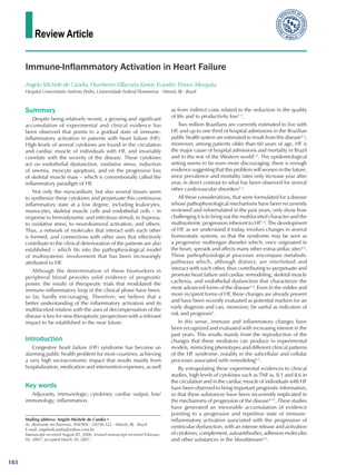
Fig. 1 - Hypothesis of myocardial cytokine production in HF.
INCREASED LV
WALL STRESS
PRODUCTION OF TNFα
DYSFUNCTION
DILATION
184
3. Review Article
Candia et al
Immune-inflammatory activation in heart failure
Arq Bras Cardiol 2007; 89(3) : 183-190
tissue hypoxia, and skeletal muscle apoptosis even more, and
this would serve as a stimulus for the systemic synthesis of
cytokines and oxidative stress, thus creating a vicious cycle of
perpetuation of the disease and promotion of cachexia that is
faithfully close to the model of multisystemic progression of
HF12,13
. These relations are shown in Figure 2.
Intestinal production: the endotoxin-cytokine hypothesis
When Anker et al14
observed a concomitant TNF-α and
soluble CD14 receptor (sCD14) elevation in the peripheral
blood of patients with advanced HF, they conceived a model
of cytokine activation in HF that would necessarily involve
the production of endotoxins derived from intestinal bacteria.
They analyzed the peripheral levels of these substances in 47
patients with advanced HF (29 of ischemic etiology) and in
17 healthy controls without structural heart disease or acute/
chronic inflammatory conditions14
. The levels of sCD14 were
increased in patients with HF, especially in cachectic ones,
and a strong correlation between the levels of sCD14 and of
TNF-α, sTNF-R1 and sTNF-R2 was observed – thus suggesting
that endotoxins (ETX) were somehow involved in the immune-
inflammatory activation of HF14
.
Thus, Anker et al14
hypothesized that, in patients with HF,
the interaction between CD14 receptors of immunocompetent
cells and ETX released by gram-negative bacteria (GNB),
possibly from the gastrointestinal tract, would result in the signal
transduction required for the production of IL6, TNF-α, and
other proinflammatory cytokines. This interaction is actually
documented as the most potent endogenous reaction capable
of releasing TNF-α4,14,15
. With the purpose of corroborating the
initial hypothesis, these investigators later demonstrated that
patients with HF and peripheral edema have higher levels of
sCD14, TNFα and ETX than those without edema, and the
latter present higher levels than healthy controls without the
disease15
. Moreover, after a mean 40-day treatment with
diuretics, a significant decrease in ETX levels and a tendency
of decrease in TNF-α levels were observed15
.
Anker et al14
suggest that their results support the hypothesis
Fig. 2 - Hypothesis of extramyocardial cytokine production in HF.
ENDOTHELIAL
DYSFUNCTION
OXIDATIVE STRESS
TISSUE ISCHEMIA/
HYPOXIA
INCREASED LV
WALL STRESS
SYSTEMIC
PRODUCTION OF TNFα
that the congestion of the intestinal wall (deemed present in
patients with systemic venous congestion) would induce a
proliferation of indigenous bacteria, with translocation and/or
ETX release in the bloodstream.
The indigenous microflora of the human gastrointestinal
tract comprises a colony of more than 1016
microorganisms
of more than 400 different species in a complex, yet stable,
symbiotic relationship with the cells from the mucosal layer16
.
This usually stable pattern of colonization may undergo
significant changes in several diseases, such as liver failure and
HF, in food deprivation states and in the critically ill patient in
general17
. There is a consensus, resulting mainly from clinical
studies conducted in intensive care settings, that translocation
of bacteria and/or their products through the intestine plays a
key role in starting or sustaining the clinical failure not only via
the systemic dissemination of bacteria, but also fundamentally
via the local production of proinflammatory factors in the
lymphoid tissue and in the bloodstream17,18
. How far we can
extrapolate these observations on intestinal translocation in
severely ill patients and experimental shock models to the
context of HF remains a source of endless debate.
Thus, in Anker et al’s hypothesis14
, after translocation there
would be immune activation via binding of circulating ETX to
CD14 receptors, with release of sCD14 into the bloodstream,
which can be detected in plasma15,18,19
. Despite the small
sample used, Anker et al’s studies14
represent, to date, the main
basis for what has been conventionally called the hypothesis
of bacterial-endotoxin-induced cytokine production, or
simply “endotoxin-cytokine hypothesis”18
. The cellular and
subcellular mechanisms of this hypothesis are shown in Figures
3A and 3B.
In an excellent pilot study, Conraads et al20
were pioneers
in evaluating the therapeutic potential of selective intestinal
decontamination (SID) on inflammatory activation in advanced
HF. The authors studied 10 patients with NYHA FC III and IV
undergoing a SID regimen with nonabsorbable antibiotics
(polymyxin B and tobramycin for eight weeks) and observed
that the treatment was able to eradicate intestinal GNB and
185
4. Review Article
Candia et al
Immune-inflammatory activation in heart failure
Arq Bras Cardiol 2007; 89(3) : 183-190
Fig. 3 - Cellular (A) and subcellular (B) mechanisms related to the immune-
inflammatory activation by serum ETX in HF. ETX - endotoxin; ECP - ETX carrier
protein; rTL-4 - tool-like receptor 4; rCD14 - CD14 receptor; sCD14 - soluble
portion of CD14 receptor.
MESENTERIC VENOUS CONGESTION
BACTERIAL OR ETX TRANSLOCATION
BLOOD VESSEL
ETX-ECP complex
MONOCYTE
ECP
INTERACTION
ETX-ECP - CD-14 receptor - TL-14 receptor
sCD-14 release to
circulation
TNF-α
production
MONOCYTE
ETX-EPC
to significantly reduce blood and fecal levels of ETX, as well as
serum levels of IL1, IL6 and TNF-α. With these results, these
authors open the possibility for further studies on the intestinal
bacteria approach to prove, with a higher power and better
statistical design, their initial favorable impressions. Our team
is conducting a randomized, double-blind, placebo-controlled
study in patients with advanced HF – who are those with
the highest level of inflammatory activity among those with
the disease, and we intend to confirm whether the judicious
eradication of ETX-producing enterobacteria is actually able
to reduce the degree of inflammatory activity in HF.
Main cytokines implied in inflammatory
activation of heart failure
Tumor necrosis factor alpha
The main TNF-α production site is the activated
macrophage, but many other cell types, such as fibroblasts,
neutrophils, endothelial cells, vascular smooth muscle, and
cardiomyocytes themselves have also already been implicated
as sources under stimuli of hypoxia, mechanical stress and
endotoxins5,6
. TNF-α is released as a stable homotrimer; under
this form, the molecule has an extremely short half-life of
approximately 30 minutes, and is measured in the serum by
using both immunoreactive and cytotoxic assays6
.
The biological actions of TNF-α are mediated by two
types of cell receptors located all over the body, known as
receptors 1 and 2 (TNF-R1 and TNF-R2)8-10
. Among them,
TNF-R1 is the most important because it mediates the main
cell effects, initiating a cascade of cytotoxic and apoptotic
responses6,10
. Experimental evidence suggests that it is via
TNF-R1 that cytokines also determine a direct stimulus on
fibroblast proliferation and synthesis of prostaglandin E2 and
superoxide dismutase, in addition to antiviral activity and
bacterial resistance8
.
Many of the clinical patterns observed in patients with HF
are reproduced in pre-clinical models by the direct action of
TNF-α4,6
. Several studies corroborate a very close experimental
relationship between TNF-α and cardiac muscle hypertrophy
and necrosis, in addition to extracellular membrane (ECM)
disarrangement and intramyocytic calcium mobilization6
.
TNF-α also induces increased baseline catabolism by
stimulating apoptosis, both in vivo and in vitro, by activating
the caspases pathway – which could contribute to a very
particular aspect of the HF syndrome, the one that has been
conventionally named cardiac cachexia12
. TNF-α also induces
myocyte necrosis via a cytotoxic mechanism related to the
complement pathway, to the induction of NO synthase, and
to the increased local production of free radicals6,12
.
Nuclear factor κ-β
NFκ-β is a transcription factor that regulates several
proinflammatory substances and may be activated by
multiple stimuli such as hypoxia, reactive O2
species, bacterial
endotoxins, cytokines and others6,21
. The myocardial tissue of
patients with HF of different etiologies exhibits overexpression
of this molecule and of the genes that it regulates, such as
that associated with the synthesis of TNF-α, NO, leukocyte-
adhesion molecules, and metalloproteinases21
. It has already
been demonstrated that many cell types such as endothelial
cells, macrophages, leukocytes, and cardiomyocytes in culture
synthesize NFκ-β in response to some cytokine stimuli in
a positive feedback pattern that sustains the inflammatory
activation per se21
.
Interleukin-6
IL-6 is a multifunctional cytokine which, thanks to the
accumulation of experimental and clinical evidence, is linked
to the progression of cardiac dysfunction because of the
worsening of functional limitation and rehospitalizations due
to decompensated disease13,22
. IL-6 promotes lymphocyte
proliferation and maturation, cardiomyocyte hypertrophy, and
stimulates the synthesis of caspases and of hepatic mediators
of the acute response, such as CRP6,22
. IL-6 also proved to
be able to induce in vitro muscle proteolysis, thus leading to
wasting and weight loss13
.
In a recent study, Plenz et al22
observed hearts of patients
with advanced HF at the moment of organ explantation for
transplantation and found a mRNA, IL-6 and IL-6 receptor
186
5. Review Article
Candia et al
Immune-inflammatory activation in heart failure
Arq Bras Cardiol 2007; 89(3) : 183-190
expression inside the myocardial tissue significantly greater
than that found in RV biopsy specimens of individuals with no
structural disease undergoing electrophysiological study22
.
AlthoughmyocardialproductionofIL-6mayseemsignificant,
an equally or even more significant peripheral synthesis is
suggested. A prognostic study showed that peripheral IL-6 levels
are not only significantly increased in femoral artery and veins
of patients with HF, but also an IL-6 spillover to the circulation
would occur, represented by the arteriovenous difference of
IL-6 for one determined patient, that increases with the severity
of the disease, and is independently and significantly correlated
with a worse prognosis13
.
Munger et al23
conducted a retrospective study with 78
patients with FC III and IV HF and found a significant increase
in IL-6 levels in patients with more advanced disease and
worse progression, regardless of the etiology23
. Lommi et al24
observed that IL-6 levels are directly related to filling pressures,
and inversely related to cardiac output, thus apparently
reflecting a hemodynamic deterioration.
Inanintriguingstudy,Kelletal25
analyzedplasmaconcentrations
of IL-1, IL-6, IL-10, IL-12, TNF-α, and s-CD-14 in 91 patients with
NYHAFCIIIHF,andEF<40%.Aftera22±13-monthfollow-up,
and using the multivariate regression analysis, IL-6 proved to be
the prognostic marker of survival with the greatest independent
predictive power in one year. Moreover, the combination of EF
and VO2
values increased the risk prediction.
Interleukin-1
Allmammalianspeciesareabletoexpresstwogenesassociated
with IL-1 synthesis in monocytes; in humans, most of the body
cell types can produce IL-1 under optimal conditions6
.
The mechanism through which IL-1 triggers its
proinflammatory effects seems to involve prostaglandin synthesis
and, perhaps, a direct action on beta-receptor uncoupling6
.
Thaik et al26
studied rat cardiomyocyte cultures and observed
that the IL-1β stimulus is able to cause hypertrophy via a NO-
independent mechanism, with induction of fetal gene synthesis
and downregulation of genes that regulate intramyocytic
calcium dynamics. Francis et al27
attributed a negative inotropic
effect to IL-1 that could depress myocardial contractility by
directly stimulating NO synthesis.
IL-1 soluble receptors have been considered the most
sensitive and reliable markers of activation of the IL-1 loop,
and are more strongly correlated with the severity of several
diseases such as HF or sepsis, where peripheral levels of IL-1
are usually low, which makes them difficult to be detected
through the currently available assays6
. Recently, peripheral
detection of a soluble form of IL-1 membrane receptor called
sT2 has been reported as predictive of events in experimental
models of HF and MI28
.
C reactive protein (CRP)
Despite all the recent technological armamentarium, liver
release of CRP seems to be a pretty sensitive, specific indicator
with a high prognostic correlation in different degrees of
inflammatory states – and this results from the CRP property of
interfering with practically all stages of the immunoinflammatory
response, once released in the circulation29
.
Several stimuli seem to be involved in the direct regulation
of the hepatic synthesis of CRP, however IL-6 seems to be
the major one22,29
. Other cytokines such as IL-1β and TNF-α
also influence directly the in vitro release of CRP, albeit to a
lower extent29
.
Sato et al30
observed high CRP levels (2.6 ± 0.8 mg/dl)
in patients with decompensated HF free of associated
ischemic or infectious events; these levels were significantly
reduced after resolution of the symptoms related to the acute
manifestations. Other authors reached similar conclusions in
groups of patients with decompensated HF – recently, Mueller
et al31
and Lamblin et al32
confirmed the prognostic impact
of CRP determination in patients with decompensated HF by
randomizing a group of almost 800 patients and confirming
a strong and independent association between serum levels
of CRP and mortality.
Our team also analyzed the prognostic value of serum levels
of CRP in patients with decompensated HF: after a mean one-
year follow-up of 119 patients with NYHA functional class III
or IV, the best cut-off point determined for CRP was 3 mg/dl,
a value close to that found by Sato et al30
in a similar group of
patients. Thus, CRP ≥ 3 mg/dl suggests a population of patients
with HF at a higher risk – whose mortality reached 49% by
the end of 12 months in our case series. These CRP levels
are much higher than those found in primary and secondary
prevention of ischemic disease. Among all the variables tested
in the study, CRP was the one that best correlated with survival
time, using Cox regression test.
Infact,whenCRPiselevatedinpatientswithdecompensated
HF, it seems to be a low-cost, easily available independent
predictor of survival; what still remains unclear is whether
CRP is elevated as a mere passive marker of the process,
or whether it actually works as a direct effector in the
inflammatory component associated with both the instability
of atherosclerosis and the endothelial dysfunction that
characterizes the progression of HF32,33
.
Uric acid
Serum uric acid is significantly elevated in patients with
HF in relation to individuals without the disease, regardless of
renal function or diuretic treatment, and is directly correlated
with the functional limitation and survival, mainly in cachectic
patients34,35
. Hyperuricemia is a quite reliable marker of
inflammatory cytokine activation, of endothelial dysfunction,
and of oxidative stress; in fact, uric acid synthesis catalyzed by
xanthine-oxidase also generates superoxide radicals, hydroxyl,
and hydrogen peroxide35
.
In addition to the oxidative potential, uric acid could also
induce several direct harmful effects, such as the stimulation
of smooth muscle proliferation, renin synthesis, reduction
of intramyocardial NO synthesis, and reduction of calcium
release by the sarcoplasmic reticulum in cardiomyocytes34
.
Anker et al34
studied 294 patients hospitalized for
decompensated NYHA FC II and III HF, and observed that the
best value predictive of mortality for uric acid was 9.50 mg/dl
at 12 and 18 months of follow-up; uric acid levels above this
value were related to a survival of 52% and 36% at 12 and
18 months, respectively.
187
6. Review Article
Candia et al
Immune-inflammatory activation in heart failure
Arq Bras Cardiol 2007; 89(3) : 183-190
The group of patients with uric acid ≤ 9.50 mg/dl, in
turn, had a survival of 92% and 86% at 12 and 18 months,
respectively.
Regardless of the theoretical considerations that emerge
from experimental analyses, there seems to be clear clinical
evidence that the oxidative stress is the link between uric
acid and the different alterations related to the progression
of HF, such as endothelial dysfunction, cytokine activation,
and cardiomyocyte apoptosis34,35
. It is still relatively early to
know whether hyperuricemia and the oxidative stress that it
triggers are actually key actors in this process. A large study
with allopurinol is being conducted and shall bring many
explanations in this sense36
.
Immune-inflammatory activation in Chagas heart disease
Similarly to what is seen in idiopathic cardiomyopathy,
plasma levels of TNF-α and IL-10 are only mildly elevated in the
indeterminate phase of Chagas heart disease, and increasing
levels are found in individuals with overt heart disease. Despite
the broad spectrum of myocardial involvement, alterations
in the endothelial function, in the oxidative balance, and in
the homeostasis of the immune system are already present
in asymptomatic individuals, and become marked in patients
with severe ventricular dysfunction37,38
.
In acute myocarditis caused by Trypanossoma cruzi, the
major mechanism implicated seems to be a direct autoimmune
aggression against myosin chains, as a result of mimicry of
parasite epitopes39
. This molecular mimicry is similar to
that seen, for instance, in the cross-reaction between beta-
hemolytic streptococci and cardiac tissues in patients with
rheumatic heart disease40
. In chronic Chagas heart disease,
there are evidences of a persistent aberrant cytotoxic immune
response to T. cruzi antigens that is related to the progression
of the disease, in a mechanism that is not seen in idiopathic
or ischemic dilated cardiomyopathy41
. In fact, the persistence
of the parasite that occurs in the indeterminate and advanced
phases of the disease suggests a deficiency in the suppression
mediated by T cells, in lymphocytic polyclonal activation,
and in the mechanisms of apoptotic clearance that may also
contribute directly to myocardial aggression41
.
However, the reason why only a minority of individuals
with the latent form of the disease will progress to the
spectrum of cardiac involvement still remains unclear; the
mechanisms associated with the relations of tropism between
the various strains of T. cruzi and the cardiac tissue, the
different and unforeseeable participations of T-cell-mediated
response, and polymorphisms in cytokine synthesis in
those patients are potential sources of studies for further
clarification of these questions39,41
.
Clinical evidences
Levine et al7
were the first ones to report that serum levels of
TNF-α were much higher in patients with HF (115 ± 25 U/ml)
than in healthy controls (9 ± 3 U/ml, p < 0.001), and that
individuals with higher levels of TNF-α were the most cachectic
and those with the most advanced stage of the disease. Soon
afterwards, Torre-Amione et al8
examined hearts explanted
from cardiac transplantation recipients and established that
the failing heart is able to produce cytokines, as previously
explained. Ever since, many other authors confirmed the
significant and independent correlation between peripheral
levels of TNF-α and its soluble receptors with a worse prognosis
and mortality both in the short and in the long term in patients
with advanced HF42-44
.
Rauchhaus et al43
followed 152 patients with HF for at
least 12 months and observed that high serum levels of
TNF-α, sTNF-R1, sTNF-R2, sCD14 and IL-6 proved to be
independent predictors of a poor prognosis in the long term
for all functional classes of HF, from the milder to the most
severe forms of the disease. In the multivariate analysis,
the determination of sTNF-R1 levels proved to be the most
powerful tool predictive of survival, regardless of the NYHA FC,
peak VO2
, EF or presence of cachexia.In this study, patients in
the highest quartile for sTNF-R1 had a risk of all-cause mortality
that was 12-fold higher than those in the lowest quartile of
the study. In another analysis, the authors observed that all
main results, including those on the particular importance of
sTNF-R1 as an independent risk marker, remained valid even
if cachectic patients, therefore with a more advanced disease,
were excluded from the sample (28.9% of the total)43
.
The largest study to assess the immune-inflammatory
profile in patients with HF was conducted by Deswal et al44
,
who studied almost 1200 patients with NYHA FC III and IV
from the placebo group of a multicenter clinical trial. The
levels of TNF-α, IL-6, and their respective soluble receptors
were determined prior to randomization and were considered
significantly higher in patients with NYHA FC IV than in those
with FC III. Cox univariate analysis showed that serum levels of
TNF-α, IL6, sTNF-R1 and sTNF-R2 can be used as independent
risk predictors in patients with HF; when considered together
and associated with other variables, only sTNF-R2, NYHA FC
and EF remained as significant predictors of survival44
.
Recent attempts to modulate the TNF-α loop randomized
more than 1500 patients with NYHA FC III (70%) and IV
HF and EF < 30% in the United States and Europe. All
together, the ATTACH, RECOVER and RENASSAINCE studies
conducted at the same time, albeit by different groups,
had the same primary objective of clinical and quality of
life improvement, and left survival analyses to a secondary
assessment. Unfortunately, the three groups of researchers
found disappointing negative results in all analyses, thus
limiting perspectives of further therapeutic trials with this
objective45,46
. However, we hypothesize that there are several
explanations for this fact.
The benefit of patients who could have profited more
from the anti-TNFα therapy (for instance, patients with a
higher immune activation, like those with cardiac cachexia
or disease decompensation) may have been diluted when
patients with less severe HF were included in the recruitment
(a hypothesis corroborated by the fact that subgroup analyses
were not provided by the authors of the three trials). Very
high doses of the drugs, especially in the case of infliximab,
are also supposed to have determined prohibitive serum
levels – a limitation resulting from the sudden transition of
phase I studies to the studies mentioned above, with a high
number of patients, without establishing a solid safety profile
in the smaller studies. Another possible explanation may be
188
7. Review Article
Candia et al
Immune-inflammatory activation in heart failure
Arq Bras Cardiol 2007; 89(3) : 183-190
References
1. HuntSA,AbrahamWT,ChinMH,FeldmanAM,FrancisGS,GaniatsTG,etal.
Guideline update for the diagnosis and management of chronic heart failure
in the adult. J Am Coll Cardiol. 2005; 46 (6): e1-e82.
2. Ministério da Saúde. Fundação Nacional da Saúde. DATASUS. Sistema de
informações sobre Mortalidade, 1979-1997. Dados de declaração de óbito.
[citado 2006 janeiro 15]. Disponível em: <http://www.datasus.gov.br>.
3. Albanesi Filho FM. A insuficiência cardíaca no Brasil. Arq Bras Cardiol. 1998;
71: 561-2.
4. Torre-Amione G. Immune activation in chronic heart failure. Am J Cardiol.
2005; 95 (supl.): 3C-8C.
5. Mann DL. Inflammatory mediators and the failing heart: past, present and
the foreseeable future. Circ Res. 2002; 91: 988-98.
6. AdamopoulosS,ParissisJT,KremastinosDT.Aglossaryofcirculatingcytokines
in chronic heart failure. Eur J Heart Fail. 2001; 3: 517-26.
7. Levine B, Kalman J, Mayer L, Fillit H, Packer M. Elevated circulating levels of
tumoral necrosis factor in severe chronic heart failure. N Engl J Med. 1990;
323: 236-41.
8. Torre-Amione G, Kapadia SR, Lee J, Bies R, Lebovitz R, Mann DL. Expression
and functional significance of TNF receptors in human myocardium.
Circulation. 1995; 92: 1487-93.
9. Torre-Amione G, Kapadia S, Lee J, Durand GB, Young JB, Mann DL. Tumoral
necrosis factor alpha and tumoral necrosis factor alpha receptors in the failing
human heart. Circulation. 1996; 93: 704-11.
10. Ferrari R, Bachetti T, Confortini R, Opasich C, Febo O, Corti A, et al. Tumor
necrosis factor soluble receptors in patients with various degrees of chronic
heart failure. Circulation. 1995; 92: 1479-86.
11. HasperD,HummelM,KleberFX,VolkHD.Systemicinflammationinpatients
with heart failure. Eur Heart J. 1998; 19(5): 761-5.
12. Anker SD, Sharma R. The syndrome of cardiac cachexia. Int J Cardiol. 2002;
85: 51-66.
13. Tsutamoto T, Hisanaga T, Wada A, Maeda K, Fukai D, Mabuchi N, et al.
Interleukin-6 spillover in the peripheral circulation increases with the severity
of heart failure. J Am Coll Cardiol. 1998; 3: 391-8.
14. Anker S, Egerer K, Volk H, Kox W, Poole-Wilson P, Coats A. Elevated soluble
receptors and altered cytokines in chronic heart failure. Am J Cardiol. 1997;
79: 1426-30.
15. Niebauer J, Volk H, Kemp M, Rauchhaus M, Coats A, Anker S. Endotoxin
and immune activation in chronic heart failure: a prospective cohort study.
Lancet. 1999; 353: 1838-42.
16. Marshall JC. Gastrointestinal flora and its alterations in critical illness. Curr
Opin Crit Care. 1999; 5: 119-25.
17. DeWitt RC, Kudsk KA. The gut’s role in metabolism, mucosal barrier function
and septic shock. Infec Dis Clin North Am. 1999; 13: 465-81.
18. Krack A, Sharma R, Figulla HR, Anker SD. The importance of the
gastrointestinal system in the pathogenesis of heart failure. Eur Heart J.
2005; 26 (22): 2368-74.
19. Brunkhorst FM, Clark AL, Forycki ZF, Anker SD. Pyrexia, procalcitonin,
immune activation and survival in cardiogenic shock: the potential
importance of bacterial translocation. Int J Cardiol. 1999; 72: 3-10.
20. Conraads VM, Jorens PG, Ieven MM, Rauchhaus M, Anker SD, Vrints CJ, et
al. Selective intestinal decontamination in advanced chronic heart failure: a
pilot trial. Eur Heart J. 2004; 6: 483-91.
21. Valen G, Yan ZQ, Hansson GK. Nuclear factor kappa B and the heart. J Am
Coll Cardiol. 2001; 38: 307-14.
22. Plenz G, Eschert H, Erren M, Wichter T, Bohm M, Song ZF, et al. Activation
of the cardiac interleukin-6 system in advanced heart failure. Eur J Heart Fail.
2001; 3: 415-21.
23. Munger MA, Johnson B, Amber IJ, Gilbert EM. Circulating concentrations
of proinflammatory cytokines in mild to moderate heart failure secondary
to ischemic or idiopathic dilated cardiomiopathy. Am J Cardiol. 1996; 77:
723-7.
the loss of TNFα cytotoxic action on viruses and of its effects
in amplifying leukocyte migration and margination, which
may have worsened the myocardial lesion in those patients.
However, although we hypothesize these two reasons, it
seems clear that the immune activation in HF is broad and
widespread, so that blocking a specific pathway is not enough
to abolish all the adverse effects of the complex cascade – quite
the contrary, TNFα in merely a component of a network
of molecules that stimulate each other, repress each other,
potentiate each other, and any attempt to modulate one
single element of this network seems simplistic and, perhaps,
doomed to failure45,46
.
Conclusions
Many studies in humans have confirmed the hypothesis
that peripheral levels of cytokines may be a new and potential
indicator of prognosis in patients with HF in the short, mid, and
long term, from milder to more severe forms of the disease,
especially in the latter.
The real contribution of cytokines on the pathophysiology
of HF still remains unclear – whether they are mere passive
markers or whether they actually work as effectors in the
progression of the disease. The confirmation of the prognostic
value of the immune-inflammatory activation in HF does not
assume a definitive cause-effect relationship – although many
experimental evidences support the harmful effects of cytokines,
especially of TNF-α, on the cardiovascular system.
The progressive character of HF and its growing prevalence
suggest, however, that all therapeutic advances provided
by several clinical trials on neurohumoral modulation drugs
do not yet represent the gold standard we can offer to our
patients. In fact, the growing morbidity and mortality rates
of HF in the Western world suggest that there are important
pathophysiological mechanisms related to the progression of the
disease that remain active and are under little or no influence
of the recently incorporated therapeutic modalities.
We suppose that the immune-inflammatory activation may
represent this therapeutic opportunity; trying to understand
it in its completeness of an intricate cellular and subcellular
orchestration is the challenge we intend to face, as a means
of contributing to reverse the epidemiological character that
makes HF a malignant and extremely defying disease which will
become the major cause of death in the Western world in the
coming decades, if we do not take direct measures to halt it.
189
8. Review Article
Candia et al
Immune-inflammatory activation in heart failure
Arq Bras Cardiol 2007; 89(3) : 183-190
24. Lommi J, Pulkki K, Koshinen P, Naveri H, Leinonen H, Harkonen M, et al.
Haemodynamic, neuroendocrine and metabolic correlates of circulating
cytokine concentrations in congestive heart failure. Eur Heart J. 1997; 18:
1620-5.
25. Kell R, Haunstetter A, Dengler TJ, Haass M. Do cytokines enable risk
stratification to be improved in NYHA class III patients? A comparison with
other potential predictors of prognosis. Eur Heart J. 2002; 23: 70-8.
26. Thaik CM, Colucci WS. Interleukin-1β modulates the growth and phenotype
of neonatal rat cardiac myocytes. J Clin Invest. 1995; 96: 1093-9.
27. Francis SE, Holden H, Holt CM, Duff GW. Interleukin-1 in myocardium and
coronary arteries of patients with dilated cardiomyopathy. J Moll Cell Cardiol.
1998; 30: 215-23.
28. Shimpo M, Morrow DA, Weinberg EO, Sabatine SA, Murphy SA, Antman
EM. Serum levels of the interleukin-1 receptor family member ST2 predict
mortality and clinical outcome in acute myocardial infarction. Circulation.
2004; 109: 2186-90.
29. Gabay C, Kushner I. Acute phase proteins and other systemic responses to
inflammation. N Engl J Med. 1999; 340: 448-53.
30. Sato Y, Takatsu Y, Yamada T, Kataoka K, Taniguchi R, Matsumori A, et al.
Serial circulation concentrations of C-reactive protein, interleucin-4 and
interleucin-6 in patients with acute left heart descompensation. Clin Cardiol.
1999; 22: 811-3.
31. Mueller C, Killian KL, Christ A, La Rocca HP, Perruchoud AP. Inflammation
and long-term mortality in acute congestive heart failure. Am Heart J. 2006;
151: 845-50.
32. Lamblin N, Mouquet F, Hennache B, Dagorn J, Susen S, Bauters C, et al. High-
sensitivityCreactiveprotein:potentialadjunctforriskstratificationinpatients
with stable congestive heart failure. Eur Heart J. 2005; 26: 2245-50.
33. Silva, ACM. Proteína C reativa: um novo marcador prognóstico em
portadores de insuficiência cardíaca descompensada na sala de Emergência.
[dissertação]. Niterói:. Universidade Federal Fluminense; 2000.
34. Anker SD, Doehner W, Rauchhaus M, Sharma R, Francis D, Knosalla C, et
al. Uric acid and survival in chronic heart failure: validation and application
in metabolic, functional, and hemodynamic staging. Circulation. 2003;
107(15): 1991-7.
35. Cicoira M, Zanolla L, Rossi A, Golia G, Franceschini L, Brighetti G, et al.
Elevated serum uric acid levels are associated with diastolic dysfunction in
patients with dilated cardiomyopathy. Am Heart J. 2002; 143 (6): 1107-11.
36. Freudenberger RS, Schwarz RP, Brown J, Moore A, Mann D, Givertz MM, et al.
Rationale,designandorganizationofanefficacyandsafetystudyofoxypurinol
added to standard therapy in patients with NYHA class III-IV congestive heart
failure. Expert Opin Investig Drugs. 2004; 13 (11): 1509-16.
37.PlentzRDM,IrigoyenMC,MullerAS,MadyC,IanniBM,Consolim-Colombo
F. Disfunção endotelial venosa em pacientes com doença de Chagas sem
insuficiência cardíaca. Arq Bras Cardiol. 2006; 86: 466-71.
38. Ferreira RC, Ianni BM, Abel LC, Buck P, Mady C, Cunha-Neto E. Increased
plasma levels fo tumor necrosis factor alpha in symptomatic, indeterminate
and Chagas disease cardiomyopathy patients. Mem Inst Oswaldo Cruz.
2003; 98 (3): 407-11.
39. Leon JS, Daniels MD, Toriello KM, Wang K. A cardiac myosin-specific
autoimmune response is induced by immunization with Trypanossoma cruzi
proteins. Infec Immun. 2004; 72: 4271-7.
40. Guilherme L, Cury P, Demarchi LM, Coelho V, Abel L, Lopez AP, et al.
Rheumatic heart disease: proinflammatory cytokines play a role in the
progression and maintenance of valvular lesions. Am J Pathol. 2004; 165:
1583-91.
41. Dos Reis GA, De Lima CGF, Nunes MP, Lopes MF. The importance of aberrant
T-cell responses in Chagas disease. Trends Parasitol. 2005; 21: 237-43.
42.Torre-AmioneG,KapadiaS,BenedictC,OralH,MannDL.Pro-inflammatory
cytokine levels in patients with depressed left-ventricular ejection fraction: a
report from the Studies On Left-Ventricular Dysfunction (SOLVD). J Am Coll
Cardiol. 1996; 76: 723-7.
43. Rauchhaus M, Doehner W, Niebauer J, Coats AJS, Volk HD, Anker SD, et
al. Plasma cytokine parameters and mortality in patients with chronic heart
failure. Circulation. 2000; 102: 3060-7.
44. Deswal A, Petersen NJ, Feldman AM, Young JB, White BG, Mann DL.
Cytokines and cytokine receptors in advanced heart failure - an analysis of
the cytokine database from the Vesnarinone Trial (VEST). Circulation. 2001;
103: 2055-9.
45. McMurray J, Mann DL. Effects of cytokine antagonism with etanercept on
morbidity and mortality in chronic heart failure: results of the randomized
etanercept world-wide evaluation (RENEWAL trial). In:. Annual Meeting
European Society of Cardiology; Oslo / Norway; 2002 June 10. [oral
presentation].
46. Chung ES, Packer M, Lo KH, Fasanmade AA, Willerson JT. Randomized,
double-blind, placebo-controlled, pilot trial of infliximab, a chimeric
monoclonal antibody to tumor necrosis factor alpha, in patients with
moderate to severe heart failure: results of the Anti-TNF Therapy Against
Congestive Heart failure (ATTACH) trial. Circulation. 2003; 107: 3133-40.
190