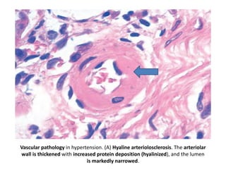
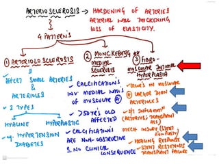
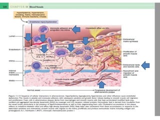
Vascular pathology inhypertension. (A) Hyaline arteriolosclerosis. The arteriolar
wall is thickened with increased protein deposition (hyalinized), and the lumen
is markedly narrowed.
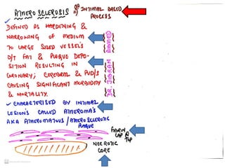
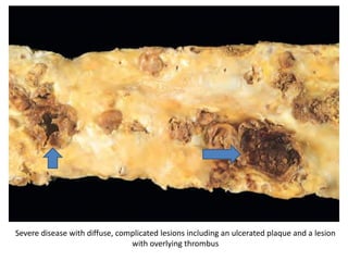
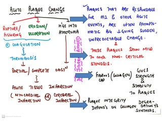
Severe disease withdiffuse, complicated lesions including an ulcerated plaque and a lesion
with overlying thrombus
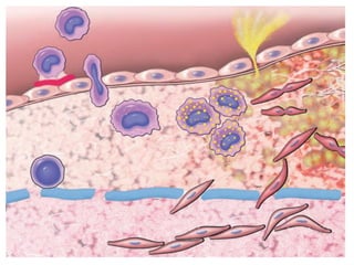
42.
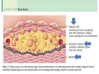
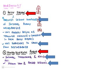
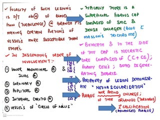
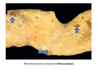
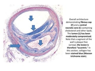
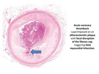
Overall architecture
demonstrating fibrouscap
(F) and a central
necrotic core (C) containing
cholesterol and other lipids.
The lumen (L) has been
moderately compromised.
Note that a segment of the
wall is plaque-free
(arrow); the lesion is
therefore “eccentric.” In
this section, collagen has
been stained blue (Masson
trichrome stain).
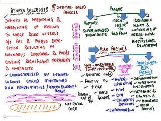
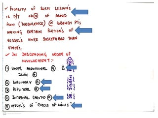
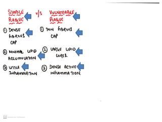
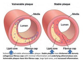
Vulnerable and stableatherosclerotic plaque. Stable plaques have thickened and densely
collagenous fibrous caps with minimal inflammation and underlying atheromatous core.
Vulnerable plaques have thin fibrous caps, large lipid cores, and increased inflammation.