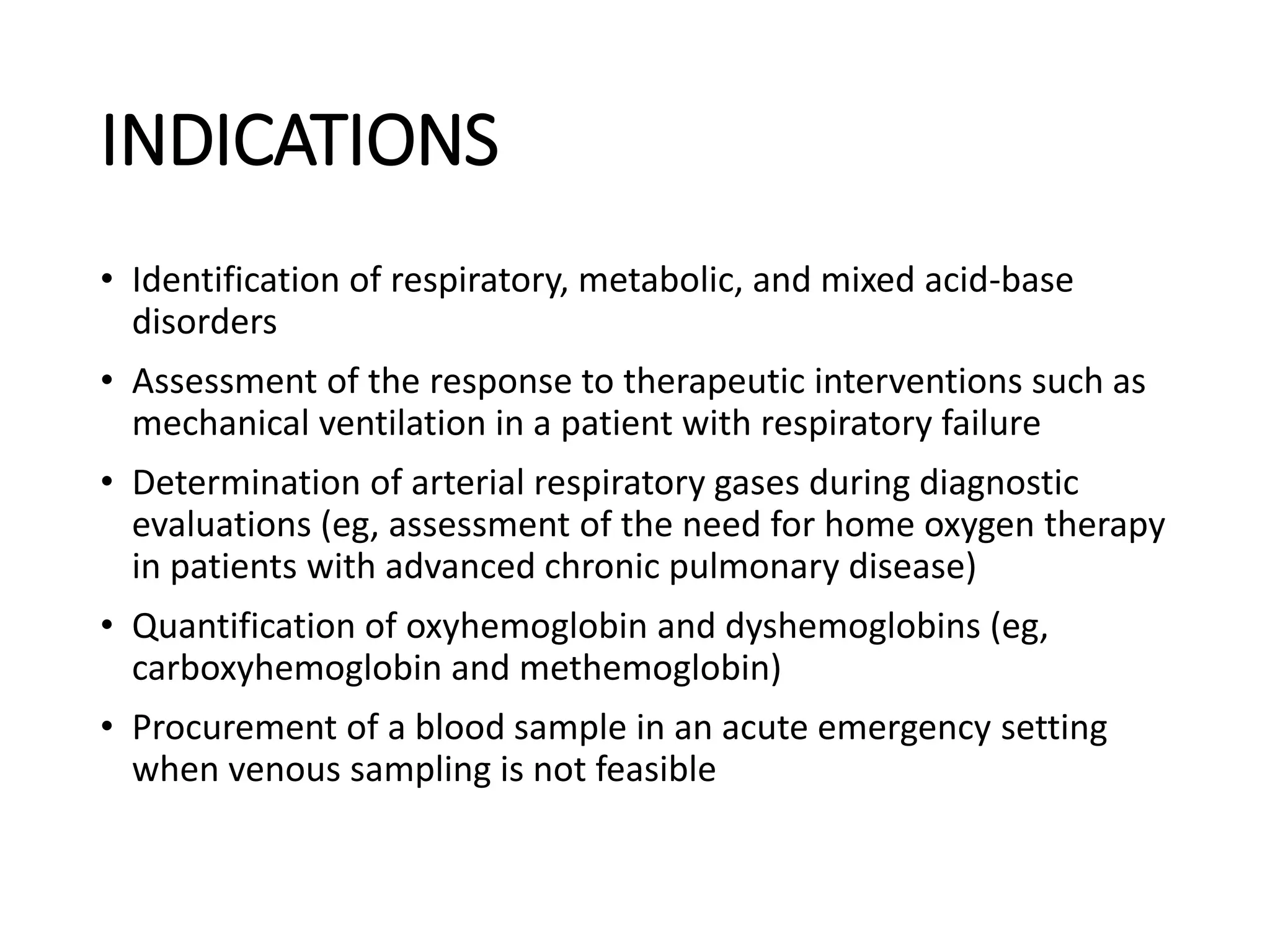
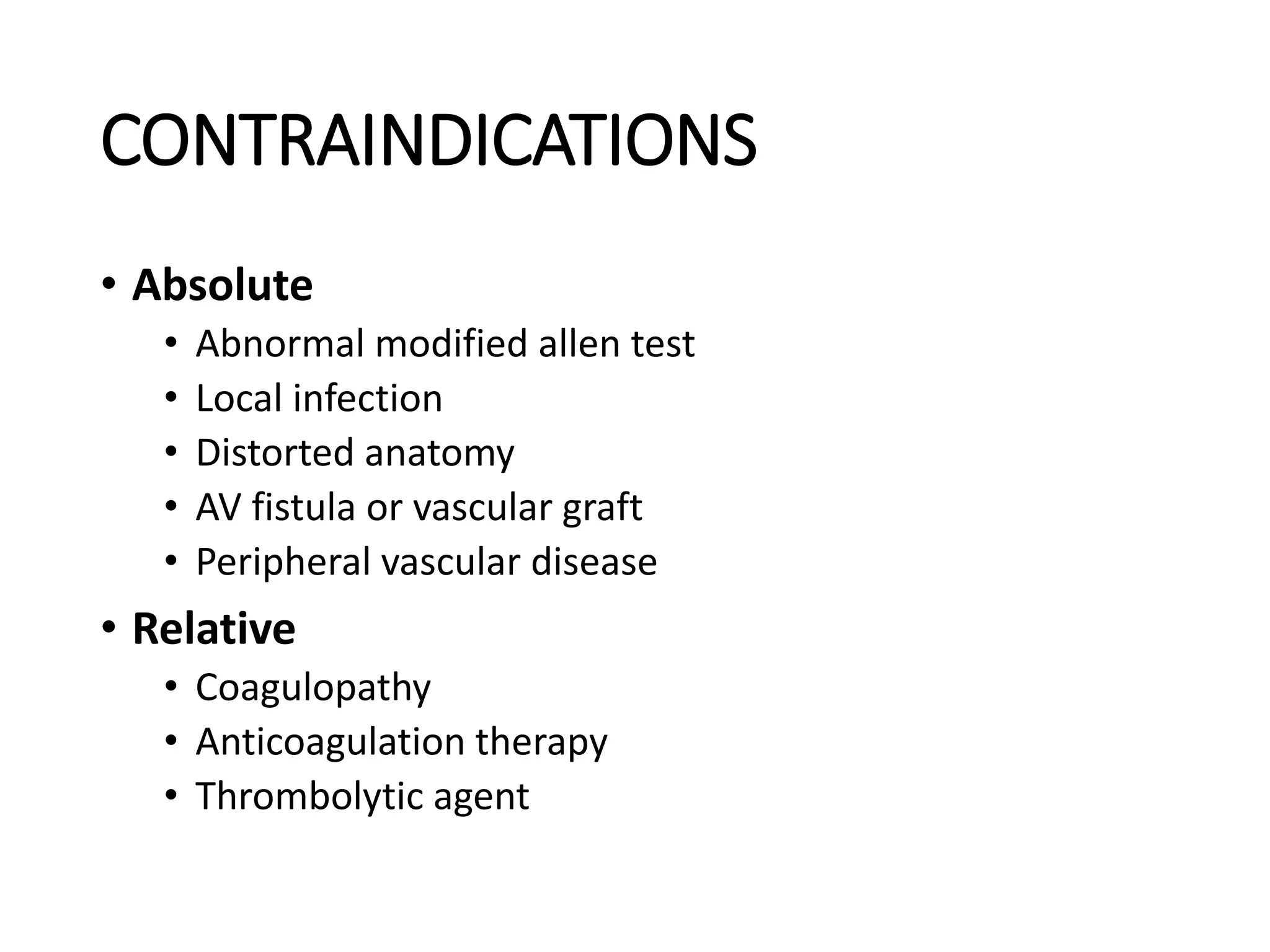
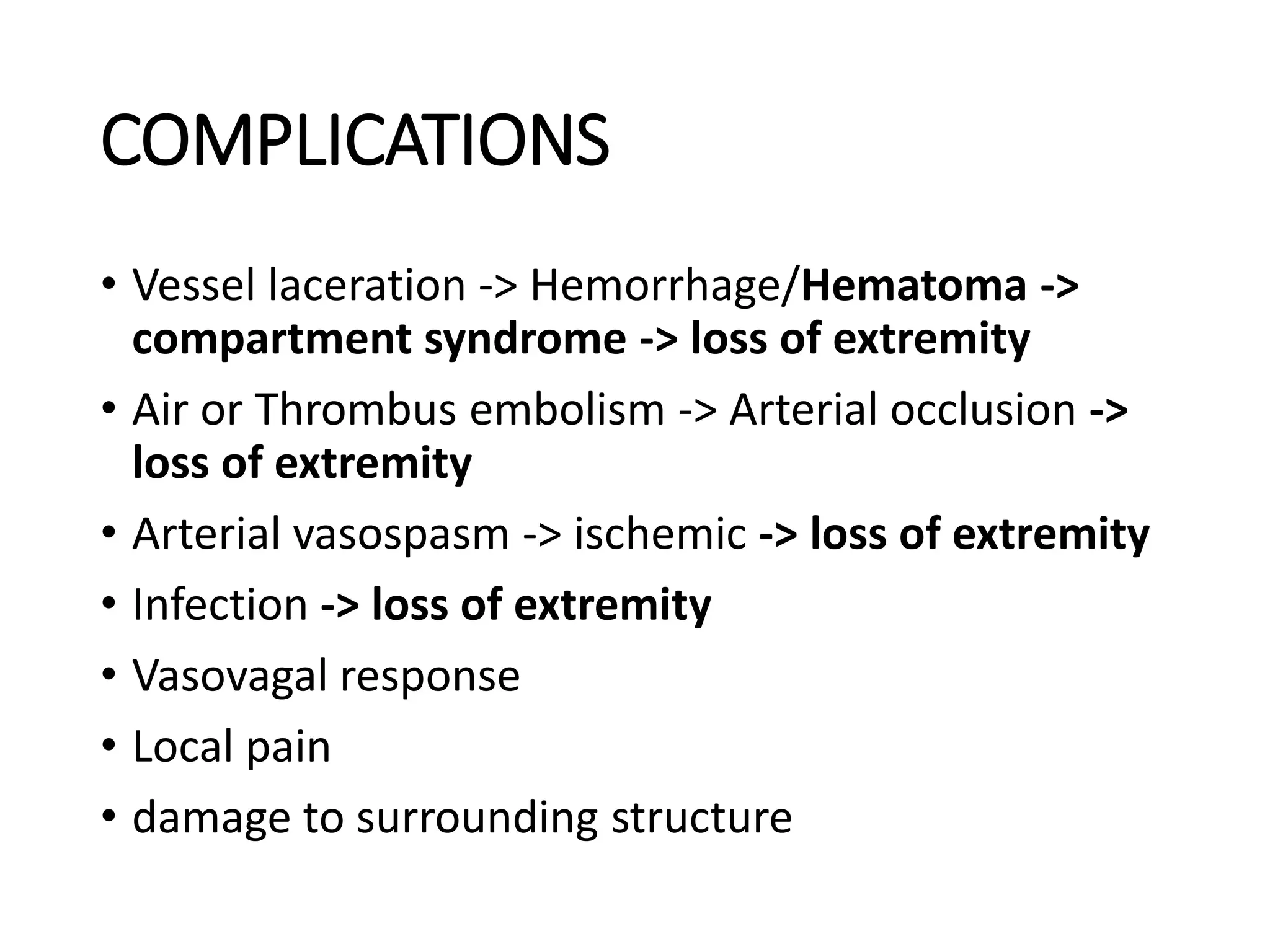
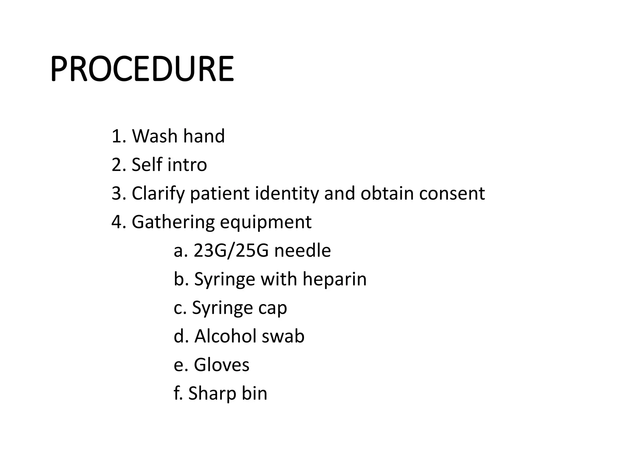
Arterial Blood Gas
Sampling
prepared by
Voon YW
Daaniyal
HKL Houseman teaching
INDICATIONS
• Identification of respiratory, metabolic, and mixed acid-base
disorders
• Assessment of the response to therapeutic interventions such as
mechanical ventilation in a patient with respiratory failure
• Determination of arterial respiratory gases during diagnostic
evaluations (eg, assessment of the need for home oxygen therapy
in patients with advanced chronic pulmonary disease)
• Quantification of oxyhemoglobin and dyshemoglobins (eg,
carboxyhemoglobin and methemoglobin)
• Procurement of a blood sample in an acute emergency setting
when venous sampling is not feasible
CONTRAINDICATIONS
• Absolute
• Abnormal modified allen test
• Local infection
• Distorted anatomy
• AV fistula or vascular graft
• Peripheral vascular disease
• Relative
• Coagulopathy
• Anticoagulation therapy
• Thrombolytic agent
COMPLICATIONS
• Vessel laceration -> Hemorrhage/Hematoma ->
compartment syndrome -> loss of extremity
• Air or Thrombus embolism -> Arterial occlusion ->
loss of extremity
• Arterial vasospasm -> ischemic -> loss of extremity
• Infection -> loss of extremity
• Vasovagal response
• Local pain
• damage to surrounding structure
PROCEDURE
1. Wash hand
2. Self intro
3. Clarify patient identity and obtain consent
4. Gathering equipment
a. 23G/25G needle
b. Syringe with heparin
c. Syringe cap
d. Alcohol swab
e. Gloves
f. Sharp bin
5. Selection of puncture
site
Radial Artery
• medial to the
radial styloid
process
• lateral to the
flexor carpi
radialis
tendon,
• 2-3 cm
proximal to
the ventral
surface of the
wrist crease
Radial artery
• Superficial & easily accessible
• Has collateral *can do allen test to ensure there's
secure of blood flow, check coagulation status also
• puncture at 30-45 degree angle
• minimizes trauma to the vessel and allows smooth-
muscle fibers to occlude the puncture site after the
procedure
Femoral Artery
• midline between the
symphysis pubis and the
anterior superior iliac
crest,
• 2-4 cm distal to the
inguinal ligament.
• medial to the femoral
nerve and lateral to the
femoral vein
• assess the distal pulses of
the lower limb prior
• 60-90º angle
• recommended only in
inpatient setting
Brachial Artery
• medial epicondyle of the
humerus and the tendon of
the biceps brachii in the
antecubital fossa.
• in the groove between the
biceps and triceps tendons
• The basilic vein and the
brachial nerve are located in
close proximity
• approach at 45-60º angle
• depper, harder to identify and
achieving hemostassis
• small caliber vessel, does not
have extensive collateral
circulation
• a/w median nerve damage
Others
• Dorsal Pedis
Artery
• Ulnar Artery
• posterior tibial
Artery
6. Compress for 5
minutes and monitor for
complication
*repeated arterial blood sampling increases risk of
complication, alternate puncture site should be considered
Thank you

Arterial Blood Gas Sampling

  • 1.
    Arterial Blood Gas Sampling preparedby Voon YW Daaniyal HKL Houseman teaching
  • 2.
    INDICATIONS • Identification ofrespiratory, metabolic, and mixed acid-base disorders • Assessment of the response to therapeutic interventions such as mechanical ventilation in a patient with respiratory failure • Determination of arterial respiratory gases during diagnostic evaluations (eg, assessment of the need for home oxygen therapy in patients with advanced chronic pulmonary disease) • Quantification of oxyhemoglobin and dyshemoglobins (eg, carboxyhemoglobin and methemoglobin) • Procurement of a blood sample in an acute emergency setting when venous sampling is not feasible
  • 3.
    CONTRAINDICATIONS • Absolute • Abnormalmodified allen test • Local infection • Distorted anatomy • AV fistula or vascular graft • Peripheral vascular disease • Relative • Coagulopathy • Anticoagulation therapy • Thrombolytic agent
  • 4.
    COMPLICATIONS • Vessel laceration-> Hemorrhage/Hematoma -> compartment syndrome -> loss of extremity • Air or Thrombus embolism -> Arterial occlusion -> loss of extremity • Arterial vasospasm -> ischemic -> loss of extremity • Infection -> loss of extremity • Vasovagal response • Local pain • damage to surrounding structure
  • 5.
    PROCEDURE 1. Wash hand 2.Self intro 3. Clarify patient identity and obtain consent 4. Gathering equipment a. 23G/25G needle b. Syringe with heparin c. Syringe cap d. Alcohol swab e. Gloves f. Sharp bin
  • 6.
    5. Selection ofpuncture site
  • 7.
    Radial Artery • medialto the radial styloid process • lateral to the flexor carpi radialis tendon, • 2-3 cm proximal to the ventral surface of the wrist crease
  • 8.
    Radial artery • Superficial& easily accessible • Has collateral *can do allen test to ensure there's secure of blood flow, check coagulation status also • puncture at 30-45 degree angle • minimizes trauma to the vessel and allows smooth- muscle fibers to occlude the puncture site after the procedure
  • 9.
    Femoral Artery • midlinebetween the symphysis pubis and the anterior superior iliac crest, • 2-4 cm distal to the inguinal ligament. • medial to the femoral nerve and lateral to the femoral vein • assess the distal pulses of the lower limb prior • 60-90º angle • recommended only in inpatient setting
  • 10.
    Brachial Artery • medialepicondyle of the humerus and the tendon of the biceps brachii in the antecubital fossa. • in the groove between the biceps and triceps tendons • The basilic vein and the brachial nerve are located in close proximity • approach at 45-60º angle • depper, harder to identify and achieving hemostassis • small caliber vessel, does not have extensive collateral circulation • a/w median nerve damage
  • 11.
    Others • Dorsal Pedis Artery •Ulnar Artery • posterior tibial Artery
  • 12.
    6. Compress for5 minutes and monitor for complication *repeated arterial blood sampling increases risk of complication, alternate puncture site should be considered
  • 13.