This study found that Sfrp5, which encodes secreted frizzled-related protein 5, is downregulated in the pancreatic islets of rats fed a cafeteria diet (obese model) and in pancreatic islets of human obese patients. The researchers demonstrated that silencing Sfrp5 increases beta cell proliferation in rats, which correlates with activation of the Wnt signaling pathway and increased proliferation markers. They also showed that expression of Sfrp5 in beta cells is modulated by IGF binding protein 3 secreted from visceral adipose tissue. Together, these findings reveal that Sfrp5 and the Wnt signaling pathway play an important role in regulating beta cell proliferation during the expansion of beta cell mass that occurs with

![Introduction
Obesity has reached epidemic proportions in western civilisa-
tion and is a predisposing factor for metabolic disorders, such
as type 2 diabetes [1, 2]. There is growing evidence suggesting
that adipose tissue influences pancreatic beta cell mass plas-
ticity, which is the capacity of the beta cell to flexibly adapt its
mass to increased insulin demands [3]. Several mechanisms
have been implicated in adult beta cell mass expansion, with
the proliferation of differentiated beta cells proving to be the
most important one, at least in rodents [4–6]. However, the
factors and mechanisms regulating the proliferation of pan-
creatic beta cells remain to be fully clarified.
Wnt signalling is critically important for organogenesis and
for the determination of cell fate. The transcription factor
7-like 2 (TCF7L2)-dependent Wnt signalling pathway (ca-
nonical pathway) is involved in pancreas development, islet
function and insulin production and secretion [7–9]. Recent
work has also suggested the participation of Wnt signalling in
beta cell proliferation [10]. Canonical Wnt signalling starts
with the binding of Wnt proteins to the frizzled receptors,
which allows the activation of β-catenin and its translocation
into the nucleus, where it interacts with transcription factors,
such as TCF7L2, to regulate the expression of several genes
[11]. The activity of Wnt signalling is often inhibited by
different modulators, such as dickkopfs, Wnt inhibitory factor
1 (WIF1) and secreted frizzled-related proteins (SFRPs). Of
these, SFRPs sequester Wnt proteins in the extracellular space
and prevent them from binding to their receptors. SFRPs have
been extensively studied in the context of cancer [11, 12] and,
more recently, in the context of adipose tissue [13]. Several
reports have implicated members of the SFRP family, includ-
ing SFRP5, in adipocyte dysfunction during obesity. For
instance, Sfrp5 was reported as highly induced in white adi-
pose tissue (WAT) during genetic and/or diet-induced obesity,
whereas others studies described the suppression of Sfrp5
under these conditions [14, 15]. In contrast, recent findings
suggest that SFRP5 is neither regulated by obesity nor active-
ly secreted from human WAT [13]. At any rate, little is known
about the role of SFRP5 in the control of Wnt signalling in
pancreatic beta cells. Only one recent study reported another
SFRP gene, SFRP4, as overexpressed in pancreatic islets
taken from type 2 diabetic patients, but the authors did not
find any changes in SFRP5 [16].
In the present study, we explored the mechanisms involved
in beta cell proliferation in the context of obesity by using a
diet-induced obese model, namely, rats fed a cafeteria diet
(CAF). As previously described, this model presents an incre-
ment in beta cell mass, which is partly ascribed to increased
beta cell proliferation [17]. By analysing global gene expres-
sion, we identified the downregulation of Sfrp5 in pancreatic
islets from CAF-fed rats relative to rats fed standard chow.
Our results show that SFRP5 knockdown promotes beta cell
proliferation, which we correlated with activation of the ca-
nonical Wnt signalling pathway. Thus, our study demonstrates
an important role for SFRP5 in pancreatic islets and provides a
link between SFRP5 and beta cell proliferation during expan-
sion of beta cell mass in obesity.
Methods
Animals The principles of laboratory animal care were
followed (European and local government guidelines), and
protocols were approved by the Animal Research Committee
of the University of Barcelona (Barcelona, Spain). Seven-
week-old male Wistar rats, Zucker rats and ob/ob mice were
purchased from Charles River (Wilmington, MA, USA).
Wistar rats were caged individually and divided into two
dietary sets: one group was fed with a CAF as previously
described, while another group was fed with standard chow
diet (STD) [17, 18]. The diet was fed for 30 days unless
otherwise stated. The animals were allowed to eat and drink
ad libitum. At the end of the experiment, animals were
anaesthetised and killed by decapitation.
Pancreatic islet isolation Pancreatic islets were isolated from
STD- and CAF-fed rats, Zucker rats and ob/ob mice by
collagenase digestion [19]. Briefly, pancreases were digested
with collagenase (Roche, Basel, Switzerland) and islets were
purified from exocrine tissue with Histopaque density gradi-
ents (Sigma-Aldrich, St Louis, MO, USA). Islets were
handpicked under a stereomicroscope and kept frozen at
−80°C until used. Pancreatic human islets were purified from
cadaver organ donors (four obese donors and six non-obese
donors) from the Transplant Services Foundation of the Hos-
pital Clinic (Barcelona, Spain) and kept at the Biobank of the
Hospital Clinic-Institut d’Investigations Biomediques August
Pi i Sunyer ([IDIBAPS], Barcelona, Spain), following in-
formed consent from donors’ families and approval by the
hospital’s ethics committee. Human islets were isolated as
previously described [20]. Inclusion criteria were 50–60 years
of age with BMI>35 kg/m2
for obese and BMI<25 kg/m2
for
non-obese donors.
RNA isolation Total RNA was extracted from frozen islets,
transfected INS1E beta cells and dispersed cells from islets
using the RNeasy MiniKit (Qiagen, Hilden, Germany),
according to the manufacturer’s instructions. RNA integrity
was analysed using a Lab-On-A-Chip in a 2100 Bioanalyzer
(Agilent Technologies, Santa Clara, CA, USA).
Global transcriptomic analysis Total RNAwas obtained from
islets of rats fed the STD or CAF for 10 and 30 days (four to
five animals per group). Total RNA, 10 μg, was converted
into cRNA, biotinylated, fragmented and hybridised to
Diabetologia (2013) 56:2446–2455 2447](https://image.slidesharecdn.com/3812da5c-7db1-4e4f-aa5e-9a8cbf706ca3-160808213930/85/art-3A10-1007-2Fs00125-013-3030-x-2-320.jpg)
![GeneChip Rat Genome 230 2.0 (Affymetrix, Santa Clara, CA,
USA). Background adjustment, normalisation and data
summarisation of raw data were performed by MAS5.0 algo-
rithm using the Simpleaffy package [21] from bioconductor
[22] on R language [23]. Samples from 10 and 30 days of diet
were analysed separately. Raw and processed data successful-
ly passed several quality controls as described previously [17].
In order to increase the sensitivity of the analysis and reduce
background noise, those genes that were called absent (calcu-
lated with the MAS5.0 algorithm from the Simpleaffy pack-
age [21]) in at least two microarrays using both experimental
groups (STD- and CAF-fed groups) were removed. Differen-
tially expressed genes were considered when presenting fold
change >1.5 or <−1.5 and Student’s t test p value ≤0.05 in
both comparisons (STD 10 days vs CAF 10 days and STD
30 days vs CAF 30 days). Raw and processed data were
deposited in the GEO database with the accession number
GSE44047.
Real-time PCR Total RNA was retrotranscribed with Super-
script III (Invitrogen, Carlsbad, CA, USA). Real-time PCR
was carried out in a 7900 HT Real Time System (Applied
Biosystems, Foster City, CA, USA) using a SYBR Green
fluorophore. A standard curve of each primer set (rat, human
and mouse Sfrp5 primers from Super Array Biosciences,
Qiagen) was generated from serial dilutions of cDNA. Ex-
pression levels obtained were normalised with a housekeeping
gene (TATA box binding protein, Tbp).
Rat Wnt signalling pathway The Rat WNT Signalling Path-
way RT2
Profiler PCR Array (Qiagen) targets 84 genes related
to WNT-mediated signal transduction. Total RNA isolated
from islets from either CAF- or STD-fed rats was reverse-
transcribed into cDNA using the RT2
First Strand Kit
(Qiagen), mixed with RT2
qPCR Mastermix containing
SYBR Green (Qiagen), and aliquoted in equal volumes to
each well of the real-time PCR arrays. The real-time PCR
cycling program was run on a Roche Light Cycler 480. The
threshold cycle (Ct) of each gene was determined and subse-
quently analysed by RT2
Profiler PCR Array Data Analysis
software (http://pcrdataanalysis.sabiosciences.com/pcr/
arrayanalysis.php). Expression profiles were obtained from
four independent experiments.
Preparation of dispersed islet cells Handpicked islets isolated
from STD-fed rats were transferred to Petri dishes and pre-
cultured overnight in RPMI 1640 medium (Gibco-BRL, Pais-
ley, UK) containing 11.1 mmol/l glucose and supplemented
with 10% FBS (vol./vol.), 2 mmol/l L-glutamine, 100 U/ml
penicillin, and 100 μg/ml streptomycin at 37°C with 5% CO2.
The protocol for the isolation of single islet cells has been
published previously [24]. Islets were digested in PBS con-
taining 0.125 mg/ml trypsin and 0.05 mg/ml EDTA (Gibco-
BRL) at 37°C and for an additional 5 min on ice to allow islets
to sediment. The cell suspension was cycled for 5 min. Then,
the supernatant fraction containing the single cells was re-
moved and placed in 1 ml FBS (Gibco-BRL). To obtain
additional single islet cells, the digestion process was repeated
a maximum of four times. Once obtained, single islet cells
were cultured in RPMI 1640 medium supplemented as de-
tailed before but containing 5.5 mmol/l glucose.
INS1E cells culture INS1E cells were maintained in RPMI
1640 containing 5.5 mmol/l glucose and supplemented with
10% FBS (vol./vol.), 1 mmol/l sodium pyruvate, 50 μmol/l 2-
mercaptoethanol, 2 mmol/l glutamine, 10 mmol/l HEPES,
100 U/ml penicillin, 100 μg/ml streptomycin and 0.1% BSA
(Sigma-Aldrich). For stimulation experiments, cells were cul-
tured on microplates for 24 h in a culture medium containing:
(1) an aliquot of peripancreatic adipose tissue secretome
(diluted 1:3 in INS1E cell medium); (2) IGF binding protein
3 (IGFBP3) antibody (Santa Cruz Biotechnology, Santa Cruz,
CA, USA) added to the culture medium at 0.1 and 10 μg/ml;
and (3) IGFBP3 protein (R&D Systems, Minneapolis, MN,
USA) added to the culture medium at 0.5 μg/ml and 10 μg/ml.
Peripancreatic adipose tissue secretome was prepared as pre-
viously described [17, 25].
Small interfering RNA transfection INS1E cells and single rat
islet cells were grown on tissue culture test plates in the media
previously described. Cells were transfected using
Metafectene Pro (Biontex, Martinsried, Germany) at a 1/2
(wt/vol.) ratio with Sfrp5 small interfering (si)RNA (silencer
select siRNA) or negative control siRNA (Ambion, Austin,
TX, USA), according to the manufacturer’s protocol. RNA
and protein were extracted at 48 h for INS1E cells and 72 h for
single islet cells, after transfection. The efficiency of Sfrp5
silencing was examined by real-time PCR using Sfrp5 primer
set (Qiagen), and by western blot analysis using primary
antibodies against SFRP5 (1:50, Santa Cruz Biotechnology,
Santa Cruz, CA, USA) and actin (1:1,000; Sigma-Aldrich).
Immunofluorescence Experiments were performed using pri-
mary antibodies against SFRP5 (1:50; Abcam, Cambridge,
UK and 1:50; Santa Cruz Biotechnology), insulin and glucagon
(1:500; Dako, Glostrup, Denmark). Anti-rabbit-phycoerythrin,
anti-guinea pig-Cy2, anti-goat-Cy3 (1:500, Santa Cruz Bio-
technology)-conjugated, and aminomethylcoumarin acetate
(AMCA) anti-guinea pig (1:200; Jackson ImmunoResearch,
Newmarket, UK) antibodies were used as secondary antibod-
ies. Fluorescence images were analysed with a Leica confocal
scanning laser microscope (Leica Microsystems, Wetzlar,
Germany).
Proliferation and cell growth assays The proliferation of
INS1E and dispersed islet cells was assessed at 48 h
2448 Diabetologia (2013) 56:2446–2455](https://image.slidesharecdn.com/3812da5c-7db1-4e4f-aa5e-9a8cbf706ca3-160808213930/85/art-3A10-1007-2Fs00125-013-3030-x-3-320.jpg)
![(INS1E) or 72 h (single islets cells) following siRNA trans-
fection, using the cell BrdU Proliferation Kit (Roche) and
following the manufacturer’s protocol. BrdU was added over
24 h. Irrelevant IgG (Dako), SFRP5 antibody (Abcam) and
IGFBP3 antibody (Santa Cruz Biotechnology) were added to
the media at 0.1 μg/ml. SFRP5 recombinant protein (R&D
Systems) was added to the media at 0.1 μg/ml. Cell growth
was measured by cell counting. Briefly, 150×103
INS1E cells
were plated on 12-well tissue culture plates, transfected with
siRNAs as previously described and counted in a Countess
automated cell counter (Invitrogen) 48 h after transfection.
Protein extraction and western blot Islets and transfected
cells were homogenised in lysis buffer containing 50 mmol/l
Tris–HCl, pH 7.3, 150 mmol/l NaCl, 5 mmol/l EDTA, 10%
glycerol, 1% Triton X-100 and protease inhibitors (Roche).
Homogenates were subjected to two freeze–thaw cycles. After
centrifugation, supernatant fractions were recovered and kept
at −80°C. Protein concentrations were determined with the
BCA protein assay (Pierce). Proteins were separated by SDS-
PAGE and transferred to polyvinylidene difluoride (PVDF)
membranes using standard protocols. The membranes were
blocked for 1 h in PBS containing 0.05% Tween-20 and 5%
skimmed milk. They were then incubated overnight at 4°C
with antibodies against SFRP5 (1:500; Santa Cruz Biotech-
nology), dephosphorylated β-catenin (1:1,000; Millipore,
Bedford, MA, USA), β-catenin (1:1,000; Cell Signaling,
Beverly, MA, USA), Akt and proliferating cell nuclear antigen
(PCNA) (1:500 and 1:1,000, respectively; Santa Cruz Biotech-
nology), cleaved caspase 3, mitogen-activated protein kinase
p44/42 (MAPK), phospho-MAPK, phospho-Akt and
phosphatidylinositide 3-kinase (PI3K) (1:1,000; Cell Signaling),
TCF7L2 (1:5,000; Abcam) and actin (1:1,000; Sigma-Aldrich).
Horseradish peroxidase-conjugated anti-rabbit and anti-mouse
antibodies were used as secondary antibodies (1:2,000;
Amersham, Biosciences, Buckinghamshire, UK). The complex
was visualised with enhanced chemiluminescence (ECL,
Amersham Biosciences) on a Luminescent Image Analyzer
(Image Quant LAS 4000). Intensity values were obtained with
Image J software, version 1.47 (http://rsbweb.nih.gov/ij/).
Statistical analysis All data are expressed as means ± SEM.
Differences were analysed by Student’s paired or unpaired
t test. A value of p<0.05 was accepted as statistically
significant.
Results
Sfrp5 is downregulated in pancreatic islets from obese rodents
and humans In order to investigate the molecular mecha-
nisms altered in pancreatic islets during the development of
obesity, we compared the islet transcriptome of CAF- and
STD-fed rats. Among the differentially expressed genes, we
identified the gene encoding SFRP5, which was
downregulated in CAF-fed relative to STD-fed rat islets (elec-
tronic supplementary material [ESM] Table 1). Decreased
SFRP5 expression in CAF-fed islets was confirmed at both
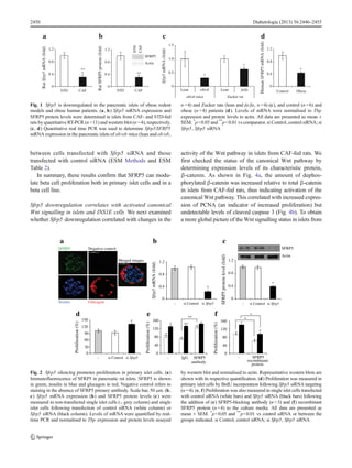
the mRNA (Fig. 1a) and protein levels (Fig. 1b). The mRNA
expression of Sfrp5 was also found to be lower in the pancre-
atic islets from two different models of obesity, ob/ob mice
and Zucker rats (Fig. 1c), further corroborating altered islet
Sfrp5 expression in obesity. Finally, we confirmed these re-
sults in isolated islets from human obese patients, which
showed decreased levels of SFRP5 mRNA compared with
control patients (Fig. 1d, 0.56±0.20 vs 1.02±0.08; p<0.01).
Taken together, these data provide evidence that islets exhibit
decreased Sfrp5 expression in obese states.
The silencing of Sfrp5 promotes proliferation in primary islet
cells and in the beta cell line INS1E SFRP5 is an inhibitor of
Wnt signalling, and the Wnt signalling pathway is known to
regulate beta cell proliferation [10, 12]. Hence, we
hypothesised that the downregulation of Sfrp5 expression
might be involved in enhanced beta cell proliferation in islets
from CAF-fed rats. As an initial step, we performed staining
with an antibody against SFRP5 in fixed pancreatic tissue
from control rats and found co-localisation of SFRP5 with
insulin (Fig. 2a), thus confirming the presence of SFRP5 in
beta cells. Next, to investigate the role of SFRP5 in prolifer-
ation, we assessed BrdU incorporation in dispersed pancreatic
islet cells transfected with an siRNA targeting Sfrp5. The
efficiency of the siRNA knockdown was validated for both
mRNA and protein (Fig. 2b, c). The percentage of cells
positive for BrdU increased significantly when we silenced
Sfrp5 (Fig. 2d, 132.40%±4.60; p<0.05 vs control siRNA)
without any detectable effect on apoptosis, as measured by
annexin Vand propidium iodide labelling (ESM Methods and
ESM Table 2). Furthermore, blocking SFRP5 protein with an
antibody induced the same increase in proliferation as seen
when Sfrp5 was silenced with siRNA (Fig. 2e). Conversely,
incubation with an SFRP5 recombinant protein resulted in
decreased proliferation (Fig. 2f).
As single islet cell cultures contain several endocrine types,
we performed additional experiments using the rat beta cell
line INS1E. INS1E cells transfected with the siRNA against
Sfrp5 exhibited lower Sfrp5 mRNA expression and decreased
SFRP5 protein levels compared with cells transfected with
control siRNA (Fig. 3a–c). As we had seen in dispersed islet
cells, SFRP5 knockdown led to a significant increase in
proliferation rates in INS1E cells (Fig. 3d, 151.13%±9.60;
p<0.05 vs control siRNA), which correlated with higher cell
number in Sfrp5 siRNA transfections compared with control
siRNA (Fig. 3e; p<0.01). We also determined the effect of
Sfrp5 silencing on beta cell apoptosis but found no difference
Diabetologia (2013) 56:2446–2455 2449](https://image.slidesharecdn.com/3812da5c-7db1-4e4f-aa5e-9a8cbf706ca3-160808213930/85/art-3A10-1007-2Fs00125-013-3030-x-4-320.jpg)
![CAF-fed rats, we examined expression of a panel of genes
related to this pathway in islets from CAF- and STD-fed rats.
We observed an increase in the expression of several genes in
CAF-fed rats (Table 1) including Wnt ligands, Wnt receptors,
Tcf7 and target genes such as cyclin D1 and Myc.
To establish a direct role of SFRP5 on the activation of the
canonical Wnt pathway in beta cells, we assessed whether
Sfrp5 silencing impacted this pathway in INSE cells. We
found increased levels of β-catenin (dephosphorylated form)
and TCF7L2 by western blot analysis in Sfrp5 siRNA-
transfected cells compared with control cells (Fig. 4c). In
addition, we observed translocation of β-catenin to the nucle-
us in SFRP5 knockdown cells, whereas β-catenin was found
mainly located in the cytoplasm of control cells (ESM
Methods and ESM Fig. 1). Together, these data link Sfrp5
downregulation with activated canonical Wnt signalling in
beta cells.
Next we evaluated whether SFRP5 could influence other
signalling pathways known to regulate beta cell proliferation.
INS1E transfected with Sfrp5 siRNA presented increased
levels of phospho-Akt, PI3K and phospho-MAPK (Fig. 5a),
thus suggesting that these other pathways could also contrib-
ute to enhanced proliferative capacity of SFRP5 knockdown
cells. Last, we determined the status of these pathways in islets
from CAF-fed rats and found similar results (Fig. 5b).
Sfrp5 expression is modulated by IGFBP3 We previously
showed that the visceral adipose tissue of CAF-fed rats exerts
pro-proliferative effects on beta cells in vitro, thus providing
evidence for the involvement of adipose-derived signals in
beta cell mass expansion in obesity [3]. To determine whether
Sfrp5 expression in pancreatic islets could be influenced by
secreted factors from adipose tissue, we investigated the effect
of the secretome obtained from the peripancreatic adipose
tissue of CAF- and STD-fed rats on INS1E cells. We observed
that Sfrp5 mRNA levels decreased in INS1E cells exposed to
Sfrp5mRNA(fold)
a
INS1E
-
INS1E
si Sfrp5
INS1E
si Control
0.0
0.4
0.8
1.2
SFRP5proteinlevel(fold)
Actin
SFRP5
b
0.0
0.4
0.8
1.2
INS1E
-
INS1E
si Sfrp5
INS1E
si Control
e
c si Sfrp5si Control
Proliferation(%)
d
INS1E
-
INS1E
si Sfrp5
INS1E
si Control
60
90
120
150
180
0
30
*
*
* *
Numberofcells(fold)
INS1E
-
INS1E
si Sfrp5
INS1E
si Control
0.0
0.5
1.0
1.5
2.0
2.5
Fig. 3 Sfrp5 silencing promotes beta cell proliferation in INS1E cells.
(a, b) Sfrp5 mRNA expression and SFRP5 protein levels were deter-
mined by quantitative RT-PCR and western blot, respectively, in non-
transfected INS1E cells (grey column) and in INS1E cells transfected
with the control siRNA (white column) or siRNA against Sfrp5 (black
column). Levels of mRNA were normalised to Tbp expression and
protein levels to actin. Representative western blots are shown and
quantified. (c) Immunofluorescence of SFRP5 in siRNA-transfected
INS1E cells. SFRP5 is shown in red (magnification, x40) (d) INS1E
cell proliferation was measured following Sfrp5 siRNA transfection by
BrdU incorporation (n =6). (e) Cell numbers before and after transfection
were determined (n =4) and values expressed as fold increase between
time 48 h and time 0. All data are presented as mean ± SEM. *
p<0.05 vs
siRNA control. si Control, control siRNA; si Sfrp5, Sfrp5 siRNA
STD
CAF
0
1
2
3
PCNA
Proteinlevel(fold)
caspase 3
*
Cleaved
Total β-catenin
Actin
0.0
1.0
2.0
3.0
STD CAF
Dephosphorylated
/totalβ-catenin
a
b
*
c
0.0
1.5
3.0
4.5
siControl
siSfrp5
Dephosphorylated β-catenin
Actin
Total β-catenin
siControlsiSfrp5
*
STD
CAF
Cleaved caspase 3
Actin
PCNA
TCF7L2
Actin
*
Dephosphorylated β-catenin
Dephosphorylated
/totalβ-catenin
Fig. 4 The canonical Wnt signalling pathway is activated in islets from
CAF-fed rats. (a, c) Levels of dephosphorylated and total β-catenin were
analysed in pancreatic islets isolated from CAF- and STD-fed rats (n =3–5)
and in INS1E cells transfected with control siRNA and Sfrp5
siRNA (n =4). Bands were quantified by densitometry and values
expressed as ratio of dephosphorylated β-catenin relative to total β-catenin.
(b) PCNA and cleaved caspase 3 levels were determined in CAF-fed
(black bars) vs STD-fed (white bars) islets. Protein levels were normalised
to actin. All data are presented as mean ± SEM. *
p <0.05 vs STD-fed or
siRNA control. si Control, control siRNA; si Sfrp5, Sfrp5 siRNA
Diabetologia (2013) 56:2446–2455 2451](https://image.slidesharecdn.com/3812da5c-7db1-4e4f-aa5e-9a8cbf706ca3-160808213930/85/art-3A10-1007-2Fs00125-013-3030-x-6-320.jpg)
![the adipose-derived secretome from CAF-fed rats but not that
from STD-fed rats (Fig. 6a), indicating that adipose-derived
signals can influence beta cell expression of this Wnt compo-
nent. IGFBP3 is decreased in the secretome of peripancreatic
adipose tissue and may be involved in pancreatic beta cell
proliferation in obese states [17]. Thus, here we investigated if
IGFBP3 modulated the expression of Sfrp5 in beta cells. To
this aim, we cultured INS1E cells in the presence of IGFBP3
recombinant protein or an antibody against IGFBP3 (which
sequesters IGFBP3 protein). Exogenous IGFP3 significantly
increased Sfrp5 mRNA levels, whereas the IGFBP3 antibody
lowered Sfrp5 mRNA expression in INS1E cells (Fig. 6a).
Finally, we measured the effects of recombinant IGFBP3 and
the IGFBP3 antibody on cell proliferation in single islet cell
cultures transfected with the siRNA against Sfrp5 or the
control siRNA. We observed further enhancement in prolifer-
ation when Sfrp5 was silenced in the presence of IGFBP3
antibody compared with cells that were transfected with con-
trol siRNA and treated with the same antibody (Fig. 6b).
Therefore, our data demonstrate that IGFBP3 can modulate
Sfrp5 expression and beta cell proliferation and thus reveal a
possible link between adipose-derived signals and intrinsic
beta cell proliferative pathways.
Discussion
Lifelong maintenance of appropriate beta cell mass is essential
for normal glucose homeostasis. Consequently, there is grow-
ing interest in understanding the mechanisms that control beta
cell expansion during times of increased metabolic demand
such as during obesity [26]. Several studies have demonstrated
that beta cell mass is increased in rodent obese models, mainly
due to increased beta cell proliferation. However, the mecha-
nisms underlining enhanced proliferation in obesity remain
poorly understood. Here, we show that the Sfrp5 gene is
downregulated in the islets of CAF-fed rats as well as in other
obese rodent models. Importantly, we find that SFRP5 mRNA
levels are also decreased in pancreatic islets from obese human
donors. Using cell-based assays, we demonstrate that Sfrp5
silencing promotes beta cell proliferation and activation of the
canonical Wnt signalling pathway. Finally, we provide evi-
dence of the modulation of Sfrp5 expression and beta cell
proliferation through IGFBP3 availability.
Table 1 Expression changes in Wnt-pathway-related genes in islets from
CAF- and STD-fed rats
Gene Fold change (CAF vs STD)
Downregulated
Apc 2.25
Csnk1α1 1.35
Ep300 1.50
Wif1 2.17
Sfrp5 1.45
Upregulated
Dvl2 1.35
Wisp1 1.31
Tcf7 1.31
Ccnd1 1.39
Myc 1.33
Fzd1 1.77
Fzd2 1.45
Wnt1 1.56
Wnt11 2.02
Wnt2b 2.32
Wnt5a 1.33
Wnt5b 1.62
Wnt7a 1.34
Wnt7b 1.36
Wnt9a 2.05
Wnt-signalling-related gene expression was studied in isolated islets from
STD- and CAF-fed rats. Genes shown in this table were differentially
expressed between STD- and CAF-fed islets with a fold change > ± 1.3
and p value <0.05 (n =4)
STD
CAF
0
2
4
6
8
Proteinlevel(fold)
*
*
*
b
a
p-Akt PI3K p-MAPK
MAPK
pathway
PI3K/Akt
pathway
-
siSfrp5
siControl
p-Akt
PI3K
pathway
PI3K/Akt
pathway
MAPK p-MAPK
Actin
Actin
MAPK
p-MAPK
p-Akt
PI3K
Akt
0
1
2
3
4
5
6
Proteinlevel(fold)
p-Akt
pPI3K
MAPK
p-MAPK
Akt
*
*
*
INS1E
Fig. 5 SFRP5 downregulation stimulates several signalling pathways
involved in cell proliferation. Levels of the indicated proteins were
assayed by western blot analysis in (a) INS1E cells transfected with
control siRNA and Sfrp5 siRNA (n =5) and (b) pancreatic islets isolated
from CAF- and STD-fed rats (n =3). Representative western blots are
shown. Bands were quantified and protein levels were normalised to actin
expression. *
p<0.05 vs STD-fed or siRNA control. si Control, control
siRNA; si Sfrp5, Sfrp5 siRNA
2452 Diabetologia (2013) 56:2446–2455](https://image.slidesharecdn.com/3812da5c-7db1-4e4f-aa5e-9a8cbf706ca3-160808213930/85/art-3A10-1007-2Fs00125-013-3030-x-7-320.jpg)
![There is strong evidence that the Wnt signalling pathway
regulates prenatal and postnatal beta cell development in mice
as well as glucose sensing in pancreatic beta cells [27, 28].
Also, the activation of Wnt signalling in beta cell lines or in
isolated islets has been shown to enhance beta cell prolifera-
tion [10, 29–31]. Thus, increased β-catenin levels in islets
caused an expansion of beta cell mass, whereas the depletion
of TCF7L2 reduced proliferation in human islets [10, 30]. In
the current study, we show that Sfrp5 silencing activates
proliferation in INS1E and dispersed islet cells, which is
accompanied by increased β-catenin and TCF7L2 levels in
both cellular models. Together, this evidence supports the
notion that Sfrp5 can regulate the canonical Wnt pathway in
beta cells. Correlating with these data, islets from CAF-fed
rats exhibit upregulation of several genes involved in the Wnt
pathway, as well as of well-known Wnt targets implicated in
cell cycle progression such as cyclin D1 and Myc [32].
Recent observations have offered a new perspective on
SFRP family functions and mechanisms of action in both
pancreatic development and disease [11, 12]. Ouchi et al
reported that two independent mouse models of obesity
exhibited reduced SFRP5 in adipose tissue, suggesting that
this molecule could be involved in the development of obesity
[15]. According to Schulte et al, SFRP5 represents a new
regulatory system in low-grade inflammation in obesity,
which could be influenced by nutritional therapy [33]. Re-
cently, in contrast to these findings, SFRP5 expression was
found unchanged in human WAT [13]. In agreement with this
study, we found no changes in circulating SFRP5 levels or in
Sfrp5 expression in peripancreatic adipose tissue between
CAF- and STD-fed rats (data not shown). Therefore, its im-
plication in metabolic dysfunction remains controversial
[34–36]. Importantly, our study demonstrates a novel auto-
crine role for SFRP5 in beta cell proliferation.
Several Wnt proteins appear to have both canonical and
non-canonical properties [12]. Our results show that, in addi-
tion to β-catenin-dependent signalling, SFRP5 has effects on
the MAPK and PI3K pathways. Information available on
SFRP5 is limited, thus we cannot rule out multiple effects of
this protein. However, it is possible that these additional
activation events are consequent of the crosstalk between the
Wnt pathway and MAPK or PI3K, described elsewhere [37,
38]. Recently, Gherzi et al reported that the half-life of
β-catenin mRNA is prolonged not only by Wnt but also by
PI3K–Akt signalling, adding further complexity to the poten-
tial mechanisms by which insulin/IGF-1 could interact with
Wnt signalling [39]. With the data available, we cannot dis-
miss the possibility that these different signalling events could
converge to activate beta cell proliferation.
Recently, we described that reduced IGFBP3 secretion by
the peripancreatic adipose tissue of CAF-fed rats induced beta
cell proliferation. Our data reveal that decreased IGFBP3
leads to a reduction in Sfrp5 mRNA expression. These find-
ings are in line with the increased beta cell proliferation
previously described in CAF-fed rats and provide additional
proof of the existence of a crosstalk between adipose tissue
(IGFBP3) and beta cells (SFRP5). Remarkably, the increment
in beta cell proliferation observed in SFRP5-knockdown beta
cells can be further enhanced by blocking exogenous IGFBP3
levels, which suggests a synergic effect of decreased IGFBP3
and SFRP5 on beta cell proliferation. Little is known regard-
ing the regulation of Sfrp5 expression. Of note, insulin, IGF-1
and other hormonal factors have been shown to activate the
canonical Wnt signalling by increasing nuclear β-catenin
content and binding of β-catenin/TCF to Wnt target gene
promoters [39–41]. Thus, it is plausible that IGFBP3 and
SFRP5 are two of the molecules mediating the crosstalk
between the Wnt and insulin/IGF-1 signalling pathways
-
Sfrp5mRNA(fold)
a
3.5
3.0
2.5
2.0
1.5
1.0
0.5
0
(0.5 µg/ml) (1 µg/ml) (0.1 µg/ml) (1 µg/ml)
**
*
*
*
**
b
protein
IGFBP3 antibodyCAF
Secretome
STD
**
*
0
40
80
120
160
200
IgG
Proliferation(%)
**
IGFBP3
antibody
IGFBP3 recombinant
**
Fig. 6 Effect of IGFBP3-blocking antibody on Sfrp5 mRNA expression
and beta cell proliferation. (a) Sfrp5 mRNA expression was measured in
INS1E cells after 24 h in normal medium alone (-) or supplemented with
secretome obtained from peripancreatic adipose tissue of STD- or CAF-
fed rats (diluted 1:3 in culture medium). The modulation of Sfrp5 mRNA
expression was also detected after stimulation with IGFBP3 recombinant
protein (0.5 and 1 μg/ml) or IGFBP3 antibody (0.1 and 1 μg/ml) added to
the culture medium. (b) Measurement of proliferation in Sfrp5 siRNA-
transfected primary islet cells (black bars) compared with control siRNA-
transfected cells (white bars) in the presence of IGFBP3 antibody. Data
are presented as mean ± SEM of three independent experiments. *
p<0.05,
**
p<0.01 compared to cells cultivated in normal medium alone in (a) or
between the groups indicated in (b)
Diabetologia (2013) 56:2446–2455 2453](https://image.slidesharecdn.com/3812da5c-7db1-4e4f-aa5e-9a8cbf706ca3-160808213930/85/art-3A10-1007-2Fs00125-013-3030-x-8-320.jpg)