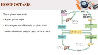
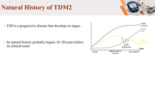
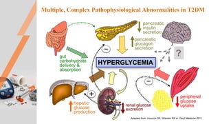
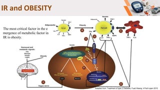
Type 2 diabetes pathogenesis
Diletta Rosati
Samaneh Gholami
Kristina Zguro
Ali Hakimzadeh
INTRODUCTION
EPIDEMIOLOGY
 T2D is one of the major challenges to human
health in the 21st century.
 Has a great economic impact on countries and
national health systems.
 The majority of countries spend 5-20% of
their total health expenditure on diabetes.
Normal glucose homeostasis:
• Hepatic glucose output
• Glucose uptake and utilization by peripheral tissues
• Action of insulin and glucagon on glucose metabolism
HOMEOSTASIS
Insulin
• Immediate release of insulin
Insulin signaling
 Insulin increase the rate of glucose transport into certain cells in the body
Natural History of TDM2
• T2D is a progressive disease that develops in stages.
• Its natural history probably begins 10–20 years before
its clinical onset
Factors
• Multiple factors involved in the genesis of T2D.
INSULIN RESISTANCE
Multiple, Complex Pathophysiological Abnormalities in T2DM
peripheral
glucose
uptake
hepatic
glucose
production
pancreatic
insulin
secretion
pancreatic
glucagon
secretion
gut
carbohydrate
delivery &
absorption
HYPERGLYCEMIA
?
Adapted from: Inzucchi SE, Sherwin RS in: Cecil Medicine 2011
_
_
+
renal glucose
excretion
INSULIN RESISTANCE
Lower biological activity of
Insulin in its different
metabolic actions for a
certain concentration;
Inability of tissues to respond
to normal levels of insulin
All processes malfunctions result in IR;
Receptor phosphorylation,
receptor number,
GLUT-4,
IRS-1 phosphorylation, etc
INSULIN RESISTANCE
Cause of IR
Strong genetic component
o Identical twins >90% concordant
o Contribution of Epigenetics
Environment Important
o Increased incidence over last century
o Lifestyle, exercise, habits
o Diet
o Most commonly associated with obesity (>80% of cases)
IR genes
• Directly associated with lower
glucose uptake
• Insulin receptor substrate gene
• Phosphoinositide 3-kinase gene
• Explaining the obesity-type 2
diabetes relationship
• β-3 adrenergic receptor gene
• Tumor necrosis factor alpha gene
• Peroxisome proliferator-activated
receptor gene
• Adipocytokines in obesity
• Leptin gene
• Resistin gene
• Adiponectin gene
• Lipid metabolism
• Lipoprotein lipase gene
• Fatty acid-binding protein gene
• The thermogenesis obesity relationship
• Uncoupling protein gene
IR in liver, muscle and adipose tissue
Adapted from: pathogenesis of T2DM, Pilar Durruty et al, Elsevier feb2019
Increase in FFA
Or
Lipoatrophy
IR and OBESITY
Adapted from: Treatment of type 2 Diabetes, Fuad Alseraj, InTech open 2015
The most critical factor in the e
mergence of metabolic factor in
IR is obesity.
β-CELLS DISFUNCTION
Decline of ꞵ cell function
 There is a progressive deterioration in β-cell function over time in type 2 Diabetes mellitus
 The pancreatic islet function was found to be about 50% of normal at the time of diagnosis (as
indicate by UK-PDS)
 The reduction in function probably commencing 10-12 yr before diagnosis and aggravated by
increasing fasting plasma glucose level
 With a corresponding decline in β-cell function, was found glycated haemoglobin levels to
increase over time (level of HbA1c 6.5% or higher)
Insulin secretion defect in DM2
The defect of insulin secretion in DM2 is related to two confounding components :
 Insulin deficiency
DM2 is characterized by an increased fasting ratio of proinsulin to total immunoreactive insulin indicating a
reduced processing of proinsulin, which correlates with β-cell dysfunction and is predictive of disease development
 β-cell secretory defect
Studies (BMC Biology Cantley & M.Ashcroft) with modest sample sizes (n = 5 to 17 cases) have clearly shown that
glucose-stimulated insulin secretion (GSIS) is defective in islets from T2D donors, relative to non-diabetic donors.
In two of these studies, islets from T2D donors responded normally to non-glucose stimuli ( eg. Arginine) ,
suggesting defective GSIS in these cohorts is likely due to impaired glucose-sensing (stimulus-secretion coupling)
rather than a loss of insulin content or a constitutive defect in insulin exocytosis
Insulin secretion defect in DM2
 Decreased β-cell mass
Diminished proliferation, or increased apoptosis or
both will result in lower β-cell mass
There is evidence that increased β-cell apoptosis in
the Zucker diabetic fatty rat, an animal model of
DM2, underlies the decreased β-cell mass seen in
these animals.
Insulin secretion defect in DM2
The study by Butler et al.(4)
Relative β-cell volume increased by 50%
in obese compared with lean nondiabetic
Relative β-cell volume was decreased in
obese IGT and more in obese DM2
Factors for progressive loss of β-cell function and mass
o Glucotoxicity
o Lipotoxicity
o Proinflammatory cytokines and leptin
 Adipocyte-secreted factors
Increased cell nutrients
Innate immune system and
autoimmunity
o Islet cell amyloid
o Linkage of reduced β-cell mass and
dysfunction
Factors for progressive loss of β-cell function and mass
GLUCOTOXICITY
Pathway of oxidative stress production Reduction of insulin biosynthesis and
secretion by oxidative stress
Factors for progressive loss of β-cell function and mass
 Elevated levels of free fatty acids can have many
deleterious effects on β-cells:
Decreased glucose-stimulated insulin secretion
Impaired insulin gene exepression
Increased synthesis of ceramides
Activation of the oxidative stress
Increased β-cells inflammation
β-cell apoptosis
 Adipocyte – secreted factors
Leptin
TNF-α
IL-6
IL-1Ra
Factors for progressive loss of β-cell function and mass
 Increased in cell nutrients
Increasing glucose concentrations induce β-cell production of IL-1β leading to β cell apoptosis
Both IL-1β and ROS activate NF-κB an important mediator of inflammatory responses
Increased FFAs concentration may act directly on β-cells or activate the innate immune system
 Innate immune system and autoimmunity
• In addition to the endocrine activity of the adipocytes macrophages and endothelium may contribute to
increasing serum levels of IL-1β, IL-6, TNF-α
• Increasin apoptotic cells can provoke an immune response
• Some DM2 patients may show mobilization of T cells reactive to β-cell antigens, culminating in
autoimmune destruction of β-cells
Factors for progressive loss of β-cell function and mass
 Islet cell amyloid
Amylin or Islet amyloid polypeptide (IAPP) is a 37-
residue peptide hormone cosecreted with insulin
Amylin contributes to glycemic control, functioning
as a synergistic partner to insulin.
Impaired N-terminal processing of proIAPP is an
important factor initiating amyloid formation and β-
cell death
Factors for progressive loss of β-cell function and mass
 2 possible explanations account for the impaired β-cell function consequent to
decreased β-cell mass:
Increased insulin demand on residual β-cells per se
Hyperglycemia consequent to decreased β-cell mass driving the impairment in
β-cell function
Genetics in T2D
Genetics and T2D
 Individuals with a positive family history are about 2-6 times more likely to develop
T2D than those with a negative family history
– Risk ~40% if T2D parent; ~80% if 2 T2D parents
 Higher concordance for MZ VERSUS DZ twins
 Has been difficult to find genes for T2D
− Late age at onset
− Polygenic inheritance
− Multifactorial inheritance
Challenges in Finding Genes
 Inadequate sample sizes
– Multiplex families
– Cases and controls
 Difficult to define the phenotype
 Reduced penetrance
– Influence of environmental factors
– Gene-gene interactions
 Variable age at onset
 Failure to replicate findings
 Genes identified have small effects
CAPN10 – NIDDM1
 Chromosome 2q37.3
– Encodes an intracellular calcium-dependent cytoplasmic protease that is ubiquitously
expressed
• May modulate activity of enzymes and/or apoptosis
– Likely involves insulin secretion and resistance
– Stronger influence in Mexican Americans than other ethnic groups
• Responsible for ~40% of familial clustering
– Genetic variant: A43G, Thr50Ala, Phe200Thr
PPARγ
 Chromosome 3p25
– Transcription factors that play an important role in adipocyte differentiation and
function
– Is associated with decreased insulin sensitivity
– Target for hypoglycemic drugs
– Genetic variant: Pro12Ala, Pro is risk allele (common)
– Variant is common
– May be responsible for ~25% of T2D cases
ABCC8 and KCNJ11
 ATP-binding cassette, subfamily C member 8 (chromosome
11p15.1)
 Potassium channel, inwardly rectifying, subfamily J, member 11
(chromosome 11p15.1)
– ABCC8 encodes the sulfonylurea receptor (drug target )
– Is coupled to the Kir6.2 subunit (encoded by KCNJ11 – 4.5 kb apart
& near INS )
– Part of the ATP-sensitive potassium channel
• Involved in regulating insulin and glucagon
• Mutations affect channel’s activity and insulin secretion
– Site of action of sulfonylurea drugs
– Genetic variants: Ser1369Ala & Glu23Lys, respectively
TCF7L2
 Transcription factor 7-like 2 (chromosome 10q25)
– Related to impaired insulin release of glucagon-like peptide-1 (islet
secretagogue), reduced β-cell mass or β-cell dysfunction
• Stronger among lean versus obese T2D
– 10% of individuals are homozygous have 2-fold increase in risk
relative to those with no copy of the variant
– Responsive to sulfonylureas
– Genetic variant: rs7901695 and others in LD
GWAS New Loci Identified
 FTO – chr 16q12
– Fat mass and obesity associated gene
– Governs energy balance; gene expression is regulated by feeding and fasting
 HHEX/IDE – chr 10q23-24; near TCF7L2
– HHEX - Haematopoietically expressed homeobox
• Transcription factor in liver cells
– IDE - Insulin degrading enzyme
• Has affinity for insulin; inhibits IDE-mediated degradation of other substances
 CDKAL1 – chr 6p22
– Likely plays role in CDK5 inhibition and decreased insulin secretion
 IGF2BP2 – chr 3q28
– Regulates IGF2 translation; stimulates insulin action
 CDKN2A/B – chr 9p21
– Plays role in pancreatic development and islet proliferation
MODY Genes
Type Gene Locus Protein # Mutations % MODY
MODY1 HNF4A 20q12-q13.1 Hepatocyte nuclear factor 4-
alpha
12 ~5%
MODY2 GCK 7p15-p13 Glucokinase ~200 ~15%
MODY3 HNF1A 12q24.2 Hepatocyte nuclear factor 1-
alpha
>100 ~65%
MODY4 IPF1 13q12.1 Insulin promotor factor-1 Few
MODY5 HNF1B 17cen-q21.3 Hepatocyte nuclear factor 1-
beta
Few <3%
MODY6 NEUROD1 2q32 Neurogenic differentiation
factor 1
Few
 All MODY genes are expressed in the pancreas, and play a role in:
– The metabolism of glucose
– The regulation of insulin or other genes involved in glucose transport
– The development of the fetal pancreas
 Zheng, Y., Ley, S. H., & Hu, F. B. (2018). Global a etiology and epidemiology of type 2 diabet
es mellitus and its complications. Nature Reviews Endocrinology, 14(2), 88.
 Campbell Biology (11th edition)
 Mitchell A. Lazar, Mohammad Qatanani. Mechanisms of obesity-associated insulin resistance,
genes and development, 2009
 Christopher j. Hupfeld, c. Hamish Courtney, and Jerrold M. Olefsky. Type 2 diabetes mellitus:
Etiology, Pathogenesis, and Natural History. 2014
 Gerald Reaven. Insulin Resistance, Type 2 Diabetes Mellitus, and Cardiovascular Disease, Jou
rnal of the American Heart Association, 2019
 Vandana Saini. Molecular mechanisms of insulin resistance in type 2 diabetes mellitus, World J
ournal of Diabetes, 2010
 Pilar Durruty, María Sanzana and Lilian Sanhueza, Pathogenesis of Type 2 Diabetes Mellitus,2
018
 Bernardo L, Wajchenberg, ꞵ Cell failure in Diabetes and Pereservation By Clinical Treatment
, 2017
REFRENCES
Type 2 diabetes pathogenesis

Type 2 diabetes pathogenesis

  • 1.
    Type 2 diabetespathogenesis Diletta Rosati Samaneh Gholami Kristina Zguro Ali Hakimzadeh
  • 2.
  • 3.
    EPIDEMIOLOGY  T2D isone of the major challenges to human health in the 21st century.  Has a great economic impact on countries and national health systems.  The majority of countries spend 5-20% of their total health expenditure on diabetes.
  • 4.
    Normal glucose homeostasis: •Hepatic glucose output • Glucose uptake and utilization by peripheral tissues • Action of insulin and glucagon on glucose metabolism HOMEOSTASIS
  • 5.
  • 6.
    Insulin signaling  Insulinincrease the rate of glucose transport into certain cells in the body
  • 7.
    Natural History ofTDM2 • T2D is a progressive disease that develops in stages. • Its natural history probably begins 10–20 years before its clinical onset
  • 8.
    Factors • Multiple factorsinvolved in the genesis of T2D.
  • 9.
  • 10.
    Multiple, Complex PathophysiologicalAbnormalities in T2DM peripheral glucose uptake hepatic glucose production pancreatic insulin secretion pancreatic glucagon secretion gut carbohydrate delivery & absorption HYPERGLYCEMIA ? Adapted from: Inzucchi SE, Sherwin RS in: Cecil Medicine 2011 _ _ + renal glucose excretion
  • 11.
    INSULIN RESISTANCE Lower biologicalactivity of Insulin in its different metabolic actions for a certain concentration; Inability of tissues to respond to normal levels of insulin
  • 12.
    All processes malfunctionsresult in IR; Receptor phosphorylation, receptor number, GLUT-4, IRS-1 phosphorylation, etc INSULIN RESISTANCE
  • 13.
    Cause of IR Stronggenetic component o Identical twins >90% concordant o Contribution of Epigenetics Environment Important o Increased incidence over last century o Lifestyle, exercise, habits o Diet o Most commonly associated with obesity (>80% of cases)
  • 14.
    IR genes • Directlyassociated with lower glucose uptake • Insulin receptor substrate gene • Phosphoinositide 3-kinase gene • Explaining the obesity-type 2 diabetes relationship • β-3 adrenergic receptor gene • Tumor necrosis factor alpha gene • Peroxisome proliferator-activated receptor gene • Adipocytokines in obesity • Leptin gene • Resistin gene • Adiponectin gene • Lipid metabolism • Lipoprotein lipase gene • Fatty acid-binding protein gene • The thermogenesis obesity relationship • Uncoupling protein gene
  • 15.
    IR in liver,muscle and adipose tissue Adapted from: pathogenesis of T2DM, Pilar Durruty et al, Elsevier feb2019 Increase in FFA Or Lipoatrophy
  • 16.
    IR and OBESITY Adaptedfrom: Treatment of type 2 Diabetes, Fuad Alseraj, InTech open 2015 The most critical factor in the e mergence of metabolic factor in IR is obesity.
  • 17.
  • 18.
    Decline of ꞵcell function  There is a progressive deterioration in β-cell function over time in type 2 Diabetes mellitus  The pancreatic islet function was found to be about 50% of normal at the time of diagnosis (as indicate by UK-PDS)  The reduction in function probably commencing 10-12 yr before diagnosis and aggravated by increasing fasting plasma glucose level  With a corresponding decline in β-cell function, was found glycated haemoglobin levels to increase over time (level of HbA1c 6.5% or higher)
  • 19.
    Insulin secretion defectin DM2 The defect of insulin secretion in DM2 is related to two confounding components :  Insulin deficiency DM2 is characterized by an increased fasting ratio of proinsulin to total immunoreactive insulin indicating a reduced processing of proinsulin, which correlates with β-cell dysfunction and is predictive of disease development  β-cell secretory defect Studies (BMC Biology Cantley & M.Ashcroft) with modest sample sizes (n = 5 to 17 cases) have clearly shown that glucose-stimulated insulin secretion (GSIS) is defective in islets from T2D donors, relative to non-diabetic donors. In two of these studies, islets from T2D donors responded normally to non-glucose stimuli ( eg. Arginine) , suggesting defective GSIS in these cohorts is likely due to impaired glucose-sensing (stimulus-secretion coupling) rather than a loss of insulin content or a constitutive defect in insulin exocytosis
  • 20.
    Insulin secretion defectin DM2  Decreased β-cell mass Diminished proliferation, or increased apoptosis or both will result in lower β-cell mass There is evidence that increased β-cell apoptosis in the Zucker diabetic fatty rat, an animal model of DM2, underlies the decreased β-cell mass seen in these animals.
  • 21.
    Insulin secretion defectin DM2 The study by Butler et al.(4) Relative β-cell volume increased by 50% in obese compared with lean nondiabetic Relative β-cell volume was decreased in obese IGT and more in obese DM2
  • 22.
    Factors for progressiveloss of β-cell function and mass o Glucotoxicity o Lipotoxicity o Proinflammatory cytokines and leptin  Adipocyte-secreted factors Increased cell nutrients Innate immune system and autoimmunity o Islet cell amyloid o Linkage of reduced β-cell mass and dysfunction
  • 23.
    Factors for progressiveloss of β-cell function and mass GLUCOTOXICITY Pathway of oxidative stress production Reduction of insulin biosynthesis and secretion by oxidative stress
  • 24.
    Factors for progressiveloss of β-cell function and mass  Elevated levels of free fatty acids can have many deleterious effects on β-cells: Decreased glucose-stimulated insulin secretion Impaired insulin gene exepression Increased synthesis of ceramides Activation of the oxidative stress Increased β-cells inflammation β-cell apoptosis
  • 25.
     Adipocyte –secreted factors Leptin TNF-α IL-6 IL-1Ra
  • 26.
    Factors for progressiveloss of β-cell function and mass  Increased in cell nutrients Increasing glucose concentrations induce β-cell production of IL-1β leading to β cell apoptosis Both IL-1β and ROS activate NF-κB an important mediator of inflammatory responses Increased FFAs concentration may act directly on β-cells or activate the innate immune system  Innate immune system and autoimmunity • In addition to the endocrine activity of the adipocytes macrophages and endothelium may contribute to increasing serum levels of IL-1β, IL-6, TNF-α • Increasin apoptotic cells can provoke an immune response • Some DM2 patients may show mobilization of T cells reactive to β-cell antigens, culminating in autoimmune destruction of β-cells
  • 27.
    Factors for progressiveloss of β-cell function and mass  Islet cell amyloid Amylin or Islet amyloid polypeptide (IAPP) is a 37- residue peptide hormone cosecreted with insulin Amylin contributes to glycemic control, functioning as a synergistic partner to insulin. Impaired N-terminal processing of proIAPP is an important factor initiating amyloid formation and β- cell death
  • 28.
    Factors for progressiveloss of β-cell function and mass  2 possible explanations account for the impaired β-cell function consequent to decreased β-cell mass: Increased insulin demand on residual β-cells per se Hyperglycemia consequent to decreased β-cell mass driving the impairment in β-cell function
  • 29.
  • 30.
    Genetics and T2D Individuals with a positive family history are about 2-6 times more likely to develop T2D than those with a negative family history – Risk ~40% if T2D parent; ~80% if 2 T2D parents  Higher concordance for MZ VERSUS DZ twins  Has been difficult to find genes for T2D − Late age at onset − Polygenic inheritance − Multifactorial inheritance
  • 31.
    Challenges in FindingGenes  Inadequate sample sizes – Multiplex families – Cases and controls  Difficult to define the phenotype  Reduced penetrance – Influence of environmental factors – Gene-gene interactions  Variable age at onset  Failure to replicate findings  Genes identified have small effects
  • 32.
    CAPN10 – NIDDM1 Chromosome 2q37.3 – Encodes an intracellular calcium-dependent cytoplasmic protease that is ubiquitously expressed • May modulate activity of enzymes and/or apoptosis – Likely involves insulin secretion and resistance – Stronger influence in Mexican Americans than other ethnic groups • Responsible for ~40% of familial clustering – Genetic variant: A43G, Thr50Ala, Phe200Thr PPARγ  Chromosome 3p25 – Transcription factors that play an important role in adipocyte differentiation and function – Is associated with decreased insulin sensitivity – Target for hypoglycemic drugs – Genetic variant: Pro12Ala, Pro is risk allele (common) – Variant is common – May be responsible for ~25% of T2D cases
  • 33.
    ABCC8 and KCNJ11 ATP-binding cassette, subfamily C member 8 (chromosome 11p15.1)  Potassium channel, inwardly rectifying, subfamily J, member 11 (chromosome 11p15.1) – ABCC8 encodes the sulfonylurea receptor (drug target ) – Is coupled to the Kir6.2 subunit (encoded by KCNJ11 – 4.5 kb apart & near INS ) – Part of the ATP-sensitive potassium channel • Involved in regulating insulin and glucagon • Mutations affect channel’s activity and insulin secretion – Site of action of sulfonylurea drugs – Genetic variants: Ser1369Ala & Glu23Lys, respectively
  • 34.
    TCF7L2  Transcription factor7-like 2 (chromosome 10q25) – Related to impaired insulin release of glucagon-like peptide-1 (islet secretagogue), reduced β-cell mass or β-cell dysfunction • Stronger among lean versus obese T2D – 10% of individuals are homozygous have 2-fold increase in risk relative to those with no copy of the variant – Responsive to sulfonylureas – Genetic variant: rs7901695 and others in LD
  • 35.
    GWAS New LociIdentified  FTO – chr 16q12 – Fat mass and obesity associated gene – Governs energy balance; gene expression is regulated by feeding and fasting  HHEX/IDE – chr 10q23-24; near TCF7L2 – HHEX - Haematopoietically expressed homeobox • Transcription factor in liver cells – IDE - Insulin degrading enzyme • Has affinity for insulin; inhibits IDE-mediated degradation of other substances  CDKAL1 – chr 6p22 – Likely plays role in CDK5 inhibition and decreased insulin secretion  IGF2BP2 – chr 3q28 – Regulates IGF2 translation; stimulates insulin action  CDKN2A/B – chr 9p21 – Plays role in pancreatic development and islet proliferation
  • 36.
    MODY Genes Type GeneLocus Protein # Mutations % MODY MODY1 HNF4A 20q12-q13.1 Hepatocyte nuclear factor 4- alpha 12 ~5% MODY2 GCK 7p15-p13 Glucokinase ~200 ~15% MODY3 HNF1A 12q24.2 Hepatocyte nuclear factor 1- alpha >100 ~65% MODY4 IPF1 13q12.1 Insulin promotor factor-1 Few MODY5 HNF1B 17cen-q21.3 Hepatocyte nuclear factor 1- beta Few <3% MODY6 NEUROD1 2q32 Neurogenic differentiation factor 1 Few  All MODY genes are expressed in the pancreas, and play a role in: – The metabolism of glucose – The regulation of insulin or other genes involved in glucose transport – The development of the fetal pancreas
  • 37.
     Zheng, Y.,Ley, S. H., & Hu, F. B. (2018). Global a etiology and epidemiology of type 2 diabet es mellitus and its complications. Nature Reviews Endocrinology, 14(2), 88.  Campbell Biology (11th edition)  Mitchell A. Lazar, Mohammad Qatanani. Mechanisms of obesity-associated insulin resistance, genes and development, 2009  Christopher j. Hupfeld, c. Hamish Courtney, and Jerrold M. Olefsky. Type 2 diabetes mellitus: Etiology, Pathogenesis, and Natural History. 2014  Gerald Reaven. Insulin Resistance, Type 2 Diabetes Mellitus, and Cardiovascular Disease, Jou rnal of the American Heart Association, 2019  Vandana Saini. Molecular mechanisms of insulin resistance in type 2 diabetes mellitus, World J ournal of Diabetes, 2010  Pilar Durruty, María Sanzana and Lilian Sanhueza, Pathogenesis of Type 2 Diabetes Mellitus,2 018  Bernardo L, Wajchenberg, ꞵ Cell failure in Diabetes and Pereservation By Clinical Treatment , 2017 REFRENCES

Editor's Notes

  • #11 This slide provides a broad overview of the main pathophysiological defects in type 2 diabetes. Insulin resistance is demonstrated in peripheral tissues, such as skeletal muscle and adipocytes. The pancreas develops relative insulin secretory failure and is no longer provide the amount of insulin required for normal glucose. The pancreas also demonstrates a failure of the normal suppression of glucagon after meals. Hepatic glucose production is also increased. Renal glucose output is set at a higer threshold than normal. Many of these defects may be influenced by abnormal central control of metabolism.
  • #17 Endocrine, inflammatory, and neuronal pathways link obesity to insulin resistance. ( A ) The obesity-associated increase in FAs can trigger insulin resistance through intracellular metabolites that activate PKC, leading to the activation of serine/threonine kinases that inhibit insulin signaling. ( B ) Obesity-associated changes in secretion of adipokines that modulate insulin signaling. ( C ) Obesity-associated inflammatory factors. Obesity is characterized by an increase in the accumulation of ATMs, which increase the adipose tissue production of inflammatory cytokines that inhibit insulin signaling. ( D ) Endocrine and inflammatory mediators con- verging on serine/threonine kinases that inhibit insulin signaling. ( E ) Obesity-associated activation of NF- ␬ B heightens inflammatory responses that exacerbate insulin resistance. ( F ) SOCS family proteins, induced by adipokines, induce insulin resistance either by interfering with IRS-1 and IRS-2 tyrosine phosphorylation or by targeting IRS-1 and IRS-2 for proteosomal degradation. ( G ) FAs also trigger insulin resistance by direct activation of TLR4 and the innate immune response. ( H ) Obesity-related alteration in the central response to hormonal and nutrient signals alters peripheral insulin sensitivity.