2. sity (21). This alteration in systemic energy balance is associ-
ated with elevated metabolic rate and physical activity levels
as well as disrupted circadian rhythm (28). However, it re-
mains unknown whether PGC-1␣ influences energy balance
and clock function directly through its action in peripheral
tissues or via its function in the central nervous system.
PGC-1␣ mRNA and protein expression has been observed in
several brain areas and neuronal cell types (29, 30). Whole
body PGC-1␣ null mice develop spongioform neurodegenera-
tion, most notably in the striatum and the deep layers of the
cerebral cortex. In addition, dysregulation of the PGC-1␣
pathway was recently implicated in Huntington and Parkin-
son disease (21, 31, 32). These studies strongly suggest
PGC-1␣ plays a critical role in maintaining neuronal health.
Whether neuronal PGC-1␣ is required for systemic metabolic
homeostasis has not yet been explored.
To address the role of neuronal PGC-1␣ in the regulation
of energy balance, we generated forebrain-specific PGC-1␣
null mice by crossing PGC-1␣flox/flox
mice with calcium/cal-
modulin-dependent protein kinase II␣ (CaMKII␣)-Cre trans-
genic mice. We found that inactivation of PGC-1␣ in
CaMKII␣-positive neurons leads to resistance to diet-induced
obesity and results in neurodegenerative lesions in the stria-
tum. Our studies reveal an essential role of PGC-1␣ in
CaMKII␣-positive neurons in the regulation of energy bal-
ance and neuronal function.
EXPERIMENTAL PROCEDURES
Mice—All animal experiments were performed according
to procedures approved by the University Committee on
Use and Care of Animals. Mice carrying PGC-1␣flox
alleles
were generated as described previously (21). These mice were
mated with CaMKII␣-Cre transgenic mice to generate
flox/flox control and B␣KO mice. PCR analysis was per-
formed on genomic DNA isolated from different tissues to
assess Cre-mediated deletion. Wild type allele and Cre-
mediated deletion allele was detected using primers 1 and 2
and primers 3 and 4, respectively: forward primer 1,
GTCTAAGATGTCTGCTCTTGAGG; reverse primer
2, CCAGTTTCTTCATTGGTGTG; forward primer 3,
TCCAGTAGGCAGAGATTTATGAC; reverse primer 4,
CCAACTGTCTATAATTCCAGTTC. Mice were main-
tained on a standard rodent chow or a high-fat diet con-
taining 60% fat-derived calories (D12492, Research Diets)
with 12-h light and dark cycles. For cold exposure, 11-
week-old female mice were individually housed in cages
prechilled at 4 °C with free access to food and water. Core
body temperature was monitored using a rectal thermome-
ter 3 h after the initiation of cold exposure. Brown fat was
dissected following cold exposure for gene expression and
histological analyses. For hypothalamus analysis, 3–4-
months-old control, whole body PGC-1␣ null, and B␣KO
mice were either fed or fasted for 48 h before harvest.
Metabolic Analysis—Metabolic rate and activity were mea-
sured using Comprehensive Lab Animal Monitoring System
(CLAMS), which simultaneously measures whole-body O2
consumption and physical movements (33, 34). Mice were
acclimated in the monitoring chambers for 3 days before the
experiment to minimize the effects of housing environment
changes on animal behaviors. Data were collected every 10
min for each mouse over a period of three light/dark cycles.
CLAMS study was conducted by University of Michigan Ani-
mal Phenotyping Core. Plasma concentrations of triglyceride,
insulin, and leptin were measured using commercially avail-
able assay kits. Liver triglyceride content was extracted and
measured using previously described procedures (35).
Histological Analysis—Brains were fixed in situ by intracar-
diac perfusion with 15 ml PBS followed by 15 ml 4% PFA in
PBS, post-fixed in 4% PFA in PBS overnight at 4 °C after dis-
section, dehydrated in 70% ethanol, and embedded in paraf-
fin. Coronal sections (8 m) were stained using an H&E stain-
ing method. Immunohistochemistry using antibody against
neurofilament light chain was performed as described previ-
ously (21). Other tissues were fixed directly by 4% PFA in PBS
overnight at 4 °C after dissection and underwent the same
procedure as the brain for the H&E staining. Frozen livers
were embedded in O.C.T. compound, Tissue-Tek (Sakura
Finetek) sectioned into 12-m sections, and stained using Oil
Red O method.
RNA and Protein Analysis—Total RNA was isolated from
tissues using TRIzol reagents (Invitrogen). For quantitative
real-time PCR (qPCR) analysis, RNA samples were reverse
transcribed and used in quantitative PCR reactions in the
presence of SYBR Green (Applied Biosystems). Relative abun-
dance of mRNA was normalized to ribosomal protein 36B4 or
-actin. Sequences for most qPCR primers used in this study
were described previously (21, 36). The rest of primer se-
quences are available upon request. Immunoblotting studies
were performed using specific antibodies for LC3 (LC3–5F10,
Nanotools), p62 (PW9860, Enzo Life Sciences), and ubiquitin
(catalog no. sc-8017, Santa Cruz Biotechnology).
RESULTS
Generation of Brain-specific PGC-1␣-deficient Mice—
PGC-1␣ deficiency impairs adaptive metabolic responses in
multiple tissues, including the liver, brown adipose tissue as
well as skeletal and cardiac muscle (21, 22, 24). Paradoxically,
whole body PGC-1␣ null mice are resistant to diet-induced
obesity and have elevated metabolic rate. These findings raise
the possibility that PGC-1␣ may play an important role in the
regulation of energy balance through its action in the central
nervous system. To test this possibility, we generated brain-
specific PGC-1␣ null mice using the Cre-loxP system. We
chose to use CaMKII␣-Cre transgenic mice because PGC-1␣
is abundantly expressed in forebrain, including cerebral cor-
tex, hippocampus, basal ganglia, and hypothalamus (37).
Transgenic expression of Cre recombinase under the control
of CaMKII␣ promoter has been widely used to inactivate
genes in the forebrain (38, 39). We generated PGC-1␣flox/flox
and PGC-1␣flox/flox
; CaMKII␣-Cre (B␣KO) mice for our stud-
ies (Fig. 1A). As shown in Fig. 1B, B␣KO mice have selective
deletion of exons 3–5 within the PGC-1␣ locus in several
brain areas, including cerebral cortex, striatum, olfactory
bulb, and hypothalamus, but not in any of the peripheral tis-
sues examined.
Neuronal PGC-1␣ Regulates Energy Balance
39088 JOURNAL OF BIOLOGICAL CHEMISTRY VOLUME 285•NUMBER 50•DECEMBER 10, 2010
atNYUSchoolofMedicineLibrary,onJuly17,2012www.jbc.orgDownloadedfrom
3. B␣KO Mice Have Normal Adaptive Metabolic Response—
We next examined whether adaptive metabolic responses in
peripheral tissues are affected in B␣KO mice fed chow diet. In
contrast to whole body PGC-1␣ null mice, which are cold-
sensitive due to defects in adaptive thermogenesis, B␣KO
mice maintain their core body temperature following 3-h cold
exposure at 4 °C. We observed a modest but similar decrease
in body temperature in both control and B␣KO groups (Fig.
2A). Histological analysis of brown adipose tissues reveals that
brown adipocytes appear normal in size with similar accumu-
lation of multilocular lipid droplets (Fig. 2B). Consistently,
cold-inducible expression of genes involved in adaptive ther-
mogenesis, including PGC-1␣, deiodinase 2, Ucp1 (uncou-
pling protein 1), and mitochondrial genes, are similar in con-
trol and B␣KO brown fat (Fig. 2C and data not shown).
PGC-1␣ has been demonstrated to regulate multiple aspects
of hepatic starvation response. PGC-1␣ deficient hepatocytes
have defective gluconeogenic response and heme biosyn-
thesis (18, 21). Compared with the control group, B␣KO
mice have normal induction of gluconeogenic genes fol-
lowing overnight fasting (data not shown). Plasma glucose
levels are also similar in chow-fed control and B␣KO mice
under both fed and fasted conditions. These results indi-
cate that adaptive energy metabolism in peripheral tissues
FIGURE 1. Generation of B␣KO mice. A, strategy for Cre recombinase-mediated deletion of PGC-1␣ exons 3–5 in the brain. B, PCR analysis of genomic DNA
isolated from tissues of flox/flox (Ϫ) and B␣KO (ϩ) mice. Note the deletion of PGC-1␣ exons is only detected in cortex, striatum, olfactory bulb, and hypo-
thalamus. PCR primers are indicated in A. eWAT, epididymal white adipose tissue; BAT, brown adipose tissue.
FIGURE 2. Adaptive thermogenesis in response to cold exposure. A, Rectal temperature of flox/flox (filled box) and B␣KO (open box) mice kept at room
temperature (RT) or exposed to 4 °C for 3 h. *, p Ͻ 0.05. B, H&E staining of paraffin-embedded brown fat sections. C, qPCR analysis of gene expression in
brown fat. Data represent mean Ϯ S.E. (n ϭ 3 per group). *, p Ͻ 0.004; NS, not significant.
Neuronal PGC-1␣ Regulates Energy Balance
DECEMBER 10, 2010•VOLUME 285•NUMBER 50 JOURNAL OF BIOLOGICAL CHEMISTRY 39089
atNYUSchoolofMedicineLibrary,onJuly17,2012www.jbc.orgDownloadedfrom
4. is largely unperturbed in brain-specific PGC-1␣-deficient
mice.
B␣KO Mice Are Resistant to Diet-induced Obesity—PGC-
1␣-deficient mice have elevated metabolic rate and are resis-
tant to diet-induced obesity. However, whether this can be
attributed to neuronal PGC-1␣ function remains unknown.
To determine the significance of central PGC-1␣ in energy
balance, we subjected control and B␣KO mice to high-fat diet
feeding. Although body weight of these two groups of mice
remains similar under chow-fed condition, B␣KO mice are
significantly resistant to weight gain when fed high-fat diet
(Fig. 3A). The body weight of B␣KO mice is ϳ20% lower than
control mice following 10 weeks of high-fat feeding. Trans-
genic expression of Cre recombinase alone does not affect
body weight upon high-fat feeding (data not shown). Resis-
tance to weight gain following high-fat feeding appears to be
more pronounced in whole body PGC-1␣ null group, which
weighs ϳ40% less than the wild type group. Consistently, epi-
didymal white adipose tissue weight and epididymal white
adipose tissue/body weight ratio are significantly lower in the
B␣KO and whole body PGC-1␣ null mice compared with
their respective control (Fig. 3C). In contrast to whole body
PGC-1␣ null mice, which have lower plasma glucose concen-
trations, plasma glucose remains similar in control and B␣KO
mice following high-fat feeding (Fig. 3D). Resting core body
temperature is also slightly but significantly higher in the
B␣KO mice but not whole body PGC-1␣ null mice (Fig. 3E).
These results suggest that neuronal PGC-1␣ participates in
the regulation of systemic energy balance and contributes to
weight gain resistance in whole body PGC-1␣ null mice.
A plausible explanation for the resistance to diet-induced
obesity in B␣KO mice is that they have elevated metabolic
rate. We measured metabolic rate and physical movements in
control and B␣KO mice using the CLAMS. B␣KO mice have
increased food intake when normalized to their body weight
(Fig. 4A). On a per-mouse basis, food intake is similar be-
tween these two groups. Oxygen consumption rate (VO2) is
ϳ30% higher in B␣KO group than control (Fig. 4B). When
normalized to total lean mass, VO2 remains significantly
higher in B␣KO mice. Respiratory exchange ratio, a measure-
ment of in vivo fuel preference, is slightly lower in B␣KO mice
(data not shown), suggesting that B␣KO mice may prefer fatty
acid oxidation for energy production. Surprisingly, total phys-
ical activity level and diurnal locomotor profiles appear unaf-
fected in these mice (Fig. 4C). Analyses of plasma hormones
indicate that both insulin and leptin concentrations are signif-
icantly lower in B␣KO mice, suggesting that they may have
improved insulin sensitivity. However, we were unable to ob-
serve significant improvement in glucose tolerance in insulin
and glucose tolerance tests (data not shown).
To explore whether neuronal deficiency of PGC-1␣ alters
the expression of key regulators of energy balance, we ana-
lyzed hypothalamic gene expression using qPCR. The expres-
sion of thyrotropin-releasing hormone, proopiomelanocortin,
orexin, melanin-concentrating hormone, and prohormone
convertase 2 remains similar in control and B␣KO hypothala-
mus under both fed and fasted conditions (Fig. 5 and data not
shown). In contrast, fasting induction of AgRP (Agouti-re-
lated protein) and neuropeptide Y (NPY), two factors known
to regulate energy balance, is significantly diminished in hy-
pothalamus from B␣KO mice. Similar defects in AgRP and
NPY expression were also observed in whole body PGC-1␣
null hypothalamus. These findings strongly suggest that neu-
ronal PGC-1␣ may affect diet-induced obesity through its
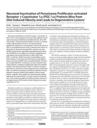
FIGURE 3. High-fat diet-induced obesity. A, body weight of flox/flox, B␣KO, WT, and whole body PGC-1␣ null (KO) mice fed high-fat diet for 10 weeks. *,
p Ͻ 0.001; **, p Ͻ 0.01 B␣KO versus KO group. B, appearance of control and B␣KO mice following high-fat diet feeding. C, epididymal fat (eWAT) weight and
epididymal white adipose tissue to body weight ratio in flox/flox, B␣KO, WT, and whole body PGC-1␣ null (KO) mice. *, p Ͻ 0.02. D and E, plasma glucose (D)
concentration and rectal temperature (E) in high-fat fed flox/flox, B␣KO, WT, and whole body PGC-1␣ null mice (KO). Data represent mean Ϯ S.E. (n ϭ 7–8
per group). *, p Ͻ 0.02; NS, not significant.
Neuronal PGC-1␣ Regulates Energy Balance
39090 JOURNAL OF BIOLOGICAL CHEMISTRY VOLUME 285•NUMBER 50•DECEMBER 10, 2010
atNYUSchoolofMedicineLibrary,onJuly17,2012www.jbc.orgDownloadedfrom
5. regulation of hypothalamic neuropeptides that control energy
balance.
High-fat Diet-fed B␣KO Mice Have Reduced Hepatic
Steatosis—We further examined the impact of neuronal
PGC-1␣ inactivation on hepatic metabolism following
high-fat feeding. Analysis of hepatic triglyceride content
indicates that B␣KO mice have significantly less lipid
accumulation in the liver (Fig. 6A). In fact, liver triglyceride
is reduced by ϳ57% in B␣KO mice. Hepatocyte swelling
and lipid accumulation is evident in high-fat-fed control
mice (Fig. 6B). In contrast, the overall liver appearance
and histology are significantly improved in the B␣KO
group. Analysis of hepatic gene expression indicates that
the expression of several lipogenic genes, including fatty
acid synthase and stearoyl-CoA desaturase as well as
Fsp27, a gene involved in lipid droplet formation, are sig-
nificantly decreased in B␣KO mouse livers (Fig. 6C). The
expression of gluconeogenic genes such as PEPCK and
glucose-6-phosphatase and fatty acid -oxidation genes
remain largely unaltered. These results suggest that
neuronal deletion of PGC-1␣ significantly improves the
metabolic profile in the liver following high-fat diet
feeding.
Region-specific Degenerative Lesions in B␣KO Mouse Brains—
To determine the effects of PGC-1␣ inactivation on neuronal
health, we performed histological analyses in several brain
regions. Consistent with previous findings (21), whole body
PGC-1␣ deficiency leads to spongiform neurodegeneration,
most readily observed in striatum and deep layers of cerebral
cortex (Fig. 7). Remarkably, striatum from B␣KO mouse brain
also contains numerous degenerative lesions that are similar
but slightly smaller than those seen in whole body PGC-1␣
null mice. B␣KO mice tend to have fewer lesions in both stri-
atum and deep cortical layers. Immunohistochemical staining
using an antibody against neurofilament light chain, a marker
for nerve fibers, indicates that the lesions correlate with ap-
parent degeneration of fibers in the striatum from B␣KO
FIGURE 4. CLAMS studies in high-fat diet fed flox/flox and B␣KO mice. A, food intake in flox/flox and B␣KO mice. Data represent food consumption per
day following normalization to body weight (left) or on a per-mouse basis (right). *, p Ͻ 0.01. B, metabolic rate in flox/flox (filled box) and B␣KO mice (open
box). The oxygen consumption rate as normalized to body weight (left) or lean mass (right) is shown. *, p Ͻ 0.05; # p Ͻ 0.09. C, total activity level during
night (N) and day (D) phases. Shown are average movement counts for flox/flox (filled box) and B␣KO (open box) mice. D and E, plasma insulin (D) and leptin
(E) concentrations in high-fat diet-fed flox/flox and B␣KO mice. Data represent mean Ϯ S.E. (n ϭ 7–8 per group). *, p Ͻ 0.02.
FIGURE 5. Hypothalamic gene expression. A, qPCR analysis of gene ex-
pression in hypothalamus of WT and whole body PGC-1␣ null (KO) mice
under fed and fasted conditions. Pooled RNA from 3–5 mice in each group
was used in the analyses. Data represent mean Ϯ S.D. *, p Ͻ 0.005. B, qPCR
analysis of hypothalamic gene expression in flox/flox and B␣KO mice under
fed and fasted conditions. Pooled RNA from 6–8 mice in each group was used.
Data represent mean Ϯ S.D. *, p Ͻ 0.005. TRH, thyrotropin-releasing hormone;
POMC, proopiomelanocortin; MCH, melanin concentrating hormone.
Neuronal PGC-1␣ Regulates Energy Balance
DECEMBER 10, 2010•VOLUME 285•NUMBER 50 JOURNAL OF BIOLOGICAL CHEMISTRY 39091
atNYUSchoolofMedicineLibrary,onJuly17,2012www.jbc.orgDownloadedfrom
6. mice (Fig. 8). Our results strongly suggest that PGC-1␣ is es-
sential for maintaining axon integrity in CaMKII␣-positive
neurons in the brain.
PGC-1␣ Deficiency Does Not Perturb Autophagy in Central
Nervous System—Autophagy is responsible for bulk degrada-
tion of cytoplasmic components in the cell and plays an im-
portant role in nutrient homeostasis during starvation (40–
42). Autophagy is also required for the clearance of organelles,
such as mitochondria (43). Inhibition of autophagy leads to
accumulation of ubiquitinated protein and results in neuronal
death and neurodegeneration (44–46). To determine
whether PGC-1␣ inactivation perturbs autophagy, we per-
formed immunoblotting analyses using antibodies against
LC3 and p62, two molecular markers of autophagy activity
(47–49). LC3 is a cytosolic protein (LC3-I) that, upon autoph-
agy induction, undergoes covalent lipid modification and
translocates to the autophagosome membrane (LC3-II) (50).
Relative abundance of these two LC3 isoforms is indicative of
autophagy activity (45). As shown in Fig. 9, LC3-I and LC3-II
levels are similar in the posterior cortex and striatum of wild
type and whole body PGC-1␣ null mouse brains. Similarly, we
did not observe differences in LC3-I and LC3-II levels in con-
trol and B␣KO mouse brains. In addition, the protein levels of
p62, an LC-3 binding protein that is involved in the formation
of ubiquitin-containing inclusions, also remain largely un-
changed (Fig. 9) (45). We further examined the expression of
genes in the autophagy pathway and found that mRNA levels
of autophagy genes are similar between control and PGC-1␣
null brains (data not shown). Together, these results suggest
that neurodegeneration in PGC-1␣ deficient neurons is un-
likely to be due to impaired neuronal autophagy.
DISCUSSION
The brain is a highly metabolically active tissue and relies
on mitochondrial oxidative metabolism for ATP production
under normal conditions. Not surprisingly, neurons are ex-
quisitely sensitive to perturbations of mitochondrial function.
Impaired mitochondrial energy metabolism has been impli-
cated in numerous heritable neurological disorders as well as
neurodegenerative diseases, including Huntington, Parkinson,
and Alzheimer disease (51–53). PGC-1␣ is abundantly ex-
pressed in the brain and its deficiency leads to degenerative
lesions in several brain regions. However, whether these le-
sions arise from cell-autonomous functions of PGC-1␣ in
neurons remains unknown. In addition, the neuronal cell
types that require PGC-1␣ for their normal function have not
been identified. In this study, we demonstrate that PGC-1␣ in
CaMKII␣-positive neurons plays an important role in main-
taining systemic energy balance and neuronal health.
FIGURE 6. Hepatic triglyceride content and gene expression. A, liver trig-
lyceride content in control (filled box) and B␣KO (open box) mice following
10 weeks of high-fat feeding. *, p Ͻ 0.01. B, H&E (top panel) and Oil Red O
staining (lower panel) of liver sections. C, qPCR analysis of gene expression.
Data represent mean Ϯ S.E. (n ϭ 7–8 per group). *, p Ͻ 0.05. FAS, fatty acid
synthase.
FIGURE 7. H&E staining of brain sections from flox/flox, B␣KO, whole body PGC-1␣ null (KO) mice. Shown are cerebral cortex (cx) and striatum (str)
regions. Lower panel, high magnification of striatum sections. Scale bar, 500 m. Note the absence of clear degenerative lesions in flox/flox mouse brain
(arrow).
Neuronal PGC-1␣ Regulates Energy Balance
39092 JOURNAL OF BIOLOGICAL CHEMISTRY VOLUME 285•NUMBER 50•DECEMBER 10, 2010
atNYUSchoolofMedicineLibrary,onJuly17,2012www.jbc.orgDownloadedfrom
7. We have previously reported that mice lacking PGC-1␣ are
resistant to high-fat diet-induced obesity and have signifi-
cantly improved insulin sensitivity (21). This lean phenotype
is associated with elevated metabolic rate and activity levels.
Because PGC-1␣ is expressed in several key metabolic tissues,
including skeletal muscle, adipose tissues, and liver (13), it
was not immediately clear which tissue(s) underlies altered
systemic energy balance in whole body PGC-1␣ null mice. To
complicate the matter further, PGC-1␣ regulates distinct
metabolic programs in different tissues, which influence sys-
temic metabolism through crosstalk and secondary effects. To
resolve these issues, we generated brain-specific PGC-1␣ null
mice using CaMKII␣-Cre transgenic line. PGC-1␣ is abun-
dantly expressed in pyramidal neurons, many of which also
express CaMKII␣ (54), as well as GABAergic neurons (29, 30).
B␣KO mice are capable of mounting normal metabolic re-
sponses in peripheral tissues. For example, the induction of
genes involved in hepatic gluconeogenesis and fatty acid
-oxidation following starvation is similar in control and
B␣KO mice. In addition, the activation of adaptive thermo-
genesis in brown fat in response to cold exposure is normal in
B␣KO mice. Unlike whole body PGC-1␣ null mice, which are
extremely cold-sensitive, B␣KO mice are indistinguishable
from control mice in maintaining their core body tempera-
ture. These observations allow us to assess the biological
function of neuronal PGC-1␣ in the absence of the confound-
ing metabolic perturbations in peripheral tissues caused by
global PGC-1␣ deficiency.
Perhaps the most remarkable outcome of neuronal PGC-1␣
deficiency is its influence on systemic energy balance when
the mice were fed a high-fat diet. Compared with control
mice, B␣KO mice gain significantly less body weight and have
lower adiposity following 10 weeks of high-fat feeding. The
resistance to weight gain in B␣KO mice appears to be quanti-
tatively less pronounced than whole body PGC-1␣ null mice,
suggesting that PGC-1␣ in CaMKII␣-negative cells in nervous
system and/or peripheral tissues may also contribute to its
effects on systemic energy balance. Additionally, in both
B␣KO and whole body PGC-1␣ null mice, fasting-induced
expression of AgRP and NPY in hypothalamus is impaired in
the absence of PGC-1␣. These observations are consistent
with previous studies that implicate FoxO1, a transcriptional
partner for PGC-1␣, in the regulation of AgRP gene expres-
sion in hypothalamus (55, 56). A recent report showed that
PGC-1␣ expression is present in NPY expressing neurons of
the dorsalmedial hypothalamic nucleus (57). Together, these
findings illustrate a potential role for the FoxO1/PGC-1␣
pathway in the regulation of hypothalamic transcription and
function. The significance of AgRP and NPY in mediating the
effects of PGC-1␣ on energy balance remains unknown at
present.
FIGURE 8. Immunohistochemical staining using antibody against neu-
rofilament light chain. Shown are cerebral cortex (cx), corpus callosum
(cc), and striatum (str) regions in forebrain sections at low (top) and high
(bottom) magnification in flox/flox and B␣KO mice. Note the presence of
small (arrowhead) and large (arrow) lesions in the striatum.
FIGURE 9. Immunoblotting analysis of proteins in the autophagy pathway. Total tissue lysates were prepared from posterior cortex and striatum dis-
sected from WT, whole body PGC-1␣ null (KO), flox/flox, and B␣KO mice. Immunoblotting was performed using indicated antibodies. Two different expo-
sure times were included for LC3. Ponceau S stain was used as loading control.
Neuronal PGC-1␣ Regulates Energy Balance
DECEMBER 10, 2010•VOLUME 285•NUMBER 50 JOURNAL OF BIOLOGICAL CHEMISTRY 39093
atNYUSchoolofMedicineLibrary,onJuly17,2012www.jbc.orgDownloadedfrom
8. Besides less weight gain under high-fat diet, B␣KO mice
also have reduced hepatic lipid content and lower plasma in-
sulin levels, suggesting that these mice are more insulin-sensi-
tive. However, B␣KO mice are similar to control group in
insulin tolerance and glucose tolerance tests. In contrast,
whole body PGC-1␣ null mice have significantly lower blood
glucose levels and improved glucose tolerance. A plausible
explanation for these differences is that hepatic glucose out-
put is impaired in whole body PGC-1␣ null mice, whereas it
remains normal in B␣KO mice. In fact, fasting glucose levels
are similar between control and B␣KO mice when kept on
chow diet. As such, impaired hepatic gluconeogenesis may
significantly contribute to improved glucose homeostasis in
whole body PGC-1␣ null mice.
Surprisingly, although B␣KO mice have increased oxygen
consumption rate in CLAMS studies, these mice appear to
have normal activity levels. In addition, diurnal regulation of
locomotor activity is apparently unaffected in these mice.
These results suggest that distinct neuronal populations
might mediate the function of PGC-1␣ in the regulation of
energy balance and circadian pacemaker. In this case,
PGC-1␣ activity in CaMKII␣-positive neurons is essential for
the control of metabolic rate and body weight homeostasis,
whereas its regulation of biological clock is mediated by a dis-
tinct population of PGC-1␣ expressing neurons. Previous
studies have shown that PGC-1␣ is also expressed in
GABAergic neurons (29). Whether PGC-1␣ in GABAergic
neurons is required for maintaining normal circadian meta-
bolic rhythms remains to be addressed. It is also possible that
certain CaMKII␣ neurons may lack Cre expression. We can-
not rule out the possibility that intact PGC-1␣ expression in a
subset of CaMKII␣-positive neurons could mediate its role in
circadian regulation.
We observed neurodenegerative lesions in the striatum of
B␣KO mouse brain. The striking similarity in the appearance
of vacuoles in B␣KO and whole body PGC-1␣ null mice
strongly suggests that the deficiency of PGC-1␣ in CaMKII␣-
positive neurons is the major neuronal population affected in
whole body PGC-1␣ null mouse brain. Interestingly, we ob-
served fewer lesions in the striatum and deep layers of cere-
bral cortex in B␣KO mice. It remains to be determined
whether this is due to partial deletion of PGC-1␣ in
CaMKII␣-positive neurons or other cell types also contribute
to neurodegeneration in whole body PGC-1␣ null mice. Be-
cause CaMKII␣ is only modestly expressed in the striatum,
our data suggest that the degenerative lesions are most likely
due to the defects of neurons that reside in other brain areas,
such as the cortex. Although autophagy is emerging as an im-
portant mechanism in neuronal homeostasis, we did not ob-
serve significant changes in autophagy activity in whole body
PGC-1␣ null and B␣KO mice compared with their respective
control. It is likely that disruption of mitochondrial function
and reactive oxygen species metabolism may be responsible
for the development of neuronal lesions in the absence of
PGC-1␣.
In summary, we have demonstrated that PGC-1␣ activity in
CaMKII␣ neurons plays a key role in the regulation of energy
balance and neuronal health. The resistance to diet-induced
obesity and brain lesions in B␣KO mice are strikingly similar
to whole body PGC-1␣ null mice. These results strongly sug-
gest that neuronal PGC-1␣ exerts profound effects on the
neural circuitry that governs systemic energy balance.
Acknowledgments—We thank Drs. Jennifer Estall and Bruce
Spiegelman for providing PGC-1␣ flox mice, Joseph Takahashi for
CaMKII␣-Cre transgenic mice, and Geoffrey Murphy for discus-
sions. We are grateful to Shengjuan Gu, Layla Yu, Fernanda Jime-
nez, and Matthew Molusky for technical assistance and other mem-
bers of the laboratory for discussions. We thank Dr. Nathan Qi and
the University of Michigan Animal Metabolic Phenotyping Core for
performing CLAMS study.
REFERENCES
1. Flier, J. S. (2004) Cell 116, 337–350
2. Spiegelman, B. M., and Flier, J. S. (2001) Cell 104, 531–543
3. Erion, D. M., and Shulman, G. I. (2010) Nat. Med. 16, 400–402
4. Beaven, S. W., and Tontonoz, P. (2006) Annu. Rev. Med. 57, 313–329
5. Chawla, A., Repa, J. J., Evans, R. M., and Mangelsdorf, D. J. (2001) Sci-
ence 294, 1866–1870
6. Feige, J. N., and Auwerx, J. (2007) Trends Cell. Biol. 17, 292–301
7. Lin, J. D. (2009) Mol. Endocrinol. 23, 2–10
8. Finck, B. N., and Kelly, D. P. (2006) J. Clin. Invest. 116, 615–622
9. Handschin, C. (2009) Trends Pharmacol Sci. 30, 322–329
10. Kelly, D. P., and Scarpulla, R. C. (2004) Genes Dev. 18, 357–368
11. Lin, J., Handschin, C., and Spiegelman, B. M. (2005) Cell. Metab. 1,
361–370
12. Lin, J., Puigserver, P., Donovan, J., Tarr, P., and Spiegelman, B. M. (2002)
J. Biol. Chem. 277, 1645–1648
13. Puigserver, P., Wu, Z., Park, C. W., Graves, R., Wright, M., and
Spiegelman, B. M. (1998) Cell 92, 829–839
14. Baar, K., Wende, A. R., Jones, T. E., Marison, M., Nolte, L. A., Chen, M.,
Kelly, D. P., and Holloszy, J. O. (2002) Faseb J. 16, 1879–1886
15. Goto, M., Terada, S., Kato, M., Katoh, M., Yokozeki, T., Tabata, I., and
Shimokawa, T. (2000) Biochem. Biophys. Res. Commun. 274, 350–354
16. Wu, Z., Puigserver, P., Andersson, U., Zhang, C., Adelmant, G., Mootha,
V., Troy, A., Cinti, S., Lowell, B., Scarpulla, R. C., and Spiegelman, B. M.
(1999) Cell 98, 115–124
17. Lin, J., Wu, H., Tarr, P. T., Zhang, C. Y., Wu, Z., Boss, O., Michael, L. F.,
Puigserver, P., Isotani, E., Olson, E. N., Lowell, B. B., Bassel-Duby, R.,
and Spiegelman, B. M. (2002) Nature 418, 797–801
18. Handschin, C., Lin, J., Rhee, J., Peyer, A. K., Chin, S., Wu, P. H., Meyer,
U. A., and Spiegelman, B. M. (2005) Cell 122, 505–515
19. Koo, S. H., Satoh, H., Herzig, S., Lee, C. H., Hedrick, S., Kulkarni, R.,
Evans, R. M., Olefsky, J., and Montminy, M. (2004) Nat. Med. 10,
530–534
20. Yoon, J. C., Puigserver, P., Chen, G., Donovan, J., Wu, Z., Rhee, J., Adel-
mant, G., Stafford, J., Kahn, C. R., Granner, D. K., Newgard, C. B., and
Spiegelman, B. M. (2001) Nature 413, 131–138
21. Lin, J., Wu, P. H., Tarr, P. T., Lindenberg, K. S., St-Pierre, J., Zhang,
C. Y., Mootha, V. K., Ja¨ger, S., Vianna, C. R., Reznick, R. M., Cui, L.,
Manieri, M., Donovan, M. X., Wu, Z., Cooper, M. P., Fan, M. C., Rohas,
L. M., Zavacki, A. M., Cinti, S., Shulman, G. I., Lowell, B. B., Krainc, D.,
and Spiegelman, B. M. (2004) Cell 119, 121–135
22. Leone, T. C., Lehman, J. J., Finck, B. N., Schaeffer, P. J., Wende, A. R.,
Boudina, S., Courtois, M., Wozniak, D. F., Sambandam, N., Bernal-
Mizrachi, C., Chen, Z., Holloszy, J. O., Medeiros, D. M., Schmidt, R. E.,
Saffitz, J. E., Abel, E. D., Semenkovich, C. F., and Kelly, D. P. (2005) PLoS
Biol 3, e101
23. Huss, J. M., Imahashi, K., Dufour, C. R., Weinheimer, C. J., Courtois, M.,
Kovacs, A., Gigue`re, V., Murphy, E., and Kelly, D. P. (2007) Cell. Metab.
6, 25–37
24. Arany, Z., He, H., Lin, J., Hoyer, K., Handschin, C., Toka, O., Ahmad, F.,
Matsui, T., Chin, S., Wu, P. H., Rybkin, II, Shelton, J. M., Manieri, M.,
Neuronal PGC-1␣ Regulates Energy Balance
39094 JOURNAL OF BIOLOGICAL CHEMISTRY VOLUME 285•NUMBER 50•DECEMBER 10, 2010
atNYUSchoolofMedicineLibrary,onJuly17,2012www.jbc.orgDownloadedfrom
9. Cinti, S., Schoen, F. J., Bassel-Duby, R., Rosenzweig, A., Ingwall, J. S., and
Spiegelman, B. M. (2005) Cell. Metab. 1, 259–271
25. Arany, Z., Foo, S. Y., Ma, Y., Ruas, J. L., Bommi-Reddy, A., Girnun, G.,
Cooper, M., Laznik, D., Chinsomboon, J., Rangwala, S. M., Baek, K. H.,
Rosenzweig, A., and Spiegelman, B. M. (2008) Nature 451, 1008–1012
26. Mootha, V. K., Lindgren, C. M., Eriksson, K. F., Subramanian, A., Sihag,
S., Lehar, J., Puigserver, P., Carlsson, E., Ridderstråle, M., Laurila, E.,
Houstis, N., Daly, M. J., Patterson, N., Mesirov, J. P., Golub, T. R.,
Tamayo, P., Spiegelman, B., Lander, E. S., Hirschhorn, J. N., Altshuler,
D., and Groop, L. C. (2003) Nat. Genet. 34, 267–273
27. Patti, M. E., Butte, A. J., Crunkhorn, S., Cusi, K., Berria, R., Kashyap, S.,
Miyazaki, Y., Kohane, I., Costello, M., Saccone, R., Landaker, E. J., Gold-
fine, A. B., Mun, E., DeFronzo, R., Finlayson, J., Kahn, C. R., and Man-
darino, L. J. (2003) Proc. Natl. Acad. Sci. U.S.A. 100, 8466–8471
28. Liu, C., Li, S., Liu, T., Borjigin, J., and Lin, J. D. (2007) Nature 447,
477–481
29. Cowell, R. M., Blake, K. R., and Russell, J. W. (2007) J. Comp. Neurol.
502, 1–18
30. Tritos, N. A., Mastaitis, J. W., Kokkotou, E. G., Puigserver, P.,
Spiegelman, B. M., and Maratos-Flier, E. (2003) Brain Res. 961, 255–260
31. Cui, L., Jeong, H., Borovecki, F., Parkhurst, C. N., Tanese, N., and
Krainc, D. (2006) Cell 127, 59–69
32. St-Pierre, J., Drori, S., Uldry, M., Silvaggi, J. M., Rhee, J., Ja¨ger, S., Hand-
schin, C., Zheng, K., Lin, J., Yang, W., Simon, D. K., Bachoo, R., and
Spiegelman, B. M. (2006) Cell 127, 397–408
33. Frayn, K. N. (1983) J. Appl. Physiol. 55, 628–634
34. Simonson, D. C., and DeFronzo, R. A. (1990) Am. J. Physiol. 258,
E399–412
35. Lin, J., Yang, R., Tarr, P. T., Wu, P. H., Handschin, C., Li, S., Yang, W.,
Pei, L., Uldry, M., Tontonoz, P., Newgard, C. B., and Spiegelman, B. M.
(2005) Cell 120, 261–273
36. Li, S., Liu, C., Li, N., Hao, T., Han, T., Hill, D. E., Vidal, M., and Lin, J. D.
(2008) Cell Metab 8, 105–117
37. Liu, X. B., and Jones, E. G. (1996) Proc. Natl. Acad. Sci. U.S.A. 93,
7332–7336
38. Tsien, J. Z., Chen, D. F., Gerber, D., Tom, C., Mercer, E. H., Anderson,
D. J., Mayford, M., Kandel, E. R., and Tonegawa, S. (1996) Cell 87,
1317–1326
39. Casanova, E., Fehsenfeld, S., Mantamadiotis, T., Lemberger, T., Greiner,
E., Stewart, A. F., and Schu¨tz, G. (2001) Genesis 31, 37–42
40. Kuma, A., Hatano, M., Matsui, M., Yamamoto, A., Nakaya, H., Yoshi-
mori, T., Ohsumi, Y., Tokuhisa, T., and Mizushima, N. (2004) Nature
432, 1032–1036
41. Mizushima, N., Yamamoto, A., Matsui, M., Yoshimori, T., and Ohsumi,
Y. (2004) Mol. Biol. Cell. 15, 1101–1111
42. Mortimore, G. E., and Schworer, C. M. (1977) Nature 270, 174–176
43. Sandoval, H., Thiagarajan, P., Dasgupta, S. K., Schumacher, A., Prchal,
J. T., Chen, M., and Wang, J. (2008) Nature 454, 232–235
44. Komatsu, M., Waguri, S., Chiba, T., Murata, S., Iwata, J., Tanida, I.,
Ueno, T., Koike, M., Uchiyama, Y., Kominami, E., and Tanaka, K. (2006)
Nature 441, 880–884
45. Komatsu, M., Waguri, S., Koike, M., Sou, Y. S., Ueno, T., Hara, T., Mi-
zushima, N., Iwata, J., Ezaki, J., Murata, S., Hamazaki, J., Nishito, Y., Ie-
mura, S., Natsume, T., Yanagawa, T., Uwayama, J., Warabi, E., Yoshida,
H., Ishii, T., Kobayashi, A., Yamamoto, M., Yue, Z., Uchiyama, Y., Komi-
nami, E., and Tanaka, K. (2007) Cell 131, 1149–1163
46. Komatsu, M., Wang, Q. J., Holstein, G. R., Friedrich, V. L., Jr., Iwata, J.,
Kominami, E., Chait, B. T., Tanaka, K., and Yue, Z. (2007) Proc. Natl.
Acad. Sci. U.S.A. 104, 14489–14494
47. Klionsky, D. J., Cuervo, A. M., and Seglen, P. O. (2007) Autophagy 3,
181–206
48. Mizushima, N. (2004) Int. J. Biochem. Cell. Biol. 36, 2491–2502
49. Mizushima, N., Yoshimori, T., and Levine, B. (2010) Cell 140, 313–326
50. Kabeya, Y., Mizushima, N., Ueno, T., Yamamoto, A., Kirisako, T., Noda,
T., Kominami, E., Ohsumi, Y., and Yoshimori, T. (2000) EMBO J. 19,
5720–5728
51. DiMauro, S., and Schon, E. A. (2008) Annu. Rev. Neurosci. 31, 91–123
52. Lin, M. T., and Beal, M. F. (2006) Nature 443, 787–795
53. Schon, E. A., and Manfredi, G. (2003) J. Clin. Invest. 111, 303–312
54. Ouimet, C. C., McGuinness, T. L., and Greengard, P. (1984) Proc. Natl.
Acad. Sci. U.S.A. 81, 5604–5608
55. Kitamura, T., Feng, Y., Kitamura, Y. I., Chua, S. C., Jr., Xu, A. W., Barsh,
G. S., Rossetti, L., and Accili, D. (2006) Nat Med 12, 534–540
56. Puigserver, P., Rhee, J., Donovan, J., Walkey, C. J., Yoon, J. C., Oriente,
F., Kitamura, Y., Altomonte, J., Dong, H., Accili, D., and Spiegelman,
B. M. (2003) Nature 423, 550–555
57. Draper, S., Kirigiti, M., Glavas, M., Grayson, B., Chong, C. N., Jiang, B.,
Smith, M. S., Zeltser, L. M., and Grove, K. L. (2010) Brain Res. 1350,
139–150
Neuronal PGC-1␣ Regulates Energy Balance
DECEMBER 10, 2010•VOLUME 285•NUMBER 50 JOURNAL OF BIOLOGICAL CHEMISTRY 39095
atNYUSchoolofMedicineLibrary,onJuly17,2012www.jbc.orgDownloadedfrom