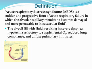
Dr. Dipali Dumbre
Ph.D Nursing
Medical Surgical Nursing
SCON
Definition
“Acute respiratory distress syndrome (ARDS) is a
sudden and progressive form of acute respiratory failure in
which the alveolar capillary membrane becomes damaged
and more permeable to intravascular fluid”.
 The alveoli fill with fluid, resulting in severe dyspnea,
hypoxemia refractory to supplemental O2, reduced lung
compliance, and diffuse pulmonary infiltrates

Etiology
 Direct lung injury may cause ARDS.
 ARDS may develop as a consequence of the systemic
inflammatory response syndrome (SIRS).
 SIRS may have an infectious or a noninfectious etiology
and is characterized by widespread inflammation or
clinical responses to inflammation following a variety of
physiologic insults, including severe trauma, lung injury,
and sepsis.
 ARDS may also develop as a consequence of multiple organ
dysfunction syndrome (MODS).
 MODS results from organ system dysfunction that
progressively increases in severity and ultimately results in
multisystem organ failure.
 An exact cause for the damage to the alveolar-capillary
membrane is not known.
 However, the pathophysiologic changes of ARDS are
thought to be due to stimulation of the inflammatory and
immune systems, which causes an attraction of neutrophils
to the pulmonary interstitium.(a collection of support tissues
within the lung that includes the alveolar epithelium, pulmonary capillary
endothelium, basement membrane, perivascular and perilymphatic tissues
 The neutrophils cause a release of biochemical, humoral,
and cellular mediators that produce changes in the lung,
including increased pulmonary capillary membrane
permeability, destruction of elastin(alveolar wall) and
collagen, formation of pulmonary micro emboli, and
pulmonary artery vasoconstriction.
Predisposing Conditions to Acute Respiratory
Distress Syndrome
DIRECT LUNG INJURY INDIRECT LUNG INJURY
Common Causes
Aspiration of gastric contents or other
substances
Viral/bacterial pneumonia
Less Common Causes
Chest trauma
Embolism: fat, air, amniotic fluid,
Thrombus
Inhalation of toxic substances
Near-drowning
O2 toxicity
Radiation pneumonitis
Common Causes
Sepsis (especially gram-negative infection)
Severe massive trauma
Less Common Causes
Acute pancreatitis
Anaphylaxis
Cardiopulmonary bypass
Disseminated intravascular coagulation
Multiple blood transfusions
Opioid drug overdose (e.g., heroin)
Nonpulmonary systemic diseases
Severe head injury
Shock states
PATHOPHYSIOLOGY
 The pathophysiologic changes in ARDS are divided
into three phases:
 (1) Injury or exudative phase,
 (2) Reparative or proliferative phase
 (3) Fibrotic phase.
Injury or Exudative Phase
 The injury or exudative phase occurs approximately 1 to 7
days (usually 24 to 48 hours) after the initial direct lung
injury .
 Neutrophils adhere to the pulmonary microcirculation,
causing damage to the vascular endothelium and
increased capillary permeability.
 In the earliest phase of injury, there is engorgement of the
peribronchial and perivascular interstitial space, which
produces interstitial edema.
 Next, fluid from the interstitial space crosses the alveolar
epithelium and enters the alveolar space.
 Intrapulmonary shunt develops because the alveoli fill
with fluid, and blood passing through them cannot be
oxygenated
 Alveolar type I and type II cells (which produce surfactant)
are damaged by the changes caused by ARDS.
 This damage, in addition to further fluid and protein
accumulation, results in surfactant dysfunction.
 The function of surfactant is to maintain alveolar stability
by decreasing alveolar surface tension and preventing
alveolar collapse.
 Decreased synthesis of surfactant and inactivation of
existing surfactant cause the alveoli to become unstable
and collapse (atelectasis).
 Widespread atelectasis further decreases lung compliance,
compromises gas exchange, and contributes to hypoxemia
 Type I alveolar cells are squamous (giving more surface
area to each cell) and cover approximately 90–95% of
the alveolar surface. Type I cells are involved in the
process of gas exchange between the alveoli and blood.
 Type II cells in the alveolar wall contain secretory
granular organelles known as lamellar bodies that fuse
with the cell membranes and secrete pulmonary
surfactant.
 This surfactant is a film of fatty substances, a group
of phospholipids that reduce alveolar surface tension.
 Also during this stage, hyaline membranes begin to line
the alveoli.
 The hyaline membrane is composed of necrotic cells,
protein, and fibrin and lies adjacent to the alveoli wall.
 These hyaline membranes are thought to result from the
exudation of high-molecular-weight substances
(particularly fibrinogen) in the edema fluid.
 Hyaline membranes contribute to the development of
fibrosis and atelectasis, leading to a decrease in gas
exchange capability and lung compliance.
 The primary pathophysiologic changes that characterize
the injury or exudative phase of ARDS are interstitial and
alveolar edema and atelectasis.
 Severe V/Q mismatch and shunting of pulmonary capillary
blood result in hypoxemia unresponsive to increasing
concentrations of O2 (termed refractory hypoxemia).
 Diffusion limitation(occurs when gas exchange across the
alveolar-capillary membrane is compromised by a process
that thickens or destroys the membrane ), caused by
hyaline membrane formation, further contributes to the
severity of the hypoxemia.
 As the lungs become less compliant because of decreased
surfactant, pulmonary edema, and atelectasis, the patient
must generate higher airway pressures to inflate “stiff”
lungs.
 Reduced lung compliance greatly increases the patient's
work of breathing.
 Progressive increase in pressures required to deliver a
controlled ventilation may occur as a result of worsening lung
compliance.
 Hypoxemia and the stimulation of juxtacapillary receptors(J-
receptors (juxtacapillary) are nerves innervating into the body
of the lung. J-receptors (respond to events such as pulmonary edema ,
pulmonary emboli pneumonia, and barotrauma, which cause a decrease in
oxygenation and thus lead to an increase in ventilation/respiration.) in the
stiff lung parenchyma (J reflex) initially cause an increase in
respiratory rate and a decrease in tidal volume.
 This breathing pattern increases CO2 removal, producing
respiratory alkalosis.
 Cardiac output increases in response to hypoxemia, a
compensatory effort to increase pulmonary blood flow.
 However, as atelectasis, pulmonary edema, and pulmonary
shunt increase, compensation fails, and hypoventilation,
decreased cardiac output, and decreased tissue O2 perfusion
eventually occur.
Reparative or Proliferative Phase
 The reparative or proliferative phase of ARDS begins 1 to 2
weeks after the initial lung injury.
 During this phase, there is an influx of neutrophils,
monocytes, and lymphocytes and fibroblast proliferation
as part of the inflammatory response.
 The injured lung has an immense regenerative capacity
after acute lung injury.
 The proliferative phase is complete when the diseased lung
becomes characterized by dense, fibrous tissue.
 Increased pulmonary vascular resistance and pulmonary
hypertension may occur in this stage because fibroblasts
and inflammatory cells destroy the pulmonary vasculature.
 Lung compliance continues to decrease as a result of
interstitial fibrosis.
 Hypoxemia worsens because of the thickened alveolar
membrane, causing diffusion limitation and shunting.
 If the reparative phase persists, widespread fibrosis
results. If the reparative phase is arrested, the lesions
resolve.
Fibrotic Phase
 The fibrotic phase of ARDS occurs approximately 2 to 3
weeks after the initial lung injury.
 This phase is also called the chronic or late phase of ARDS.
By this time, the lung is completely remodeled by sparsely
collagenous and fibrous tissues.
 There is diffuse scarring and fibrosis, resulting in
decreased lung compliance.
 In addition, the surface area for gas exchange is
significantly reduced because the interstitium is fibrotic,
and therefore hypoxemia continues.
 Pulmonary hypertension results from pulmonary vascular
destruction and fibrosis.
Clinical Manifestations
 The initial presentation of ARDS is often insidious.
 At the time of the initial injury, and for several hours to 1 to
2 days afterward, the patient may not experience
respiratory symptoms, or the patient may exhibit only
dyspnea, tachypnea, cough, and restlessness.
 Chest auscultation may be normal or reveal fine, scattered
crackles.
 ABGs usually indicate mild hypoxemia and respiratory
alkalosis caused by hyperventilation.
 Respiratory alkalosis results from hypoxemia and the
stimulation of juxtacapillary receptors.
 The chest x-ray may be normal or exhibit evidence of
minimal scattered interstitial infiltrates.
 Edema may not show on the x-ray until there is a 30%
increase in fluid content in the lung.
 As ARDS progresses, symptoms worsen because of
increased fluid accumulation and decreased lung
compliance.
 Respiratory discomfort becomes evident as the work of
breathing increases.
 Tachypnea and intercostal and suprasternal retractions
may be present.
 Pulmonary function tests in ARDS reveal decreased
compliance and decreased lung volumes, particularly a
decreased functional residual capacity (is the volume of air
present in the lungs at the end of passive expiration).
 Tachycardia, diaphoresis, changes in sensorium with
decreased mentation, cyanosis, and pallor may be present.
 Chest auscultation usually reveals scattered to diffuse
crackles and rhonchi.
 The chest x-ray demonstrates diffuse and extensive
bilateral interstitial and alveolar infiltrates.
 A pulmonary artery catheter may be inserted.
 Hypoxemia and a PaO2/FIO2 ratio below 200 (e.g., 80/0.8
= 100) despite increased FIO2 by mask, cannula, or
endotracheal tube are hallmarks of ARDS.
 ABGs may initially demonstrate a normal or decreased
PaCO2 despite severe dyspnea and hypoxemia.
 Hypercapnia signifies that hypoventilation is occurring,
and the patient is no longer able to maintain the level of
ventilation needed to provide optimum gas exchange.
 As ARDS progresses, it is associated with profound
respiratory distress requiring endotracheal intubation and
PPV.
 The chest x-ray is often termed whiteout or white lung,
because consolidation and coalescing infiltrates are
widespread throughout the lungs, leaving few recognizable
air spaces.
 Pleural effusions may also be present. Severe hypoxemia,
hypercapnia, and metabolic acidosis, with symptoms of
target organ or tissue hypoxia, may ensue if prompt
therapy is not given.
Complications
 Hospital-Acquired Pneumonia.
 A frequent complication of ARDS is hospital-acquired
pneumonia, occurring in as many as 68% of patients with
ARDS.
 Risk factors include impaired host defenses, contaminated
medical equipment, invasive monitoring devices,
aspiration of GI contents (especially in patients receiving
tube feedings), and prolonged mechanical ventilation, as
well as colonization of the respiratory tract.
 Strategies to prevent hospital-acquired pneumonia include
infection control measures (e.g., strict hand washing and
sterile technique during endotracheal suctioning) and
elevating the head of the bed 30 to 45 degrees to prevent
aspiration.
 Barotrauma.
 Barotrauma may result from rupture of overdistended alveoli
during mechanical ventilation.
 The high peak airway pressures that may be required in patients
with ARDS predispose to this complication.
 Barotrauma results in the presence of alveolar air in locations
where it is not usually found.
 This can lead to pulmonary interstitial emphysema,
pneumothorax, subcutaneous emphysema,
pneumoperitoneum, pneumomediastinum,
pneumopericardium, and tension pneumothorax.
 To avoid barotrauma and minimize risk associated with
elevated plateau and peak inspiratory pressures, the patient
with ARDS may be ventilated with smaller tidal volumes (e.g., 6
ml/kg) and varying amounts of positive end-expiratory pressure
(PEEP) in order to minimize oxygen requirements and
intrathoracic pressures.
Volu-Pressure Trauma
 Volu-pressure trauma can occur in patients with ARDS
when large tidal volumes (e.g., 10 to 15 ml/kg) are used to
ventilate noncompliant lungs.
 Volu-pressure trauma results in alveolar fractures and
movement of fluids and proteins into the alveolar spaces.
 To limit this complication, it is recommended that smaller
tidal volumes or pressure ventilation be used in patients
with ARDS.
 Physiologic Stress Ulcers.
 Critically ill patients with acute respiratory failure are at
high risk for stress ulcers.
 Bleeding from stress ulcers occurs in 30% of patients
with ARDS who require PPV, a higher incidence than
other causes of acute respiratory failure.
 Management strategies include correction of
predisposing conditions such as hypotension, shock, and
acidosis.
 Prophylactic management includes antiulcer agents
such as H2-histamine receptor antagonists (e.g.,
ranitidine [Zantac]), as well as proton pump inhibitors
(e.g., pantoprazole [Protonix]) and mucosal-protecting
agents (e.g., sucralfate [Carafate]).
 Early initiation of enteral nutrition also helps prevent
mucosal damage
Renal Failure
 Renal failure can occur from decreased renal tissue
oxygenation as a result of hypotension, hypoxemia, or
hypercapnia.
 Renal failure may also be caused by administration of
nephrotoxic drugs (e.g., aminoglycosides), which are
used to treat infections associated with ARDS.
Diagnostic Findings
Refractory Hypoxemia
 PaO2 <50 mm Hg on FIO2 >40% with PEEP >5 cm H2O
 PaO2/FIO2 ratio <200
Chest X-Ray
 New bilateral interstitial and alveolar infiltrates
Pulmonary Artery Wedge Pressure
 ≤18 mm Hg and no evidence of heart failure
Predisposing Condition
 Identification of a predisposing condition for ARDS within
48 hr of clinical manifestations
COLLABORATIVE THERAPY
Respiratory Therapy
Oxygen Administration
Mechanical Ventilation:
Endotracheal intubation and mechanical ventilation provide
additional respiratory support.
During mechanical ventilation, it is common to apply PEEP at
5 cm H2O to compensate for loss of glottic function caused by
the presence of the endotracheal tube.
In patients with ARDS, higher levels of PEEP (e.g., 10 to 20 cm
H2O) may be used.
The mechanism of action of PEEP is related to its ability to
increase FRC and recruit (open up) collapsed alveoli.
PEEP is typically applied in 3 to 5 cm H2O increments until
oxygenation is adequate with FIO2 of 60% or less.
PEEP may improve V/Q in respiratory units that collapse at low
airway pressures, thus allowing the FIO2 to be lowered.
 Positioning Strategies
 Some patients with ARDS demonstrate a marked
improvement in PaO2 when turned from the supine to the
prone position (e.g., PaO2 70 mm Hg supine, PaO2 90 mm
Hg the prone) with no change in inspired O2
concentration
 In the early phases of ARDS, fluid moves freely
throughout the lung.
 Because of gravity, this fluid pools in dependent regions of
the lung.
 As a consequence, some alveoli are fluid filled (dependent
areas), whereas others are air filled (nondependent areas).
 In addition, when the patient is supine the heart and
mediastinal contents place more pressure on the lungs
than in the prone position, which changes pleural pressure
and predisposes to atelectasis.
 If the patient is turned from supine to prone, air-filled,
nonatelectatic alveoli in the ventral (anterior) portion of
the lung become dependent.
 Perfusion may be better matched to ventilation, causing
less V/Q mismatch.
 Not all patients respond to prone positioning with an
increase in PaO2, and there is no reliable way of predicting
who will respond.
 Prone positioning is typically reserved for patients with
refractory hypoxemia who do not respond to other
strategies to increase PaO2.
 When this positioning strategy is used, there must be a
plan in place for immediate repositioning for
cardiopulmonary resuscitation in the event of a cardiac
arrest.
 Other positioning strategies that can be considered for
patients with ARDS include continuous lateral rotation
therapy (CLRT) and kinetic therapy.
 The purpose of CLRT is to provide continuous, slow, side-
to-side turning of the patient by rotating the actual bed
frame less than 40 degrees.
 The lateral movement of the bed is maintained for 18 of
every 24 hours to simulate postural drainage and to help
mobilize pulmonary secretions.

ARDS.pptx

  • 1.
    Dr. Dipali Dumbre Ph.DNursing Medical Surgical Nursing SCON
  • 2.
    Definition “Acute respiratory distresssyndrome (ARDS) is a sudden and progressive form of acute respiratory failure in which the alveolar capillary membrane becomes damaged and more permeable to intravascular fluid”.  The alveoli fill with fluid, resulting in severe dyspnea, hypoxemia refractory to supplemental O2, reduced lung compliance, and diffuse pulmonary infiltrates 
  • 3.
    Etiology  Direct lunginjury may cause ARDS.  ARDS may develop as a consequence of the systemic inflammatory response syndrome (SIRS).  SIRS may have an infectious or a noninfectious etiology and is characterized by widespread inflammation or clinical responses to inflammation following a variety of physiologic insults, including severe trauma, lung injury, and sepsis.  ARDS may also develop as a consequence of multiple organ dysfunction syndrome (MODS).  MODS results from organ system dysfunction that progressively increases in severity and ultimately results in multisystem organ failure.
  • 4.
     An exactcause for the damage to the alveolar-capillary membrane is not known.  However, the pathophysiologic changes of ARDS are thought to be due to stimulation of the inflammatory and immune systems, which causes an attraction of neutrophils to the pulmonary interstitium.(a collection of support tissues within the lung that includes the alveolar epithelium, pulmonary capillary endothelium, basement membrane, perivascular and perilymphatic tissues  The neutrophils cause a release of biochemical, humoral, and cellular mediators that produce changes in the lung, including increased pulmonary capillary membrane permeability, destruction of elastin(alveolar wall) and collagen, formation of pulmonary micro emboli, and pulmonary artery vasoconstriction.
  • 5.
    Predisposing Conditions toAcute Respiratory Distress Syndrome DIRECT LUNG INJURY INDIRECT LUNG INJURY Common Causes Aspiration of gastric contents or other substances Viral/bacterial pneumonia Less Common Causes Chest trauma Embolism: fat, air, amniotic fluid, Thrombus Inhalation of toxic substances Near-drowning O2 toxicity Radiation pneumonitis Common Causes Sepsis (especially gram-negative infection) Severe massive trauma Less Common Causes Acute pancreatitis Anaphylaxis Cardiopulmonary bypass Disseminated intravascular coagulation Multiple blood transfusions Opioid drug overdose (e.g., heroin) Nonpulmonary systemic diseases Severe head injury Shock states
  • 6.
    PATHOPHYSIOLOGY  The pathophysiologicchanges in ARDS are divided into three phases:  (1) Injury or exudative phase,  (2) Reparative or proliferative phase  (3) Fibrotic phase.
  • 7.
    Injury or ExudativePhase  The injury or exudative phase occurs approximately 1 to 7 days (usually 24 to 48 hours) after the initial direct lung injury .  Neutrophils adhere to the pulmonary microcirculation, causing damage to the vascular endothelium and increased capillary permeability.  In the earliest phase of injury, there is engorgement of the peribronchial and perivascular interstitial space, which produces interstitial edema.  Next, fluid from the interstitial space crosses the alveolar epithelium and enters the alveolar space.  Intrapulmonary shunt develops because the alveoli fill with fluid, and blood passing through them cannot be oxygenated
  • 8.
     Alveolar typeI and type II cells (which produce surfactant) are damaged by the changes caused by ARDS.  This damage, in addition to further fluid and protein accumulation, results in surfactant dysfunction.  The function of surfactant is to maintain alveolar stability by decreasing alveolar surface tension and preventing alveolar collapse.  Decreased synthesis of surfactant and inactivation of existing surfactant cause the alveoli to become unstable and collapse (atelectasis).  Widespread atelectasis further decreases lung compliance, compromises gas exchange, and contributes to hypoxemia
  • 9.
     Type Ialveolar cells are squamous (giving more surface area to each cell) and cover approximately 90–95% of the alveolar surface. Type I cells are involved in the process of gas exchange between the alveoli and blood.
  • 10.
     Type IIcells in the alveolar wall contain secretory granular organelles known as lamellar bodies that fuse with the cell membranes and secrete pulmonary surfactant.  This surfactant is a film of fatty substances, a group of phospholipids that reduce alveolar surface tension.
  • 12.
     Also duringthis stage, hyaline membranes begin to line the alveoli.  The hyaline membrane is composed of necrotic cells, protein, and fibrin and lies adjacent to the alveoli wall.  These hyaline membranes are thought to result from the exudation of high-molecular-weight substances (particularly fibrinogen) in the edema fluid.  Hyaline membranes contribute to the development of fibrosis and atelectasis, leading to a decrease in gas exchange capability and lung compliance.  The primary pathophysiologic changes that characterize the injury or exudative phase of ARDS are interstitial and alveolar edema and atelectasis.  Severe V/Q mismatch and shunting of pulmonary capillary blood result in hypoxemia unresponsive to increasing concentrations of O2 (termed refractory hypoxemia).
  • 14.
     Diffusion limitation(occurswhen gas exchange across the alveolar-capillary membrane is compromised by a process that thickens or destroys the membrane ), caused by hyaline membrane formation, further contributes to the severity of the hypoxemia.  As the lungs become less compliant because of decreased surfactant, pulmonary edema, and atelectasis, the patient must generate higher airway pressures to inflate “stiff” lungs.  Reduced lung compliance greatly increases the patient's work of breathing.
  • 15.
     Progressive increasein pressures required to deliver a controlled ventilation may occur as a result of worsening lung compliance.  Hypoxemia and the stimulation of juxtacapillary receptors(J- receptors (juxtacapillary) are nerves innervating into the body of the lung. J-receptors (respond to events such as pulmonary edema , pulmonary emboli pneumonia, and barotrauma, which cause a decrease in oxygenation and thus lead to an increase in ventilation/respiration.) in the stiff lung parenchyma (J reflex) initially cause an increase in respiratory rate and a decrease in tidal volume.  This breathing pattern increases CO2 removal, producing respiratory alkalosis.  Cardiac output increases in response to hypoxemia, a compensatory effort to increase pulmonary blood flow.  However, as atelectasis, pulmonary edema, and pulmonary shunt increase, compensation fails, and hypoventilation, decreased cardiac output, and decreased tissue O2 perfusion eventually occur.
  • 16.
    Reparative or ProliferativePhase  The reparative or proliferative phase of ARDS begins 1 to 2 weeks after the initial lung injury.  During this phase, there is an influx of neutrophils, monocytes, and lymphocytes and fibroblast proliferation as part of the inflammatory response.  The injured lung has an immense regenerative capacity after acute lung injury.  The proliferative phase is complete when the diseased lung becomes characterized by dense, fibrous tissue.  Increased pulmonary vascular resistance and pulmonary hypertension may occur in this stage because fibroblasts and inflammatory cells destroy the pulmonary vasculature.
  • 17.
     Lung compliancecontinues to decrease as a result of interstitial fibrosis.  Hypoxemia worsens because of the thickened alveolar membrane, causing diffusion limitation and shunting.  If the reparative phase persists, widespread fibrosis results. If the reparative phase is arrested, the lesions resolve.
  • 18.
    Fibrotic Phase  Thefibrotic phase of ARDS occurs approximately 2 to 3 weeks after the initial lung injury.  This phase is also called the chronic or late phase of ARDS. By this time, the lung is completely remodeled by sparsely collagenous and fibrous tissues.  There is diffuse scarring and fibrosis, resulting in decreased lung compliance.  In addition, the surface area for gas exchange is significantly reduced because the interstitium is fibrotic, and therefore hypoxemia continues.  Pulmonary hypertension results from pulmonary vascular destruction and fibrosis.
  • 20.
    Clinical Manifestations  Theinitial presentation of ARDS is often insidious.  At the time of the initial injury, and for several hours to 1 to 2 days afterward, the patient may not experience respiratory symptoms, or the patient may exhibit only dyspnea, tachypnea, cough, and restlessness.  Chest auscultation may be normal or reveal fine, scattered crackles.  ABGs usually indicate mild hypoxemia and respiratory alkalosis caused by hyperventilation.  Respiratory alkalosis results from hypoxemia and the stimulation of juxtacapillary receptors.
  • 21.
     The chestx-ray may be normal or exhibit evidence of minimal scattered interstitial infiltrates.  Edema may not show on the x-ray until there is a 30% increase in fluid content in the lung.  As ARDS progresses, symptoms worsen because of increased fluid accumulation and decreased lung compliance.  Respiratory discomfort becomes evident as the work of breathing increases.  Tachypnea and intercostal and suprasternal retractions may be present.
  • 22.
     Pulmonary functiontests in ARDS reveal decreased compliance and decreased lung volumes, particularly a decreased functional residual capacity (is the volume of air present in the lungs at the end of passive expiration).  Tachycardia, diaphoresis, changes in sensorium with decreased mentation, cyanosis, and pallor may be present.  Chest auscultation usually reveals scattered to diffuse crackles and rhonchi.  The chest x-ray demonstrates diffuse and extensive bilateral interstitial and alveolar infiltrates.  A pulmonary artery catheter may be inserted.  Hypoxemia and a PaO2/FIO2 ratio below 200 (e.g., 80/0.8 = 100) despite increased FIO2 by mask, cannula, or endotracheal tube are hallmarks of ARDS.
  • 23.
     ABGs mayinitially demonstrate a normal or decreased PaCO2 despite severe dyspnea and hypoxemia.  Hypercapnia signifies that hypoventilation is occurring, and the patient is no longer able to maintain the level of ventilation needed to provide optimum gas exchange.  As ARDS progresses, it is associated with profound respiratory distress requiring endotracheal intubation and PPV.  The chest x-ray is often termed whiteout or white lung, because consolidation and coalescing infiltrates are widespread throughout the lungs, leaving few recognizable air spaces.  Pleural effusions may also be present. Severe hypoxemia, hypercapnia, and metabolic acidosis, with symptoms of target organ or tissue hypoxia, may ensue if prompt therapy is not given.
  • 24.
    Complications  Hospital-Acquired Pneumonia. A frequent complication of ARDS is hospital-acquired pneumonia, occurring in as many as 68% of patients with ARDS.  Risk factors include impaired host defenses, contaminated medical equipment, invasive monitoring devices, aspiration of GI contents (especially in patients receiving tube feedings), and prolonged mechanical ventilation, as well as colonization of the respiratory tract.  Strategies to prevent hospital-acquired pneumonia include infection control measures (e.g., strict hand washing and sterile technique during endotracheal suctioning) and elevating the head of the bed 30 to 45 degrees to prevent aspiration.
  • 25.
     Barotrauma.  Barotraumamay result from rupture of overdistended alveoli during mechanical ventilation.  The high peak airway pressures that may be required in patients with ARDS predispose to this complication.  Barotrauma results in the presence of alveolar air in locations where it is not usually found.  This can lead to pulmonary interstitial emphysema, pneumothorax, subcutaneous emphysema, pneumoperitoneum, pneumomediastinum, pneumopericardium, and tension pneumothorax.  To avoid barotrauma and minimize risk associated with elevated plateau and peak inspiratory pressures, the patient with ARDS may be ventilated with smaller tidal volumes (e.g., 6 ml/kg) and varying amounts of positive end-expiratory pressure (PEEP) in order to minimize oxygen requirements and intrathoracic pressures.
  • 26.
    Volu-Pressure Trauma  Volu-pressuretrauma can occur in patients with ARDS when large tidal volumes (e.g., 10 to 15 ml/kg) are used to ventilate noncompliant lungs.  Volu-pressure trauma results in alveolar fractures and movement of fluids and proteins into the alveolar spaces.  To limit this complication, it is recommended that smaller tidal volumes or pressure ventilation be used in patients with ARDS.
  • 27.
     Physiologic StressUlcers.  Critically ill patients with acute respiratory failure are at high risk for stress ulcers.  Bleeding from stress ulcers occurs in 30% of patients with ARDS who require PPV, a higher incidence than other causes of acute respiratory failure.  Management strategies include correction of predisposing conditions such as hypotension, shock, and acidosis.  Prophylactic management includes antiulcer agents such as H2-histamine receptor antagonists (e.g., ranitidine [Zantac]), as well as proton pump inhibitors (e.g., pantoprazole [Protonix]) and mucosal-protecting agents (e.g., sucralfate [Carafate]).  Early initiation of enteral nutrition also helps prevent mucosal damage
  • 28.
    Renal Failure  Renalfailure can occur from decreased renal tissue oxygenation as a result of hypotension, hypoxemia, or hypercapnia.  Renal failure may also be caused by administration of nephrotoxic drugs (e.g., aminoglycosides), which are used to treat infections associated with ARDS.
  • 29.
    Diagnostic Findings Refractory Hypoxemia PaO2 <50 mm Hg on FIO2 >40% with PEEP >5 cm H2O  PaO2/FIO2 ratio <200 Chest X-Ray  New bilateral interstitial and alveolar infiltrates Pulmonary Artery Wedge Pressure  ≤18 mm Hg and no evidence of heart failure Predisposing Condition  Identification of a predisposing condition for ARDS within 48 hr of clinical manifestations
  • 30.
    COLLABORATIVE THERAPY Respiratory Therapy OxygenAdministration Mechanical Ventilation: Endotracheal intubation and mechanical ventilation provide additional respiratory support. During mechanical ventilation, it is common to apply PEEP at 5 cm H2O to compensate for loss of glottic function caused by the presence of the endotracheal tube. In patients with ARDS, higher levels of PEEP (e.g., 10 to 20 cm H2O) may be used. The mechanism of action of PEEP is related to its ability to increase FRC and recruit (open up) collapsed alveoli. PEEP is typically applied in 3 to 5 cm H2O increments until oxygenation is adequate with FIO2 of 60% or less. PEEP may improve V/Q in respiratory units that collapse at low airway pressures, thus allowing the FIO2 to be lowered.
  • 31.
     Positioning Strategies Some patients with ARDS demonstrate a marked improvement in PaO2 when turned from the supine to the prone position (e.g., PaO2 70 mm Hg supine, PaO2 90 mm Hg the prone) with no change in inspired O2 concentration
  • 32.
     In theearly phases of ARDS, fluid moves freely throughout the lung.  Because of gravity, this fluid pools in dependent regions of the lung.  As a consequence, some alveoli are fluid filled (dependent areas), whereas others are air filled (nondependent areas).  In addition, when the patient is supine the heart and mediastinal contents place more pressure on the lungs than in the prone position, which changes pleural pressure and predisposes to atelectasis.  If the patient is turned from supine to prone, air-filled, nonatelectatic alveoli in the ventral (anterior) portion of the lung become dependent.
  • 33.
     Perfusion maybe better matched to ventilation, causing less V/Q mismatch.  Not all patients respond to prone positioning with an increase in PaO2, and there is no reliable way of predicting who will respond.  Prone positioning is typically reserved for patients with refractory hypoxemia who do not respond to other strategies to increase PaO2.  When this positioning strategy is used, there must be a plan in place for immediate repositioning for cardiopulmonary resuscitation in the event of a cardiac arrest.
  • 34.
     Other positioningstrategies that can be considered for patients with ARDS include continuous lateral rotation therapy (CLRT) and kinetic therapy.  The purpose of CLRT is to provide continuous, slow, side- to-side turning of the patient by rotating the actual bed frame less than 40 degrees.  The lateral movement of the bed is maintained for 18 of every 24 hours to simulate postural drainage and to help mobilize pulmonary secretions.