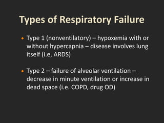
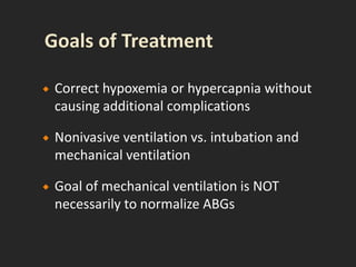
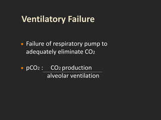
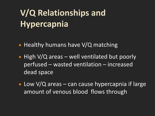
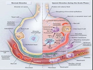
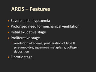
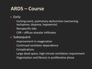
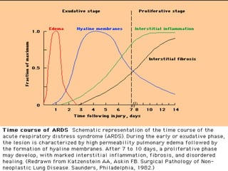
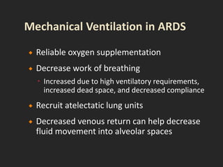
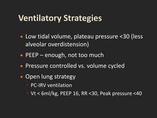
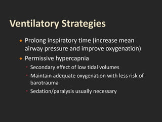
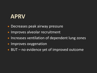
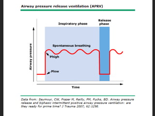
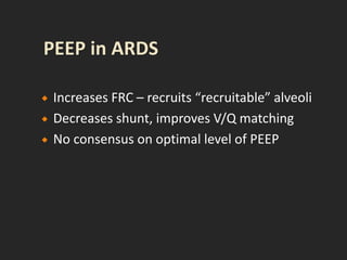
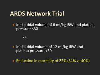
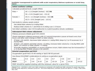
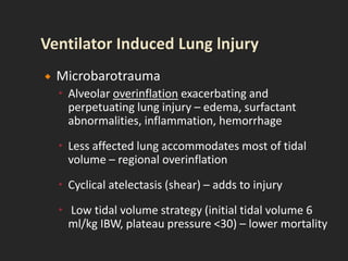
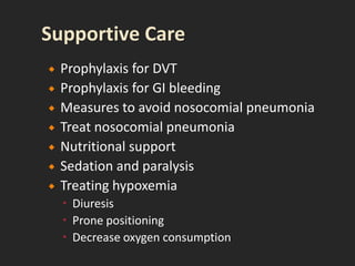
Ian B. Hoffman, MD, FCCP
Pulmonary & Critical Care Medicine
 Any disruption of function of respiratory
system – CNS, nerves, muscles, pleura, lungs
 Any process resulting in low pO2 or high pCO2 –
arbitrarily 50/50
 Acute respiratory failure can be exacerbation
of chronic disease or acute process in
previously healthy lungs
 1940’s – polio, barbiturate OD
 1960’s – blood gas analysis readily available,
aware of hypoxemia
 1970’s – decreased hypoxic mortality,
increased multiorgan failure (living longer)
 1973 – relationship between resp muscle
fatigue and resp failure
 Type 1 (nonventilatory) – hypoxemia with or
without hypercapnia – disease involves lung
itself (i.e, ARDS)
 Type 2 – failure of alveolar ventilation –
decrease in minute ventilation or increase in
dead space (i.e. COPD, drug OD)
 Correct hypoxemia or hypercapnia without
causing additional complications
 Nonivasive ventilation vs. intubation and
mechanical ventilation
 Goal of mechanical ventilation is NOT
necessarily to normalize ABGs
 Failure of respiratory pump to
adequately eliminate CO2
 pCO2 : CO2 production
alveolar ventilation
 Healthy humans have V/Q matching
 High V/Q areas – well ventilated but poorly
perfused – wasted ventilation – increased
dead space
 Low V/Q areas – can cause hypercapnia if large
amount of venous blood flows through
 Decision to mechanically ventilate is clinical
 Some criteria
 Decreased level of consciousness
 Vital capacity <15 ml/kg
 Severe hypoxemia
 Hypercarbia
 Vd/Vt >0.60
 NIF < -25 cm H20
(formerly Adult Respiratory Distress Syndrome)
 Severe end of the spectrum of acute lung injury
 Acute and persistent lung inflammation with
increased vascular permeability
 Diffuse infiltrates
 Hypoxemia – paO2/FiO2 <200
 (i.e. pO2 70 / FiO2 0.5 = 140)
 No clinical evidence of elevated left atrial
pressure (PCWP <18 if measured)
 1967 – Ashbaugh described 12 pts with acute
respiratory distress, refractory cyanosis,
decreased lung compliance, diffuse infiltrates
 1988 – 4 point lung injury score (level of PEEP,
pO2/FiO2, lung compliance, degree of
infiltrates)
 1994 – acute onset, bilat infiltrates, no direct
or clinical evidence of LV failure, pO2/FiO2)
 Annual incidence 75 per 100,000
 9% of American critical care beds occupied by
patients with ARDS
 Clinically and radiographically resembles
cardiogenic pulmonary edema
 PCWP can be misleading – high or low
 20% of pts with ARDS may have LV dysfunction
 Direct injury to the lung
 Indirect injury to the lung in setting of a systemic
process
 Multiple predisposing disorders substantially
increase risk
 Increased risk with alcohol abuse, chronic lung
disease, acidemia
 Direct Lung Injury
 Pneumonia
 Gastric aspiration
 Lung contusion
 Fat emboli
 Near drowning
 Inhalation injury
 Reperfusion injury
 Indirect Lung Injury
 Sepsis
 Multiple trauma
 Cardiopulmonary bypass
 Drug overdose
 Acute pancreatitis
 Blood transfusion
 Inflammatory injury to alveoli producing diffuse
alveolar damage
 Proinflammatory cytokines (TNF, IL-1, IL-8)
 Neutrophils recruited – release toxic mediators
 Normal barriers to alveolar edema are lost, protein
and fluid flow into air spaces, surfactant lost,
alveoli collapse
 Impaired gas exchange
 Impaired compliance
 Pulmonary hypertension
 Severe initial hypoxemia
 Prolonged need for mechanical ventilation
 Initial exudative stage
 Proliferative stage
 resolution of edema, proliferation of type II
pneumocytes, squamous metaplasia, collagen
deposition
 Fibrotic stage
 Early
 Inciting event, pulmonary dysfunction (worsening
tachypnea, dyspnea, hypoxemia)
 Nonspecific labs
 CXR – diffuse alveolar infiltrates
 Subsequent
 Improvement in oxygenation
 Continued ventilator dependence
 Complications
 Large dead space, high minute ventilation requirement
 Organization and fibrosis in proliferative phase
 Ventilator induced lung injury
 Sedation and neuromuscular blockade
 Nosocomial infection
 Pulmonary emboli
 Multiple organ dysfunction
 Improved survival in recent years – mortality was 50-60%
for many years, now 25-40%
 Improvements in supportive care, newer ventilatory
strategies
 Early deaths (3 days) usually from underlying cause of
ARDS
 Later deaths from nosocomial infections, sepsis, MOSF
 Severity of gas exchange at admission does not correlate
with mortality
 Respiratory failure only responsible for ~16% of fatalities
 Long-term survivors usually show mild abnormalities in
pulmonary function (DLCO), impaired neurocognitive
function
 Failure to improve over 1st few days
 Initially increased dead space
 Advanced age
 Sepsis
 Multiple organ dysfunction (higher APACHE)
 Steroids given prior to onset of ARDS
 Blood transfusion
 Not managed by Intensivist
 Provide adequate oxygenation without causing
damage related to:
 Oxygen toxicity
 Hemodynamic compromise
 Barotrauma
 Alveolar overdistension
 Reliable oxygen supplementation
 Decrease work of breathing
 Increased due to high ventilatory requirements,
increased dead space, and decreased compliance
 Recruit atelectatic lung units
 Decreased venous return can help decrease
fluid movement into alveolar spaces
 Low tidal volume, plateau pressure <30 (less
alveolar overdistension)
 PEEP – enough, not too much
 Pressure controlled vs. volume cycled
 Open lung strategy
 PC-IRV ventilation
 Vt < 6ml/kg, PEEP 16, RR <30, Peak pressure <40
 Prolong inspiratory time (increase mean
airway pressure and improve oxygenation)
 Permissive hypercapnia
 Secondary effect of low tidal volumes
 Maintain adequate oxygenation with less risk of
barotrauma
 Sedation/paralysis usually necessary
 Decreases peak airway pressure
 Improves alveolar recruitment
 Increases ventilation of dependent lung zones
 Improves oxygenation
 BUT – no evidence yet of improved outcome
 Increases FRC – recruits “recruitable” alveoli
 Decreases shunt, improves V/Q matching
 No consensus on optimal level of PEEP
 Initial tidal volume of 6 ml/kg IBW and plateau
pressure <30
vs.
 Initial tidal volume of 12 ml/kg IBW and
plateau pressure <50
 Reduction in mortality of 22% (31% vs 40%)
 APRV
 High-frequency ventilation
 ECMO
 Beta agonists
 Nitric Oxide
 Surfactant
 Steroids (possible benefit if given early -or- in
late fibroproliferative phase)
 ?benefit from tube feeds containing
combination of eicosapentaenoic acid and
gamma-linolenic acid (?antiinflammatory
effects)
 Selectively dilates vessels that perfuse better
ventilated lung zones, resulting in improved
V/Q matching, improved oxygenation,
reduction of pulmonary hypertension
 Less benefit in septic patients
 No clear improvement in mortality
 Known for decades that high levels of positive
pressure ventilation can rupture alveolar units
 In 1950’s became apparent that high FiO2 can
produce lung injury
 Macrobarotrauma
 Pneumothorax, interstitial emphysema,
pneumomediastinum, SQ emphysema,
pneumoperitoneum, air embolism
 ? resulting from high airway pressures, or just a
marker of severe lung injury
 Higher PEEP predicts barotrauma
 Microbarotrauma
 Alveolar overinflation exacerbating and
perpetuating lung injury – edema, surfactant
abnormalities, inflammation, hemorrhage
 Less affected lung accommodates most of tidal
volume – regional overinflation
 Cyclical atelectasis (shear) – adds to injury
 Low tidal volume strategy (initial tidal volume 6
ml/kg IBW, plateau pressure <30) – lower mortality
 Prophylaxis for DVT
 Prophylaxis for GI bleeding
 Measures to avoid nosocomial pneumonia
 Treat nosocomial pneumonia
 Nutritional support
 Sedation and paralysis
 Treating hypoxemia
 Diuresis
 Prone positioning
 Decrease oxygen consumption
ARDS.ppt
ARDS.ppt

ARDS.ppt

  • 1.
    Ian B. Hoffman,MD, FCCP Pulmonary & Critical Care Medicine
  • 2.
     Any disruptionof function of respiratory system – CNS, nerves, muscles, pleura, lungs  Any process resulting in low pO2 or high pCO2 – arbitrarily 50/50  Acute respiratory failure can be exacerbation of chronic disease or acute process in previously healthy lungs
  • 3.
     1940’s –polio, barbiturate OD  1960’s – blood gas analysis readily available, aware of hypoxemia  1970’s – decreased hypoxic mortality, increased multiorgan failure (living longer)  1973 – relationship between resp muscle fatigue and resp failure
  • 4.
     Type 1(nonventilatory) – hypoxemia with or without hypercapnia – disease involves lung itself (i.e, ARDS)  Type 2 – failure of alveolar ventilation – decrease in minute ventilation or increase in dead space (i.e. COPD, drug OD)
  • 5.
     Correct hypoxemiaor hypercapnia without causing additional complications  Nonivasive ventilation vs. intubation and mechanical ventilation  Goal of mechanical ventilation is NOT necessarily to normalize ABGs
  • 6.
     Failure ofrespiratory pump to adequately eliminate CO2  pCO2 : CO2 production alveolar ventilation
  • 7.
     Healthy humanshave V/Q matching  High V/Q areas – well ventilated but poorly perfused – wasted ventilation – increased dead space  Low V/Q areas – can cause hypercapnia if large amount of venous blood flows through
  • 8.
     Decision tomechanically ventilate is clinical  Some criteria  Decreased level of consciousness  Vital capacity <15 ml/kg  Severe hypoxemia  Hypercarbia  Vd/Vt >0.60  NIF < -25 cm H20
  • 9.
    (formerly Adult RespiratoryDistress Syndrome)
  • 10.
     Severe endof the spectrum of acute lung injury  Acute and persistent lung inflammation with increased vascular permeability  Diffuse infiltrates  Hypoxemia – paO2/FiO2 <200  (i.e. pO2 70 / FiO2 0.5 = 140)  No clinical evidence of elevated left atrial pressure (PCWP <18 if measured)
  • 11.
     1967 –Ashbaugh described 12 pts with acute respiratory distress, refractory cyanosis, decreased lung compliance, diffuse infiltrates  1988 – 4 point lung injury score (level of PEEP, pO2/FiO2, lung compliance, degree of infiltrates)  1994 – acute onset, bilat infiltrates, no direct or clinical evidence of LV failure, pO2/FiO2)
  • 12.
     Annual incidence75 per 100,000  9% of American critical care beds occupied by patients with ARDS
  • 13.
     Clinically andradiographically resembles cardiogenic pulmonary edema  PCWP can be misleading – high or low  20% of pts with ARDS may have LV dysfunction
  • 17.
     Direct injuryto the lung  Indirect injury to the lung in setting of a systemic process  Multiple predisposing disorders substantially increase risk  Increased risk with alcohol abuse, chronic lung disease, acidemia
  • 18.
     Direct LungInjury  Pneumonia  Gastric aspiration  Lung contusion  Fat emboli  Near drowning  Inhalation injury  Reperfusion injury  Indirect Lung Injury  Sepsis  Multiple trauma  Cardiopulmonary bypass  Drug overdose  Acute pancreatitis  Blood transfusion
  • 19.
     Inflammatory injuryto alveoli producing diffuse alveolar damage  Proinflammatory cytokines (TNF, IL-1, IL-8)  Neutrophils recruited – release toxic mediators  Normal barriers to alveolar edema are lost, protein and fluid flow into air spaces, surfactant lost, alveoli collapse  Impaired gas exchange  Impaired compliance  Pulmonary hypertension
  • 21.
     Severe initialhypoxemia  Prolonged need for mechanical ventilation  Initial exudative stage  Proliferative stage  resolution of edema, proliferation of type II pneumocytes, squamous metaplasia, collagen deposition  Fibrotic stage
  • 22.
     Early  Incitingevent, pulmonary dysfunction (worsening tachypnea, dyspnea, hypoxemia)  Nonspecific labs  CXR – diffuse alveolar infiltrates  Subsequent  Improvement in oxygenation  Continued ventilator dependence  Complications  Large dead space, high minute ventilation requirement  Organization and fibrosis in proliferative phase
  • 24.
     Ventilator inducedlung injury  Sedation and neuromuscular blockade  Nosocomial infection  Pulmonary emboli  Multiple organ dysfunction
  • 25.
     Improved survivalin recent years – mortality was 50-60% for many years, now 25-40%  Improvements in supportive care, newer ventilatory strategies  Early deaths (3 days) usually from underlying cause of ARDS  Later deaths from nosocomial infections, sepsis, MOSF  Severity of gas exchange at admission does not correlate with mortality  Respiratory failure only responsible for ~16% of fatalities  Long-term survivors usually show mild abnormalities in pulmonary function (DLCO), impaired neurocognitive function
  • 26.
     Failure toimprove over 1st few days  Initially increased dead space  Advanced age  Sepsis  Multiple organ dysfunction (higher APACHE)  Steroids given prior to onset of ARDS  Blood transfusion  Not managed by Intensivist
  • 27.
     Provide adequateoxygenation without causing damage related to:  Oxygen toxicity  Hemodynamic compromise  Barotrauma  Alveolar overdistension
  • 28.
     Reliable oxygensupplementation  Decrease work of breathing  Increased due to high ventilatory requirements, increased dead space, and decreased compliance  Recruit atelectatic lung units  Decreased venous return can help decrease fluid movement into alveolar spaces
  • 29.
     Low tidalvolume, plateau pressure <30 (less alveolar overdistension)  PEEP – enough, not too much  Pressure controlled vs. volume cycled  Open lung strategy  PC-IRV ventilation  Vt < 6ml/kg, PEEP 16, RR <30, Peak pressure <40
  • 30.
     Prolong inspiratorytime (increase mean airway pressure and improve oxygenation)  Permissive hypercapnia  Secondary effect of low tidal volumes  Maintain adequate oxygenation with less risk of barotrauma  Sedation/paralysis usually necessary
  • 31.
     Decreases peakairway pressure  Improves alveolar recruitment  Increases ventilation of dependent lung zones  Improves oxygenation  BUT – no evidence yet of improved outcome
  • 33.
     Increases FRC– recruits “recruitable” alveoli  Decreases shunt, improves V/Q matching  No consensus on optimal level of PEEP
  • 34.
     Initial tidalvolume of 6 ml/kg IBW and plateau pressure <30 vs.  Initial tidal volume of 12 ml/kg IBW and plateau pressure <50  Reduction in mortality of 22% (31% vs 40%)
  • 36.
     APRV  High-frequencyventilation  ECMO  Beta agonists  Nitric Oxide  Surfactant  Steroids (possible benefit if given early -or- in late fibroproliferative phase)  ?benefit from tube feeds containing combination of eicosapentaenoic acid and gamma-linolenic acid (?antiinflammatory effects)
  • 37.
     Selectively dilatesvessels that perfuse better ventilated lung zones, resulting in improved V/Q matching, improved oxygenation, reduction of pulmonary hypertension  Less benefit in septic patients  No clear improvement in mortality
  • 38.
     Known fordecades that high levels of positive pressure ventilation can rupture alveolar units  In 1950’s became apparent that high FiO2 can produce lung injury
  • 39.
     Macrobarotrauma  Pneumothorax,interstitial emphysema, pneumomediastinum, SQ emphysema, pneumoperitoneum, air embolism  ? resulting from high airway pressures, or just a marker of severe lung injury  Higher PEEP predicts barotrauma
  • 40.
     Microbarotrauma  Alveolaroverinflation exacerbating and perpetuating lung injury – edema, surfactant abnormalities, inflammation, hemorrhage  Less affected lung accommodates most of tidal volume – regional overinflation  Cyclical atelectasis (shear) – adds to injury  Low tidal volume strategy (initial tidal volume 6 ml/kg IBW, plateau pressure <30) – lower mortality
  • 41.
     Prophylaxis forDVT  Prophylaxis for GI bleeding  Measures to avoid nosocomial pneumonia  Treat nosocomial pneumonia  Nutritional support  Sedation and paralysis  Treating hypoxemia  Diuresis  Prone positioning  Decrease oxygen consumption