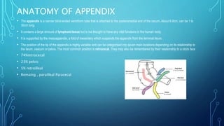
APPENDICITIS
BY KWAKU ANTWI ADJEI
OUTLINE
• INTRODUCTION
• ETIOLOGY
• EPIDEMIOLOGY AND PROGNOSIS
• ANATOMY
• PATHOPHYSIOLOGY
• MANAGEMENT
• COMPLICATIONS
• DIFFERENTIAL DIAGNOSIS
INTRODUCTION
• Appendicitis is the inflammation of the vermiform appendix. The appendix is usually
located 2.5cm below the ileocecal joint or the confluence of Tenia. However it can
have a different positions in the body due to Pregnancy, Midgut Malrotation and
previous abdominal surgeries.
• It is most often a disease of acute presentation, usually within 24 hours, but it can
also present as a more chronic condition. Perforation is likely to occur between 24
and 48hours.
• The exact function of the appendix has been a debated topic. Today it is accepted that
this organ may have an immunoprotective function and acts as a lymphoid organ,
especially in the younger person. Other theories contend that the appendix acts as a
storage vessel for "good" colonic bacteria. Still, others argue that it is a mere
developmental remnant and has no real function
ETIOLOGY
The aetiology of It most commonly affects those in their second or third decade and there is an
overall lifetime risk of 7-8%. In the young, it is mostly due to an increase in lymphoid tissue size, which
occludes the lumen. From 30 years old onwards, it is more likely to be blocked due a faecolith.
Appendicitis rarely happens in the extremes of ages, ages before 2 having a wide lumen whereas the
elderly having an obliterated lumen. Causes are;
• Fecolith
• Lymphoid hyperplasia
• Worm Infestation
• Foreign bodies like seeds
• Appendicolith
• Cancer ( Carcinoid tumor of the appendix, Appendiceal adenocarcinoma )
• Appendicitis rarely happens in the extremes of ages, ages before 2 having a wide lumen whereas the
elderly having an obliterated lumen. The cause of appendicitis is usually unknown.
ANATOMY OF APPENDIX
• The appendix is a narrow blind-ended vermiform tube that is attached to the posteromedial end of the cecum. About 6-9cm, can be 1 to
30cm long.
• It contains a large amount of lymphoid tissue but is not thought to have any vital functions in the human body.
• It is supported by the mesoappendix, a fold of mesentery which suspends the appendix from the terminal ileum.
• The position of the tip of the appendix is highly variable and can be categorised into seven main locations depending on its relationship to
the ileum, caecum or pelvis. The most common position is retrocecal. They may also be remembered by their relationship to a clock face
• 74%retrocecal
• 23% pelvic
• 5% retroilleal
• Remaing , parailleal Paracecal
ANOMALIES OF THE APPENDIX
• Anomalies of the appendix are extremely uncommon.
• Cases of complete agenesia have been reported only few times.
• Abnormal development of the appendix usually takes the form of a double appendix.
• Type A is described as a single appendix with the body or tip branching, or, alternatively, completely divided like a
double-barreled gun.
• Type Bis described as occurrence of completely separated appendices with bases also being located on different sites
of the cecum (the avian type) or with both bases springing from the intestinal tenia (tenia-coli type).
• Type C is a doubled cecum, each containing its own appendix.
• Type D is a horseshoe appendix with two openings at the common cecum.
• All these anomalies are of great practical importance, and a surgeon has to bear them in mind during an operation,
since in case he overlooks them the operated patient may experience grave consequences. They also may be the
forensic issue in cases when repeated explorative laparotomy reveals" previously removed" vermiform appendix.
VASCULATURE OF APPENDIX
• The appendix is derived from the embryologic midgut. Therefore, the vascular supply is via branches of the superior mesenteric vessels.
• Arterial supply is from the appendicular artery (derived from the ileocolic artery, a branch of the superior mesenteric artery ) and venous
drainage is via the corresponding appendicular vein. Both are contained within the mesoappendix.
• Sympathetic and parasympathetic branches of the autonomic nervous system innervate the appendix. This is achieved by the ileocolic
branch of the superior mesenteric plexus. It accompanies the ileocolic artery to reach the appendix.
• Note: Of clinical relevance, the sympathetic afferent fibres of the appendix arise from T10 of the spinal cord – thus explaining why the
visceral pain of early appendicitis is felt centrally within the abdomen.
• Lymphatic fluid from the appendix drains into lymph nodes within the mesoappendix and into the ileocolic lymph nodes (which surround
the ileocolic artery).
EMBRYOLOGY
• The appendix develops embryonically in the fifth week. During this time,
there is a rotation of the midgut to the external umbilical cord with the
eventual return to the abdomen and rotation of the cecum. This results in the
usual retrocecal location of the appendix.
EPIDEMIOLOGY
Family history
• Twin studies suggest that genetics account for 30% of risk*
• Ethnicity
• More common in Caucasians, yet ethnic minorities like blacks are at greater risk of perforation if they do get
appendicitis. This finding can be attributed to more fibre diets amongst underdeveloped countries and minorities
leading to greater defaecation-frequency and lesser transit-time. This also explains an increased incidence in
developing countries especially in urbanized areas.
• Environmental
• Seasonal presentation during the summer
• *No specific gene has been identified specifically, but the risk is roughly three times higher in
members of families with a positive history
• Acute appendicitis is the commonest general surgical emergency in Accra, being twice as common in
males as in females below 60 years. Its incidence has remained relatively stable over the past 30
years. A prospective study of all patients admitted with acute
appendicitis at the Korle Bu Teaching Hospital (KBTH) . There were 1409 cases of appendicitis, 72%
male and 28% female, an overall male to female ratio of 2.6:1. The yearly incidence of appendicitis in
Accra was 3.18/10,000, only slightly more than three decades ago. The peak incidence was in the 20
to 24 age group. In almost all age groups it was twice as common in males as in females. In KBTH,
appendicitis constituted 31% of emergencies and 14% of general surgical operations. Ninety one
percent received prophylactic antibiotics. The appendix was retrocaecal at operation in 55% of cases.
The wound infection rate following appendicectomy in KBTH was 8.75% with a total complication rate
of 17%. The mean duration of stay in hospital was 6 days. The 30-day mortality was 1.3%.
• The yearly incidence of acute appendicitis in Kumasi was 1.8 per 10,000 of the
population, as of March 2009. There were a total of 1266 patients 869 (68.6%)
males and 397 (31.4%) females with an overall male to female ratio of 2.2:1. The
peak incidence was in the 25-29 year age groups in both sexes. Appendicitis was
twice as common in the male and in all age groups. The admission rates at the start
of the study were 1.7/10,000 and 0.8/10,000 for male and females respectively and
3.4/10,000 and 1.4/10.000 for male and females at the conclusion of the study.
IN CHILDREN
Perforation rate is nearly 100% in infants less than a year old; between 70 and 80% in infants under
2years of age, and above 50% in children up to 5years of age.Because of the high rate of perforation,
the mortality rate is also high, approaching 10%
• The high mortality is also in part attributed to the inability of the poorly developed omentum in this
age group to prevent the rapid development of generalized peritonitis.
• Another factor is late diagnosis due to a low index of suspicion.Unexplained abdominal pain suspect
appendicitis
IN ELDERLY
More than 30% of elderly patients have a perforated appendix at the time they arrive in theatre.
• The early perforation is attributed to impaired blood supply and structural weakness of the appendix
in the elderly as well as diagnostic delay
IN IN PREGNANCY
Acute appendicitis occurs during pregnancy just as in non-pregnant females, with an
incidence of about 1 in every 1000 pregnancies. It is the most common extrauterine condition
requiring an abdominal operation during pregnancy. The frequency of appendicitis during the
first two trimesters is greater than during the third trimester. But the mortality rate during the
last trimester is five times as high as during the first and second trimesters
• If performed before the appendix ruptures, the appendicectomy does not disturb the foetus,
and even if it turns to be a negative laparotomy, the effects are minor enough to warrant the
risk.
• DURING the THIRD TRIMESTER, the following changes occur in the clinical picture of
appendicitis: Displacement of the caecum and appendix by the enlarged uterus leads to
localization of pain higher in the abdomen or in the right flank. The appendicitis tends to be
more serious though less frequent. The displaced omentum is not able to reach the area of
the inflamed appendix to help contain the infection.
• Premature labor occurs in about half of women who develop appendicitis during the third
trimester. Early appendicectomy is the treatment of choice for appendicitis at all stages of
pregnancy.
PATHOPHYSIOLOGY
• It is typically caused by direct luminal obstruction, usually secondary to a faecolith or lymphoid
hyperplasia, impacted stool or, rarely, an appendiceal or caecal tumour.
• When obstructed, commensal bacteria in the appendix can multiply, resulting in acute inflammation.
Reduced venous drainage and localised inflammation can result in increased pressure within the
appendix, in turn can result in ischaemia.
• If left untreated, ischaemia within the appendiceal wall can result in necrosis, which in turn can cause
the appendix to perforate.
• Inflammation leads to edema leading to extravasation of fluids. Mucosa which produces mucinnous
fluids increases its fluids , intraluminal pressure increases . Causes lymphoid irritation and
Hypertrophied , majority being at the base. Causes the veins draining the appendix to collapse due to
increased intraluminal pressure leading to more fluid retention. Continues to involve arterial
involvement. Blood stasis, is a good medium of bacteria to over produce
• If caught at this time, leads to resolution..but if not controlled, perforation and rupture. Later cause
abscess with pus。Appendix is supplied by the appendiceal artery which is a branch of illeocolic
artery. It's a terminal artery and small too, so with little pressure it's overcomed. Then due to low
blood there is ischemia leading to necrosis and gangrene, sloughing off leading to perforation. If not
localized by post immunity or omentum, it becomes generalized.
• Catarrhal Appendicitis
• Phlegmonous Appendicitis
• Necrotic Appendicitis
• Gangrenous Appendicitis
• Perforated Appendicitis
• Recurrent Appendicitis
• Spontaneous resolving Appendicitis
CLINICAL PRESENTATION
Abdominal pain ( 55% typical, 45% atypical )
• In established appendicitis, the abdomen is most tender at McBurney’s point – situated one third of the
distance from the right anterior superior iliac spine to the umbilicus. This corresponds to the location of
the tip of the appendix when it lies in a retrocecal position. Initially, pain from the appendix and its
visceral peritoneum is referred to the umbilical region. As the appendix becomes increasingly
inflamed, it irritates the parietal peritoneum, causing the pain to localise to the right lower quadrant.
Anorexia and Nausea
Vomiting
Constipation ( diarrhea in Pelvic )
Dysuria, frequency ( Pelvic )
PHYSICAL EXAMINATION
• There are different scoring systems used in Appendicitis. Eg RIPASA,
MODIFIED MANTRELS SCORE etc. In Ghana, the Modified Alvarado
score is mostly used
MANTRELS:
SYMPTOM/SIGN SCORE
• M(migrating pain) 1
• A(anorexia) 1
• N(nausea) 1
• T( right lower tenderness) 2
• MANTRELS
• SYMPTOM/SIGN SCORE
• R(rebound tenderness) 1
• E(elevated body temperature; mostly mild) 1
• L(leucocytosis) 2
• S(left shift in the WBC differentials) 1*
• Omitted in the revised version*
The scores are summed up for each patient and interpreted as follows:
Score < 4: Appendicitis unlikely
Score 5-6: Possible acute appendicitis
Score7-8: Probable acute appendicitis
Score 9-10: Very probable acute appendicitis
• Signs include:Right lower quadrant guarding and rebound tenderness
over McBurney's point (1.5 to 2 inches from the anterior superior iliac
spine (ASIS) on a straight line from the ASIS to the
umbilicus)Rovsing's sign (right lower quadrant pain elicited by
palpation of the left lower quadrant)Dunphy's sign (increased
abdominal pain with coughing)
• Other associated signs such as the psoas sign (pain on external
rotation or passive extension of the right hip suggesting retrocecal
appendicitis) or obturator sign (pain on internal rotation of the right
hip suggesting pelvic appendicitis) are rare
• Sherrens Triangle (Anterior Superior iliac spine, Pubic Tubercle
and Umbilicus
• Pelvic tenderness on the right side on DRE
INVESTIGATIONS
• Appendicitis is clinically diagnosed, even though FBC for leukocytosis can be
helpful. Abdominal CT or USG may also be helpful in Advanced Appendicitis.
• The Symptoms and its chronological order is essential in diagnosing as its
symptoms are closely similar to other conditions. The different positions of
the tip of the appendix with also individual variations in presentation can
also cloud diagnosis. Hence other investigations can be done to rule out
other diseases. Investigations Blood C/S for Typhoid fever, Urine R/E for UTI.
Plain Abdominal Xray for xray indications as contrasts can actually rupture
an appendicitis. Barium enema is contraindicated.
• In an elderly with symptoms of appendicitis, and low HB , it's more likely to
be a colorectal cancer
• Laparoscopy is the diagnostic investigation.
MANAGEMENT
• Primary Survey ( ABC, NPO, vitals, Urretheral catheter,
analgesics, antibiotics)
• Secondary Survey (History, Exams)
• Investigations
• Informed Consent
• Anaesthesic review
• Appendicectomy
TREATMENT
Acute Appendicitis
· Appendicectomy
Peritonitis
• Laparotomy+ appendicectomy
Appendiceal mass
• Conservative
• Interval appendicectomy
Appendicoal abscess
• Drainage
• Interval appendicectomy
COMPLICATIONS
• 1. Generalized peritonitis
• 2. Appendix mass
• 3. Pelvic abscess
• 4. Subhepatic/subphrenic abscess
• 5. Pylephlebitis(inflammation of the portal vein or any of its branches)
• 6.Acute intestinal obstruction
• 7. Septicaemia
REFERENCES
• Incidence of Appendicitis in Kumasi, March 2009 by Ohene Yeboah and Francis Abantanga
• Appendicitis in Accra; A contemporary appraisal, Joe Nat A Clegg Lamptey and Simon Naaeder
• Association between Appendectomy and Subsequent Colorectal Cancer Development: An Asian Population Study
• Incidence and odds ratio of appendicitis as first manifestation of colon cancer: a retrospective analysis of 1873 patients .Hung-Wen Lai et al. J
Gastroenterol Hepatol. 2006 Nov.
• What causes appendicitis? AR Walker, I Segal
• Acute appendicitis. Mark D Stringer
The use of computed tomography versus clinical acumen in diagnosing appendicitis in children: A two-institution international study
• Role of the faecolith in modern-day appendicitis JP Singh, JG Mariadason
• Appendicitis Jones MW, Lopez RA, Deppen JG.

APPENDICITIS

  • 1.
  • 2.
    OUTLINE • INTRODUCTION • ETIOLOGY •EPIDEMIOLOGY AND PROGNOSIS • ANATOMY • PATHOPHYSIOLOGY • MANAGEMENT • COMPLICATIONS • DIFFERENTIAL DIAGNOSIS
  • 3.
    INTRODUCTION • Appendicitis isthe inflammation of the vermiform appendix. The appendix is usually located 2.5cm below the ileocecal joint or the confluence of Tenia. However it can have a different positions in the body due to Pregnancy, Midgut Malrotation and previous abdominal surgeries. • It is most often a disease of acute presentation, usually within 24 hours, but it can also present as a more chronic condition. Perforation is likely to occur between 24 and 48hours. • The exact function of the appendix has been a debated topic. Today it is accepted that this organ may have an immunoprotective function and acts as a lymphoid organ, especially in the younger person. Other theories contend that the appendix acts as a storage vessel for "good" colonic bacteria. Still, others argue that it is a mere developmental remnant and has no real function
  • 4.
    ETIOLOGY The aetiology ofIt most commonly affects those in their second or third decade and there is an overall lifetime risk of 7-8%. In the young, it is mostly due to an increase in lymphoid tissue size, which occludes the lumen. From 30 years old onwards, it is more likely to be blocked due a faecolith. Appendicitis rarely happens in the extremes of ages, ages before 2 having a wide lumen whereas the elderly having an obliterated lumen. Causes are; • Fecolith • Lymphoid hyperplasia • Worm Infestation • Foreign bodies like seeds • Appendicolith • Cancer ( Carcinoid tumor of the appendix, Appendiceal adenocarcinoma ) • Appendicitis rarely happens in the extremes of ages, ages before 2 having a wide lumen whereas the elderly having an obliterated lumen. The cause of appendicitis is usually unknown.
  • 5.
    ANATOMY OF APPENDIX •The appendix is a narrow blind-ended vermiform tube that is attached to the posteromedial end of the cecum. About 6-9cm, can be 1 to 30cm long. • It contains a large amount of lymphoid tissue but is not thought to have any vital functions in the human body. • It is supported by the mesoappendix, a fold of mesentery which suspends the appendix from the terminal ileum. • The position of the tip of the appendix is highly variable and can be categorised into seven main locations depending on its relationship to the ileum, caecum or pelvis. The most common position is retrocecal. They may also be remembered by their relationship to a clock face • 74%retrocecal • 23% pelvic • 5% retroilleal • Remaing , parailleal Paracecal
  • 6.
    ANOMALIES OF THEAPPENDIX • Anomalies of the appendix are extremely uncommon. • Cases of complete agenesia have been reported only few times. • Abnormal development of the appendix usually takes the form of a double appendix. • Type A is described as a single appendix with the body or tip branching, or, alternatively, completely divided like a double-barreled gun. • Type Bis described as occurrence of completely separated appendices with bases also being located on different sites of the cecum (the avian type) or with both bases springing from the intestinal tenia (tenia-coli type). • Type C is a doubled cecum, each containing its own appendix. • Type D is a horseshoe appendix with two openings at the common cecum. • All these anomalies are of great practical importance, and a surgeon has to bear them in mind during an operation, since in case he overlooks them the operated patient may experience grave consequences. They also may be the forensic issue in cases when repeated explorative laparotomy reveals" previously removed" vermiform appendix.
  • 7.
    VASCULATURE OF APPENDIX •The appendix is derived from the embryologic midgut. Therefore, the vascular supply is via branches of the superior mesenteric vessels. • Arterial supply is from the appendicular artery (derived from the ileocolic artery, a branch of the superior mesenteric artery ) and venous drainage is via the corresponding appendicular vein. Both are contained within the mesoappendix. • Sympathetic and parasympathetic branches of the autonomic nervous system innervate the appendix. This is achieved by the ileocolic branch of the superior mesenteric plexus. It accompanies the ileocolic artery to reach the appendix. • Note: Of clinical relevance, the sympathetic afferent fibres of the appendix arise from T10 of the spinal cord – thus explaining why the visceral pain of early appendicitis is felt centrally within the abdomen. • Lymphatic fluid from the appendix drains into lymph nodes within the mesoappendix and into the ileocolic lymph nodes (which surround the ileocolic artery).
  • 8.
    EMBRYOLOGY • The appendixdevelops embryonically in the fifth week. During this time, there is a rotation of the midgut to the external umbilical cord with the eventual return to the abdomen and rotation of the cecum. This results in the usual retrocecal location of the appendix.
  • 9.
    EPIDEMIOLOGY Family history • Twinstudies suggest that genetics account for 30% of risk* • Ethnicity • More common in Caucasians, yet ethnic minorities like blacks are at greater risk of perforation if they do get appendicitis. This finding can be attributed to more fibre diets amongst underdeveloped countries and minorities leading to greater defaecation-frequency and lesser transit-time. This also explains an increased incidence in developing countries especially in urbanized areas. • Environmental • Seasonal presentation during the summer • *No specific gene has been identified specifically, but the risk is roughly three times higher in members of families with a positive history
  • 10.
    • Acute appendicitisis the commonest general surgical emergency in Accra, being twice as common in males as in females below 60 years. Its incidence has remained relatively stable over the past 30 years. A prospective study of all patients admitted with acute appendicitis at the Korle Bu Teaching Hospital (KBTH) . There were 1409 cases of appendicitis, 72% male and 28% female, an overall male to female ratio of 2.6:1. The yearly incidence of appendicitis in Accra was 3.18/10,000, only slightly more than three decades ago. The peak incidence was in the 20 to 24 age group. In almost all age groups it was twice as common in males as in females. In KBTH, appendicitis constituted 31% of emergencies and 14% of general surgical operations. Ninety one percent received prophylactic antibiotics. The appendix was retrocaecal at operation in 55% of cases. The wound infection rate following appendicectomy in KBTH was 8.75% with a total complication rate of 17%. The mean duration of stay in hospital was 6 days. The 30-day mortality was 1.3%. • The yearly incidence of acute appendicitis in Kumasi was 1.8 per 10,000 of the population, as of March 2009. There were a total of 1266 patients 869 (68.6%) males and 397 (31.4%) females with an overall male to female ratio of 2.2:1. The peak incidence was in the 25-29 year age groups in both sexes. Appendicitis was twice as common in the male and in all age groups. The admission rates at the start of the study were 1.7/10,000 and 0.8/10,000 for male and females respectively and 3.4/10,000 and 1.4/10.000 for male and females at the conclusion of the study.
  • 11.
    IN CHILDREN Perforation rateis nearly 100% in infants less than a year old; between 70 and 80% in infants under 2years of age, and above 50% in children up to 5years of age.Because of the high rate of perforation, the mortality rate is also high, approaching 10% • The high mortality is also in part attributed to the inability of the poorly developed omentum in this age group to prevent the rapid development of generalized peritonitis. • Another factor is late diagnosis due to a low index of suspicion.Unexplained abdominal pain suspect appendicitis IN ELDERLY More than 30% of elderly patients have a perforated appendix at the time they arrive in theatre. • The early perforation is attributed to impaired blood supply and structural weakness of the appendix in the elderly as well as diagnostic delay
  • 12.
    IN IN PREGNANCY Acuteappendicitis occurs during pregnancy just as in non-pregnant females, with an incidence of about 1 in every 1000 pregnancies. It is the most common extrauterine condition requiring an abdominal operation during pregnancy. The frequency of appendicitis during the first two trimesters is greater than during the third trimester. But the mortality rate during the last trimester is five times as high as during the first and second trimesters • If performed before the appendix ruptures, the appendicectomy does not disturb the foetus, and even if it turns to be a negative laparotomy, the effects are minor enough to warrant the risk. • DURING the THIRD TRIMESTER, the following changes occur in the clinical picture of appendicitis: Displacement of the caecum and appendix by the enlarged uterus leads to localization of pain higher in the abdomen or in the right flank. The appendicitis tends to be more serious though less frequent. The displaced omentum is not able to reach the area of the inflamed appendix to help contain the infection. • Premature labor occurs in about half of women who develop appendicitis during the third trimester. Early appendicectomy is the treatment of choice for appendicitis at all stages of pregnancy.
  • 13.
    PATHOPHYSIOLOGY • It istypically caused by direct luminal obstruction, usually secondary to a faecolith or lymphoid hyperplasia, impacted stool or, rarely, an appendiceal or caecal tumour. • When obstructed, commensal bacteria in the appendix can multiply, resulting in acute inflammation. Reduced venous drainage and localised inflammation can result in increased pressure within the appendix, in turn can result in ischaemia. • If left untreated, ischaemia within the appendiceal wall can result in necrosis, which in turn can cause the appendix to perforate.
  • 14.
    • Inflammation leadsto edema leading to extravasation of fluids. Mucosa which produces mucinnous fluids increases its fluids , intraluminal pressure increases . Causes lymphoid irritation and Hypertrophied , majority being at the base. Causes the veins draining the appendix to collapse due to increased intraluminal pressure leading to more fluid retention. Continues to involve arterial involvement. Blood stasis, is a good medium of bacteria to over produce • If caught at this time, leads to resolution..but if not controlled, perforation and rupture. Later cause abscess with pus。Appendix is supplied by the appendiceal artery which is a branch of illeocolic artery. It's a terminal artery and small too, so with little pressure it's overcomed. Then due to low blood there is ischemia leading to necrosis and gangrene, sloughing off leading to perforation. If not localized by post immunity or omentum, it becomes generalized.
  • 15.
    • Catarrhal Appendicitis •Phlegmonous Appendicitis • Necrotic Appendicitis • Gangrenous Appendicitis • Perforated Appendicitis • Recurrent Appendicitis • Spontaneous resolving Appendicitis
  • 16.
    CLINICAL PRESENTATION Abdominal pain( 55% typical, 45% atypical ) • In established appendicitis, the abdomen is most tender at McBurney’s point – situated one third of the distance from the right anterior superior iliac spine to the umbilicus. This corresponds to the location of the tip of the appendix when it lies in a retrocecal position. Initially, pain from the appendix and its visceral peritoneum is referred to the umbilical region. As the appendix becomes increasingly inflamed, it irritates the parietal peritoneum, causing the pain to localise to the right lower quadrant. Anorexia and Nausea Vomiting Constipation ( diarrhea in Pelvic ) Dysuria, frequency ( Pelvic )
  • 17.
    PHYSICAL EXAMINATION • Thereare different scoring systems used in Appendicitis. Eg RIPASA, MODIFIED MANTRELS SCORE etc. In Ghana, the Modified Alvarado score is mostly used MANTRELS: SYMPTOM/SIGN SCORE • M(migrating pain) 1 • A(anorexia) 1 • N(nausea) 1 • T( right lower tenderness) 2
  • 18.
    • MANTRELS • SYMPTOM/SIGNSCORE • R(rebound tenderness) 1 • E(elevated body temperature; mostly mild) 1 • L(leucocytosis) 2 • S(left shift in the WBC differentials) 1* • Omitted in the revised version* The scores are summed up for each patient and interpreted as follows: Score < 4: Appendicitis unlikely Score 5-6: Possible acute appendicitis Score7-8: Probable acute appendicitis Score 9-10: Very probable acute appendicitis
  • 19.
    • Signs include:Rightlower quadrant guarding and rebound tenderness over McBurney's point (1.5 to 2 inches from the anterior superior iliac spine (ASIS) on a straight line from the ASIS to the umbilicus)Rovsing's sign (right lower quadrant pain elicited by palpation of the left lower quadrant)Dunphy's sign (increased abdominal pain with coughing) • Other associated signs such as the psoas sign (pain on external rotation or passive extension of the right hip suggesting retrocecal appendicitis) or obturator sign (pain on internal rotation of the right hip suggesting pelvic appendicitis) are rare
  • 20.
    • Sherrens Triangle(Anterior Superior iliac spine, Pubic Tubercle and Umbilicus • Pelvic tenderness on the right side on DRE
  • 21.
    INVESTIGATIONS • Appendicitis isclinically diagnosed, even though FBC for leukocytosis can be helpful. Abdominal CT or USG may also be helpful in Advanced Appendicitis. • The Symptoms and its chronological order is essential in diagnosing as its symptoms are closely similar to other conditions. The different positions of the tip of the appendix with also individual variations in presentation can also cloud diagnosis. Hence other investigations can be done to rule out other diseases. Investigations Blood C/S for Typhoid fever, Urine R/E for UTI. Plain Abdominal Xray for xray indications as contrasts can actually rupture an appendicitis. Barium enema is contraindicated. • In an elderly with symptoms of appendicitis, and low HB , it's more likely to be a colorectal cancer • Laparoscopy is the diagnostic investigation.
  • 22.
    MANAGEMENT • Primary Survey( ABC, NPO, vitals, Urretheral catheter, analgesics, antibiotics) • Secondary Survey (History, Exams) • Investigations • Informed Consent • Anaesthesic review • Appendicectomy
  • 23.
    TREATMENT Acute Appendicitis · Appendicectomy Peritonitis •Laparotomy+ appendicectomy Appendiceal mass • Conservative • Interval appendicectomy Appendicoal abscess • Drainage • Interval appendicectomy
  • 24.
    COMPLICATIONS • 1. Generalizedperitonitis • 2. Appendix mass • 3. Pelvic abscess • 4. Subhepatic/subphrenic abscess • 5. Pylephlebitis(inflammation of the portal vein or any of its branches) • 6.Acute intestinal obstruction • 7. Septicaemia
  • 25.
    REFERENCES • Incidence ofAppendicitis in Kumasi, March 2009 by Ohene Yeboah and Francis Abantanga • Appendicitis in Accra; A contemporary appraisal, Joe Nat A Clegg Lamptey and Simon Naaeder • Association between Appendectomy and Subsequent Colorectal Cancer Development: An Asian Population Study • Incidence and odds ratio of appendicitis as first manifestation of colon cancer: a retrospective analysis of 1873 patients .Hung-Wen Lai et al. J Gastroenterol Hepatol. 2006 Nov. • What causes appendicitis? AR Walker, I Segal • Acute appendicitis. Mark D Stringer The use of computed tomography versus clinical acumen in diagnosing appendicitis in children: A two-institution international study • Role of the faecolith in modern-day appendicitis JP Singh, JG Mariadason • Appendicitis Jones MW, Lopez RA, Deppen JG.

Editor's Notes

  • #6 Pre-ileal – anterior to the terminal ileum – 1 or 2 o’clock. Post-ileal – posterior to the terminal ileum – 1 or 2 o’clock. Sub-ileal – parallel with the terminal ileum – 3 o’clock. Pelvic – descending over the pelvic brim – 5 o’clock. Subcecal – below the cecum – 6 o’clock. Paracecal – alongside the lateral border of the cecum – 10 o’clock. Retrocecal – behind the cecum – 11 o’clock.
  • #8 Anomalies of the appendix are extremely uncommon. Cases of complete agenesia have been reported only few times. Abnormal development of the appendix usually takes the form of a double appendix. Type A is described as a single appendix with the body or tip branching, or, alternatively, completely divided like a double-barreled gun. Type Bis described as occurrence of completely separated appendices with bases also being located on different sites of the cecum (the avian type) or with both bases springing from the intestinal tenia (tenia-coli type). Type C is a doubled cecum, each containing its own appendix. Type D is a horseshoe appendix with two openings at the common cecum All these anomalies are of great practical importance, and a surgeon has to bear them in mind during an operation, since in case he overlooks them the operated patient may experience grave consequences. They also may be the forensic issue in cases when repeated explorative laparotomy reveals" previously removed" vermiform appendix.