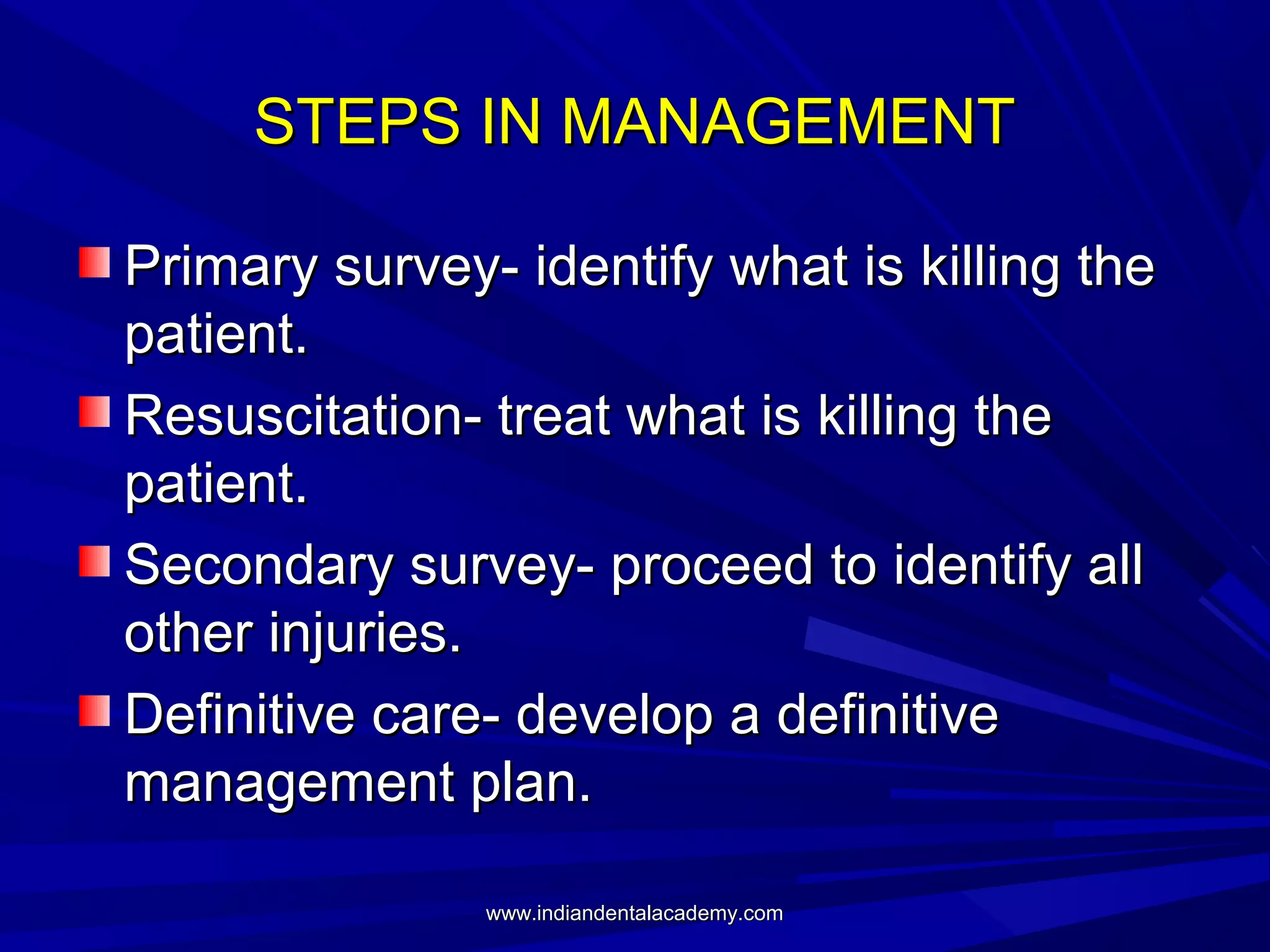
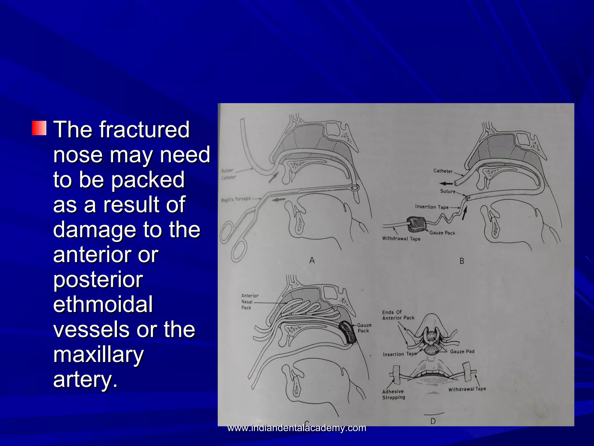
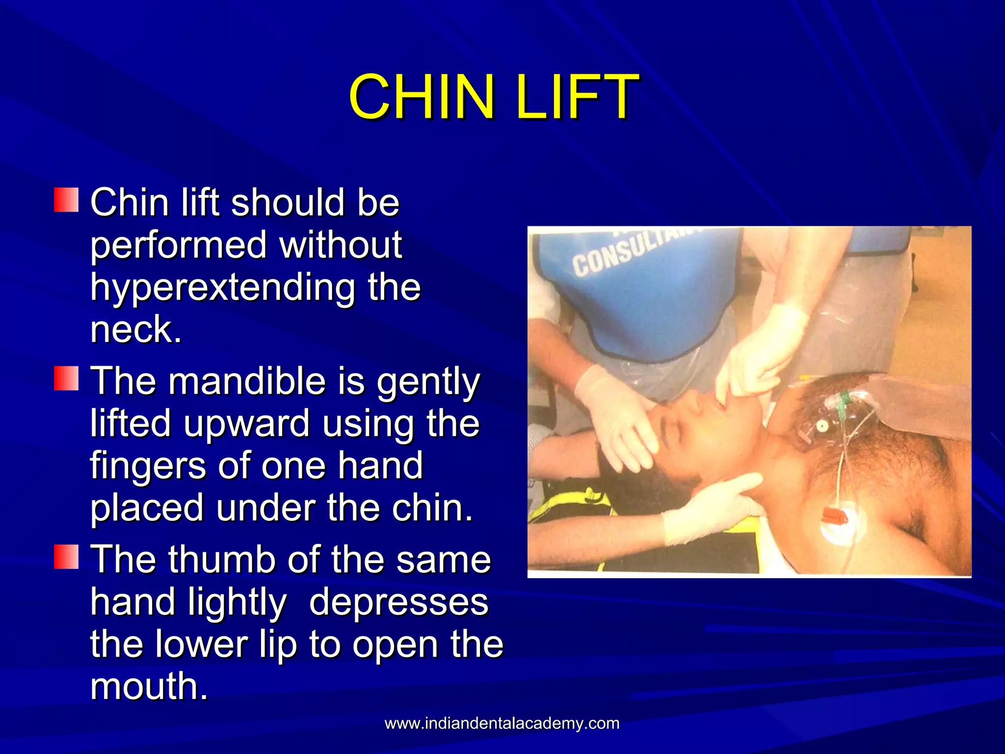
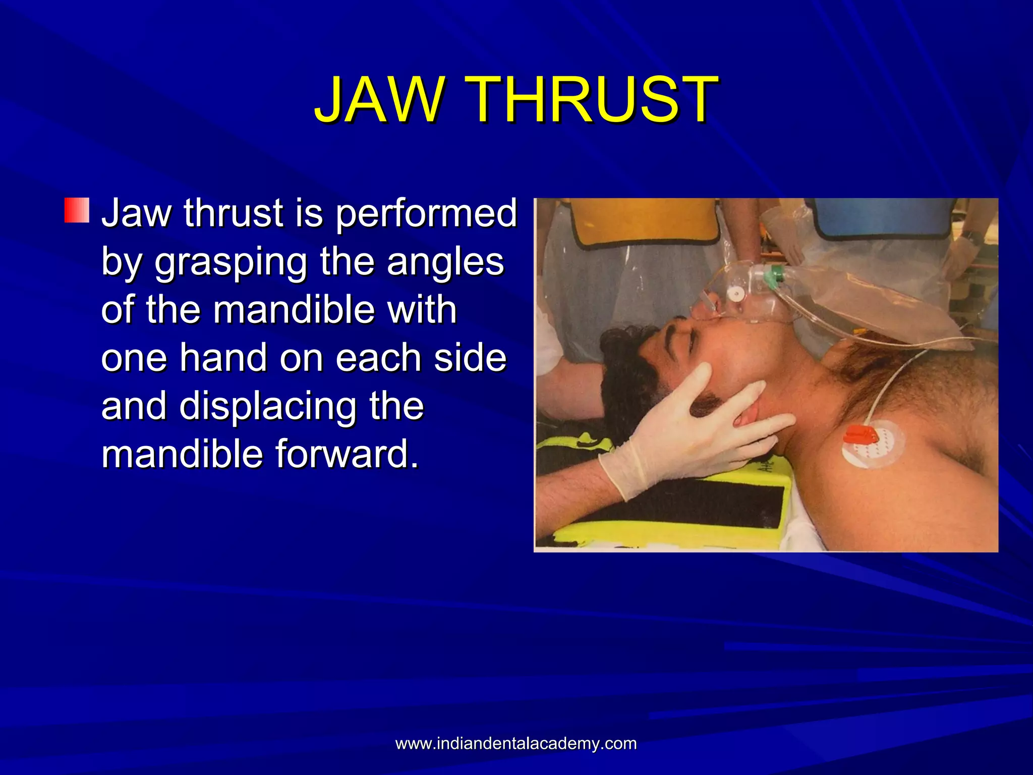
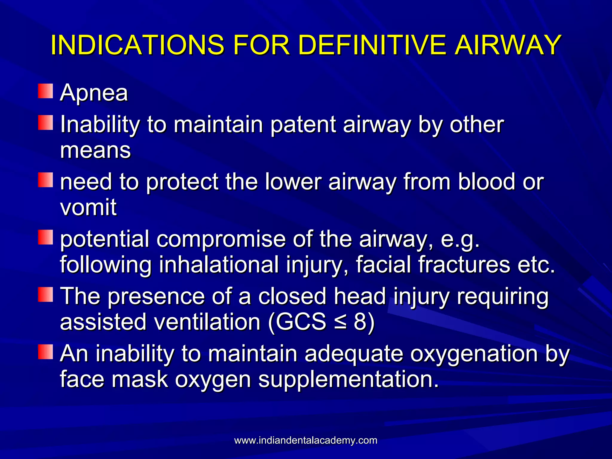
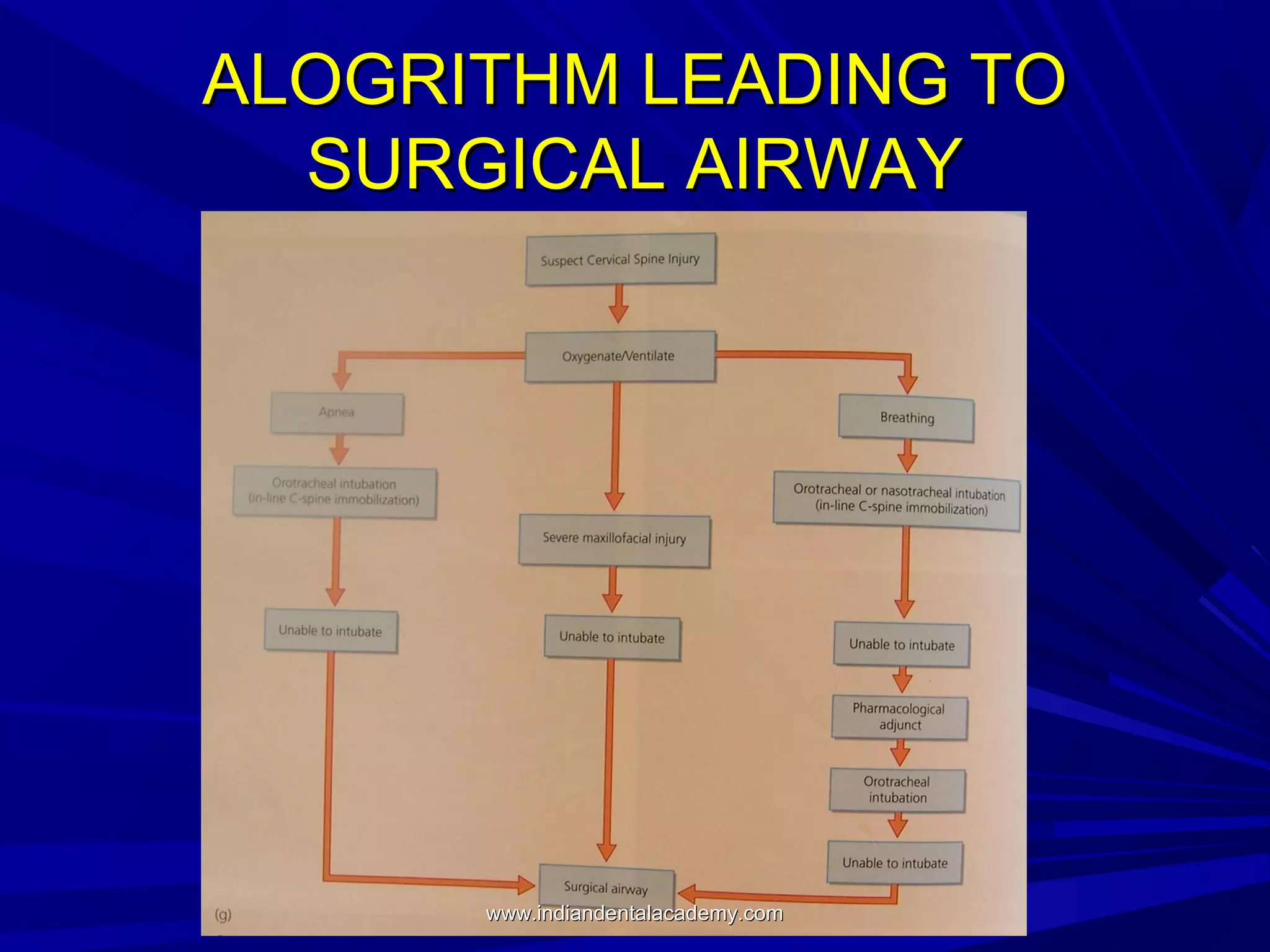
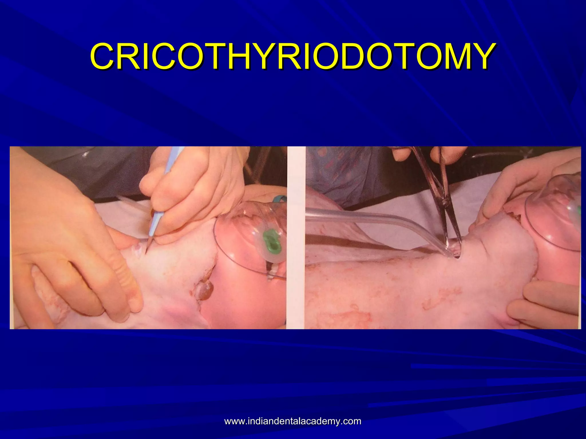
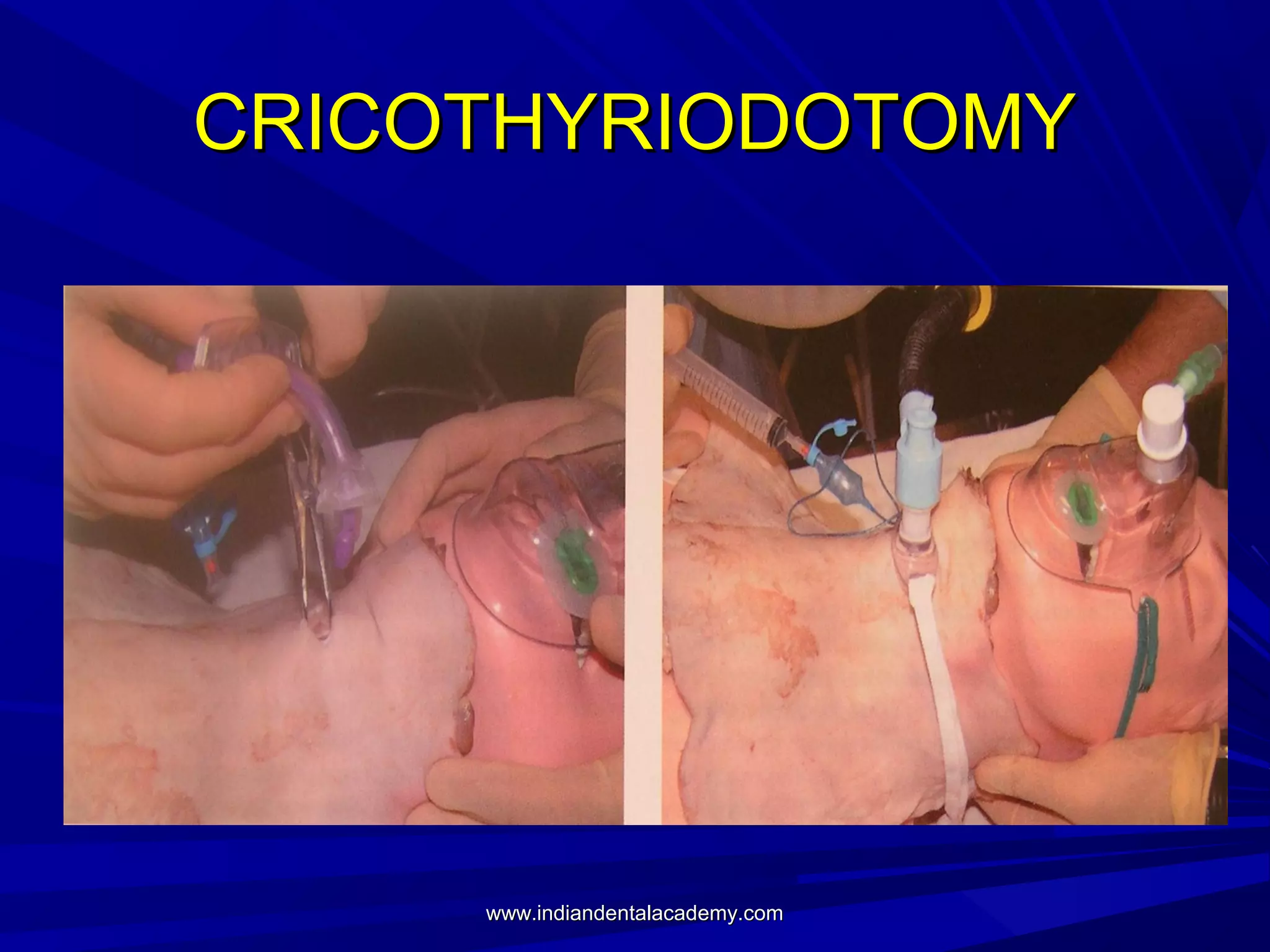
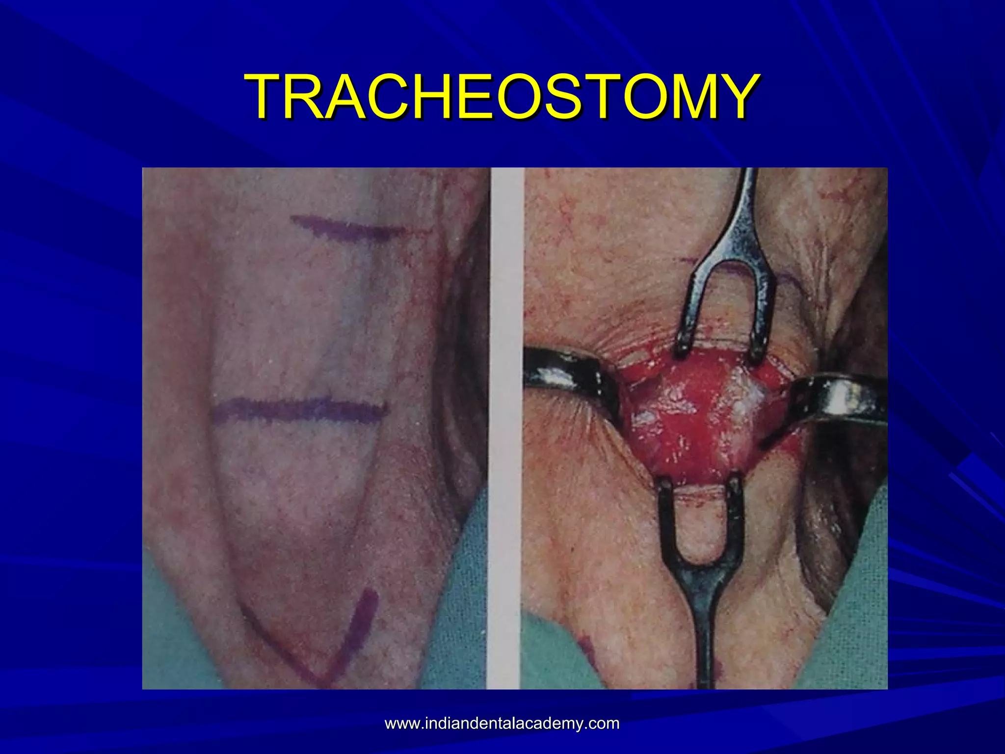
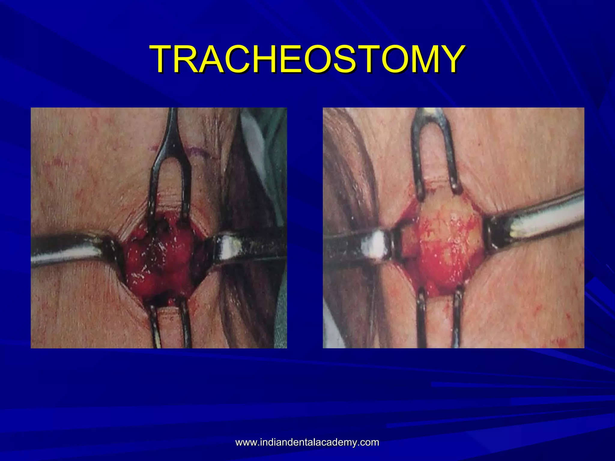
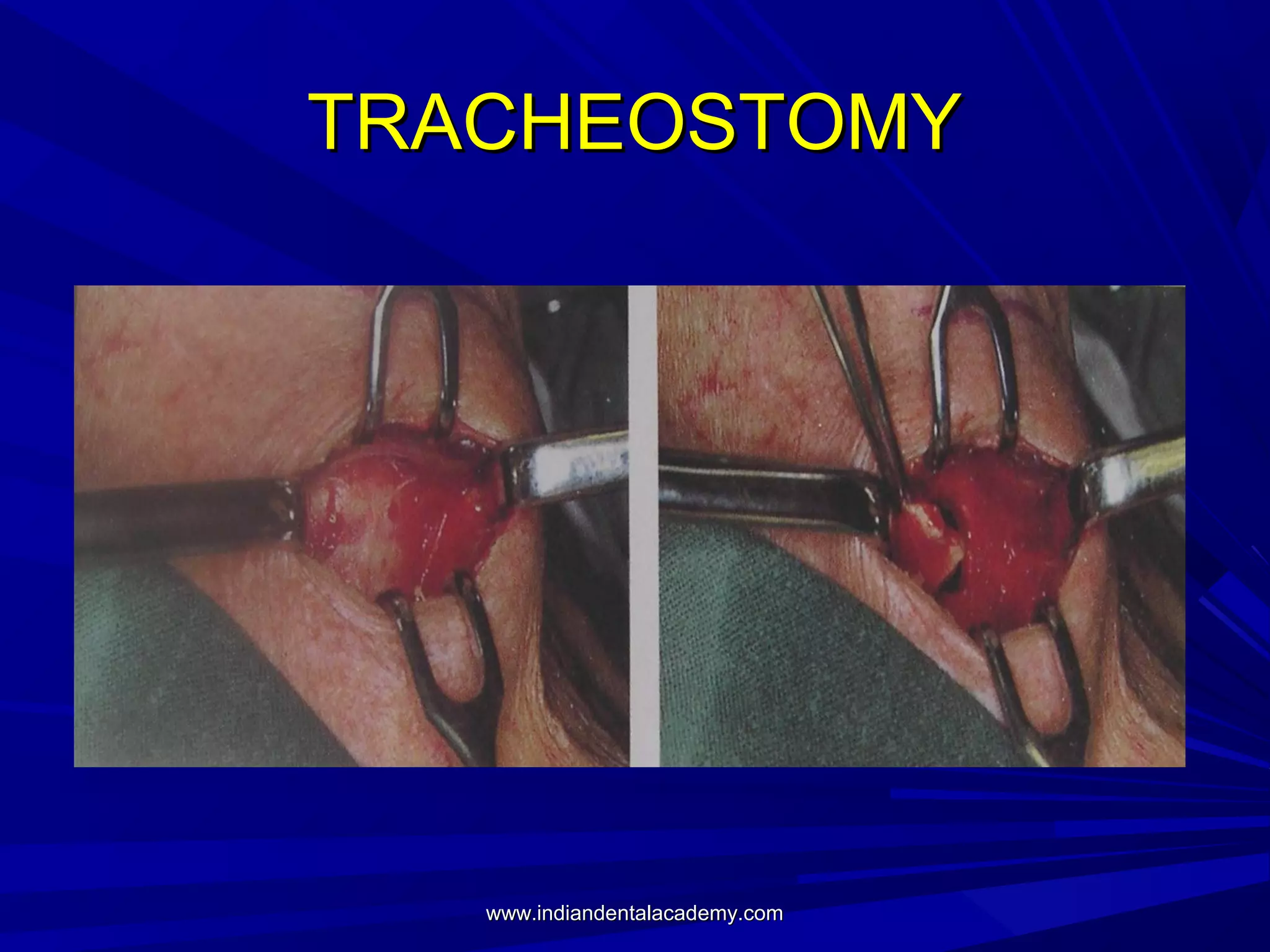
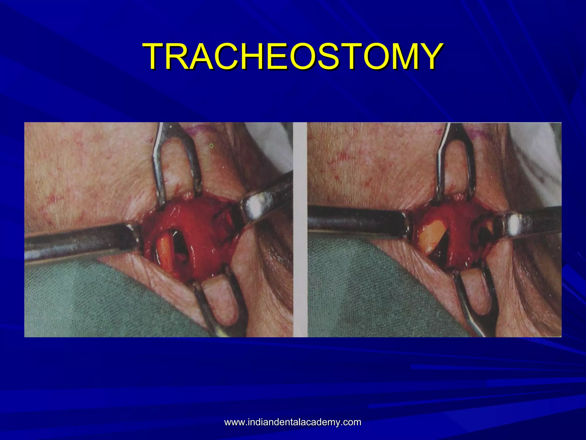
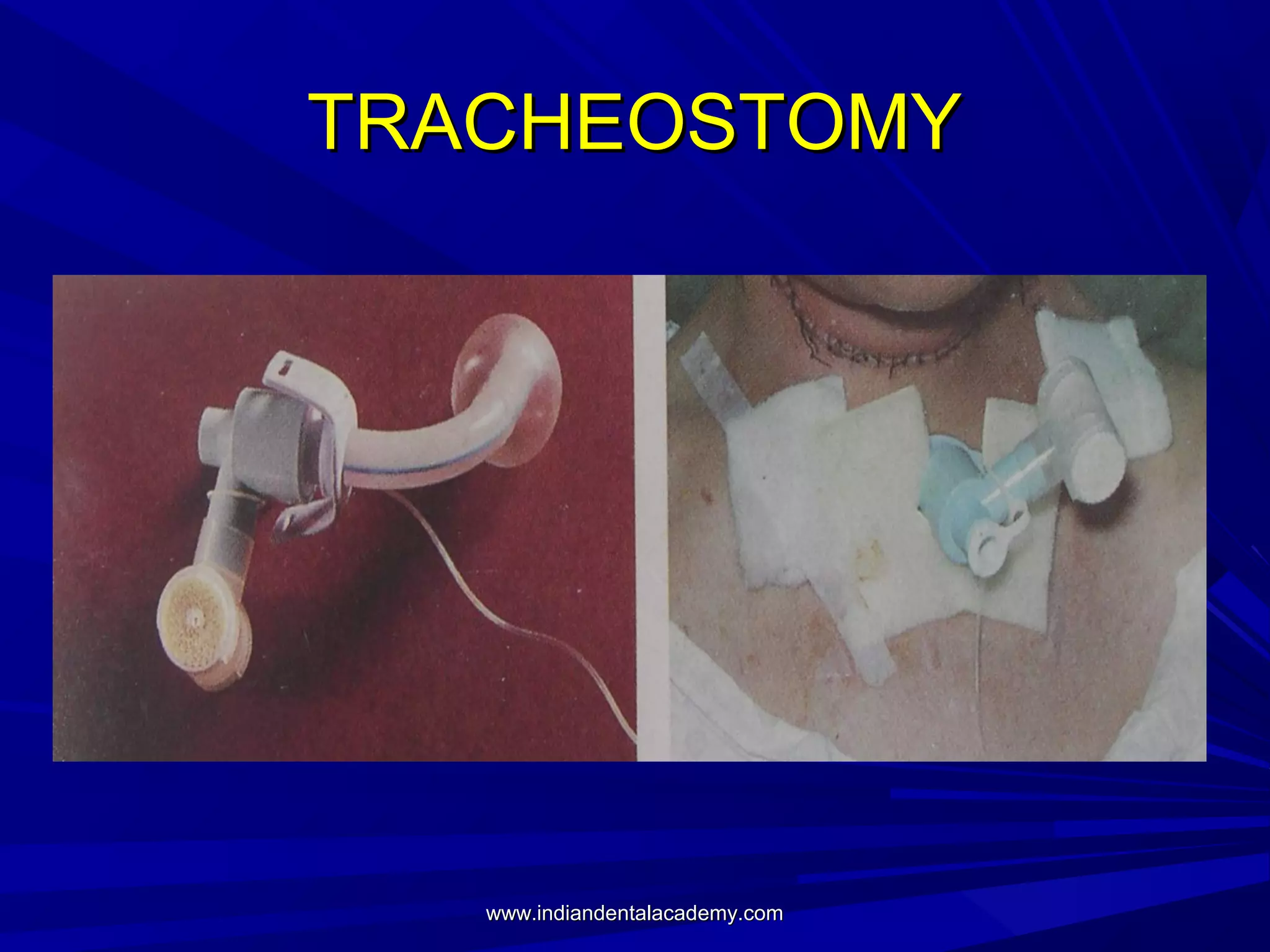
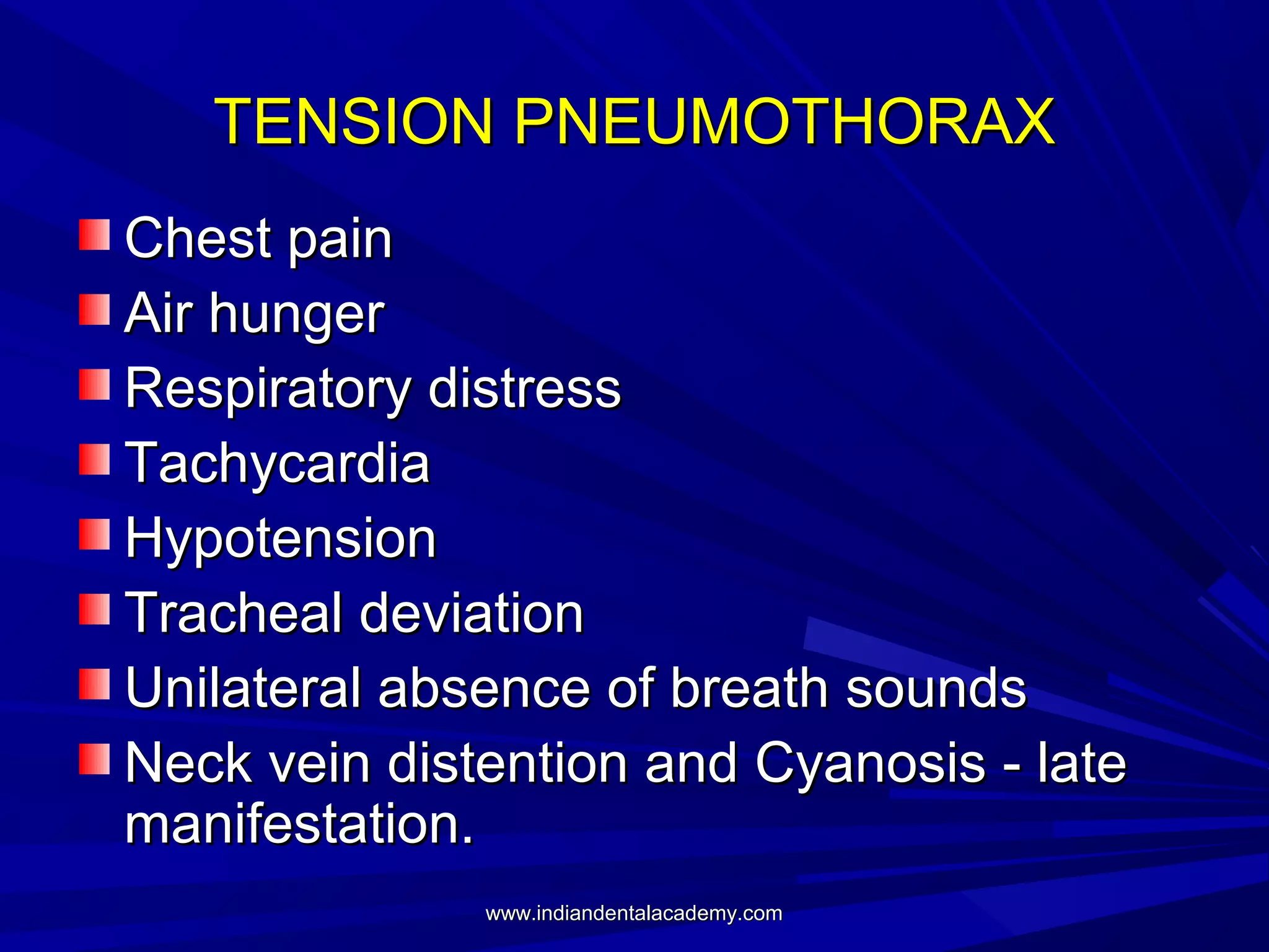
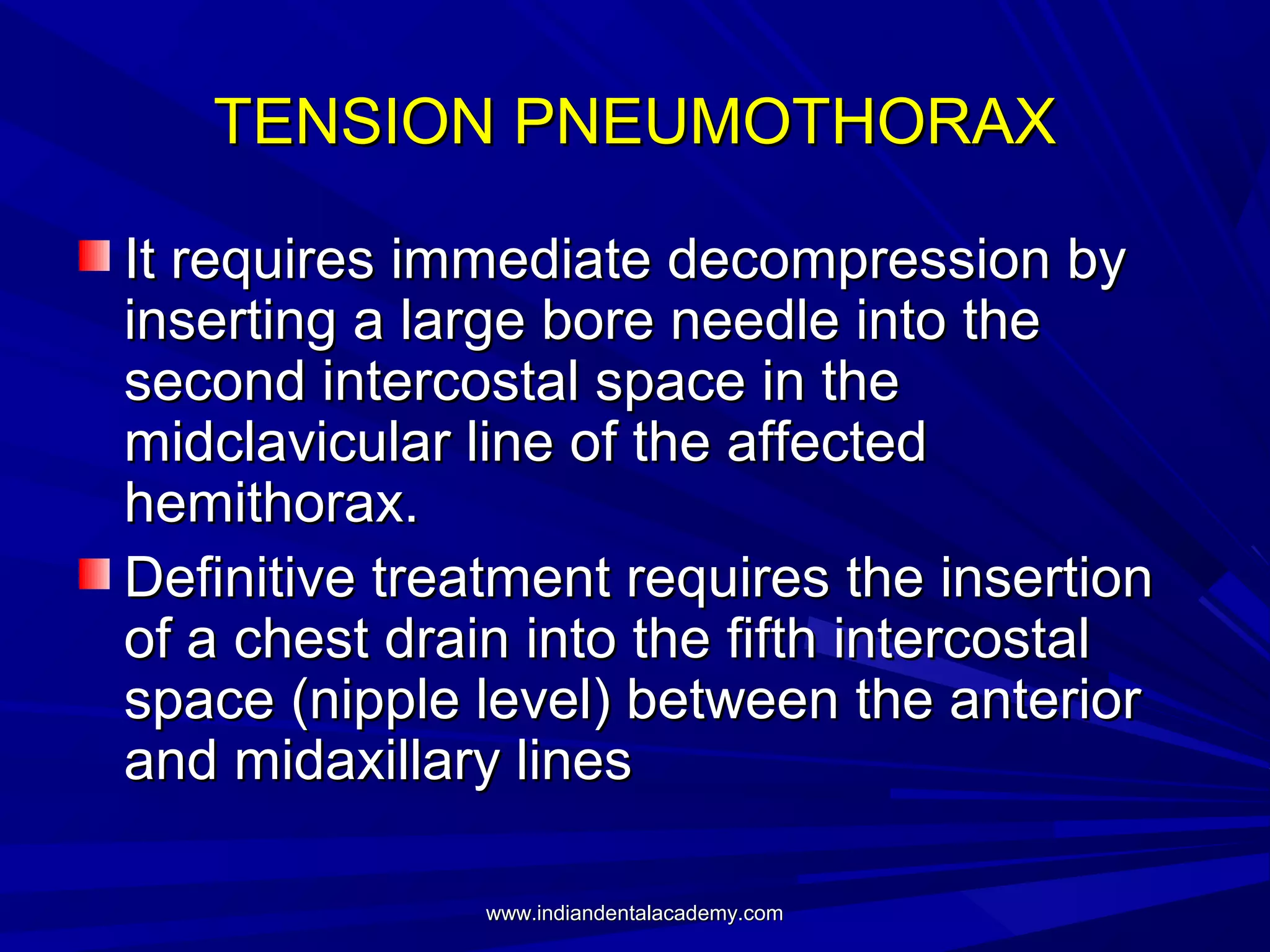
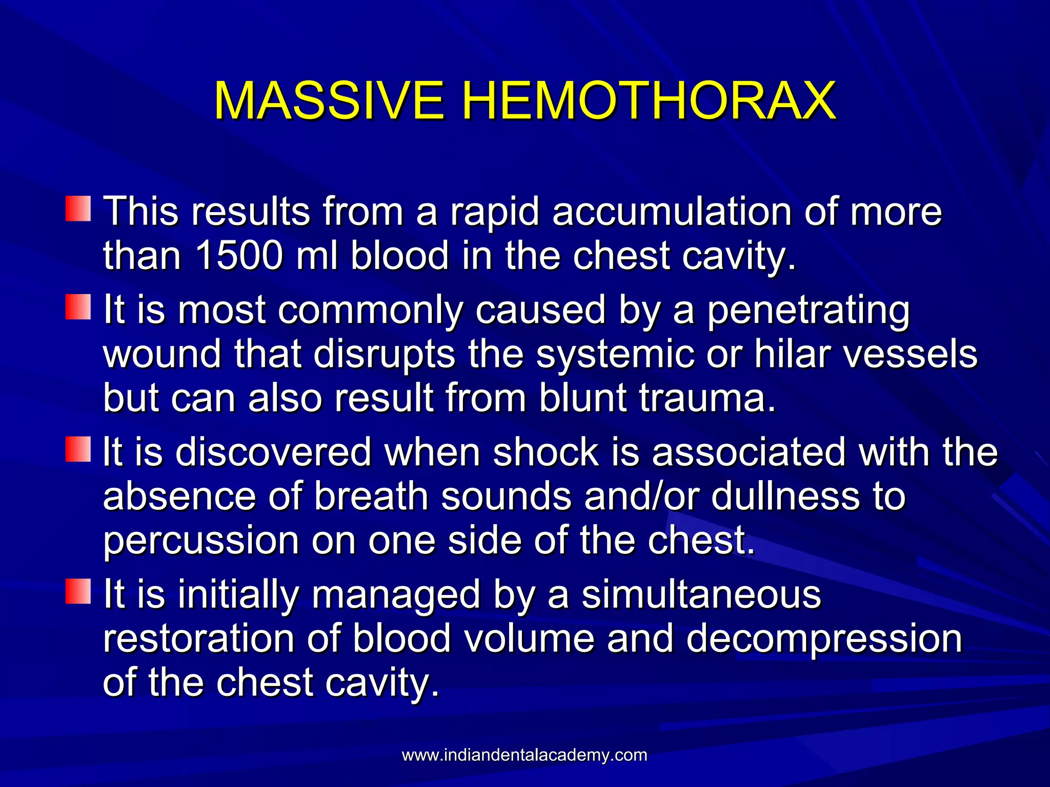
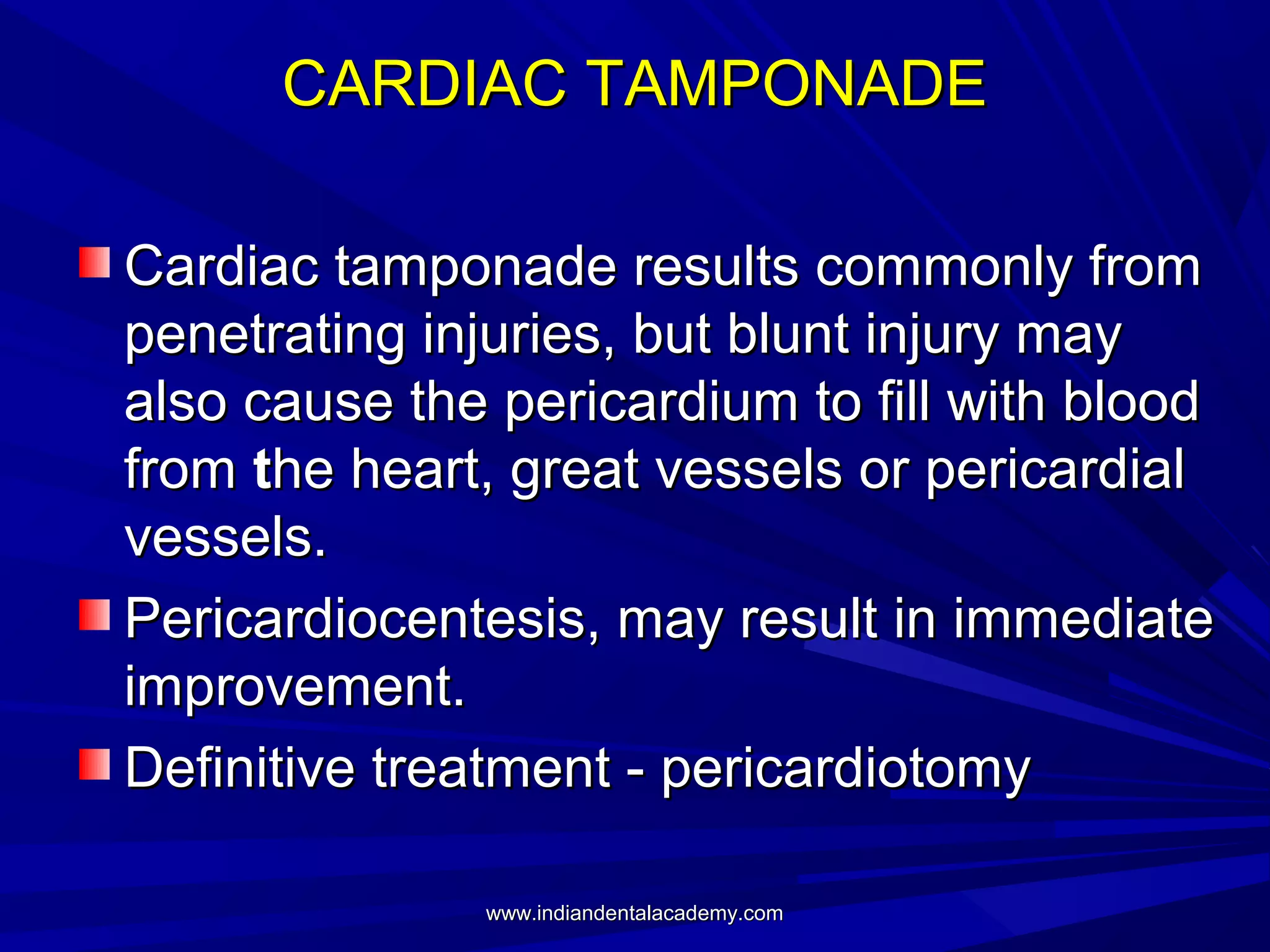
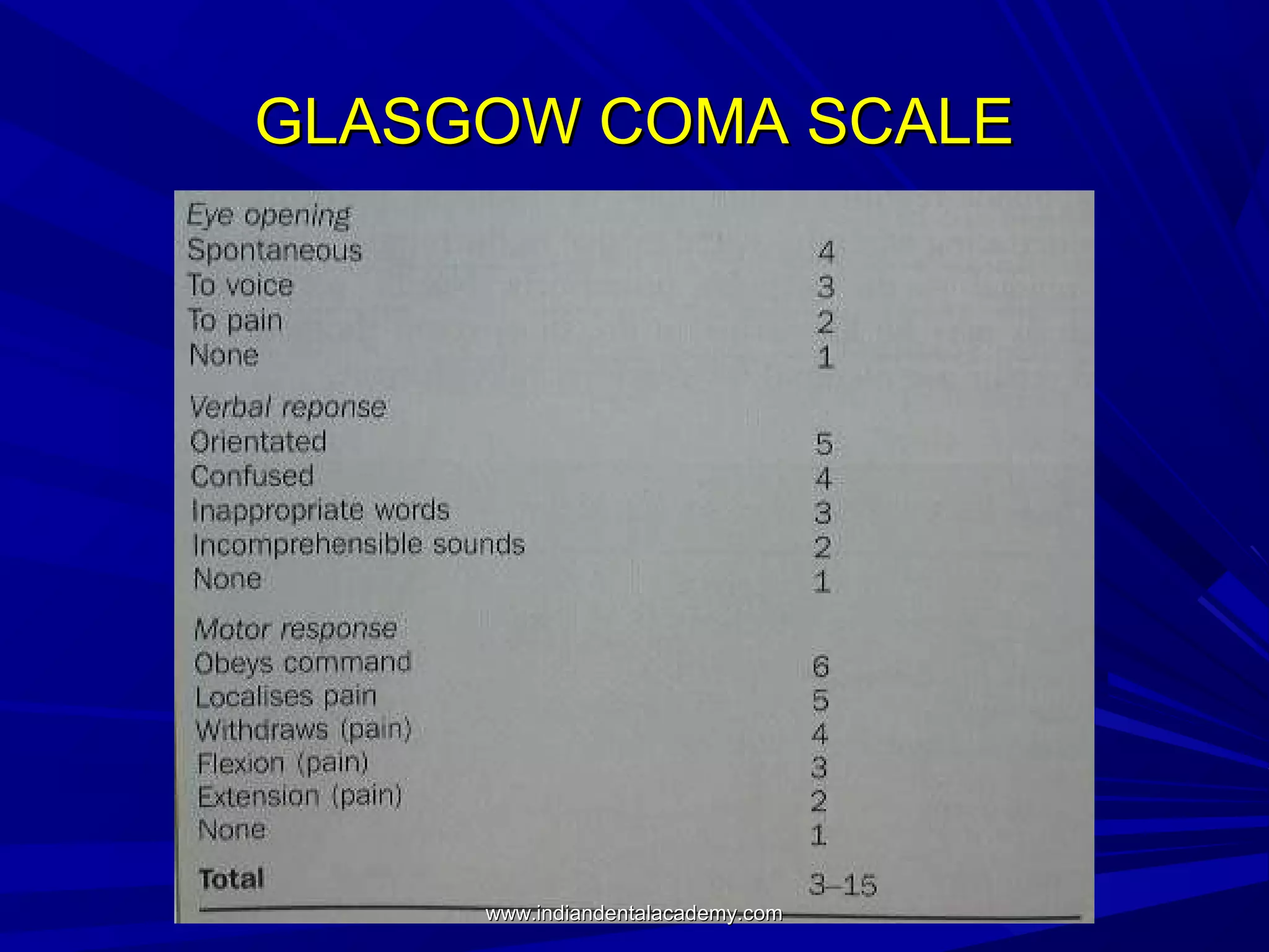
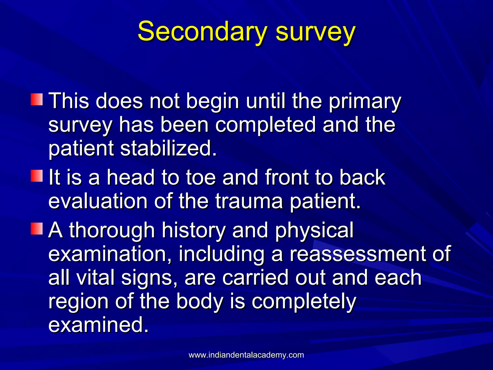
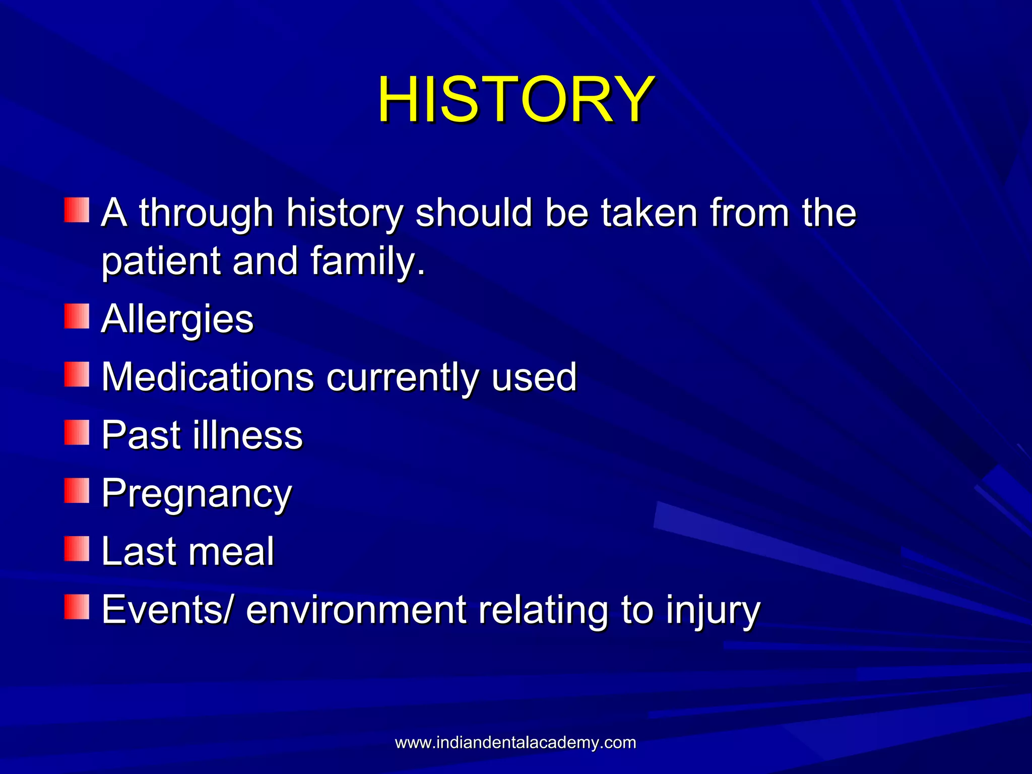
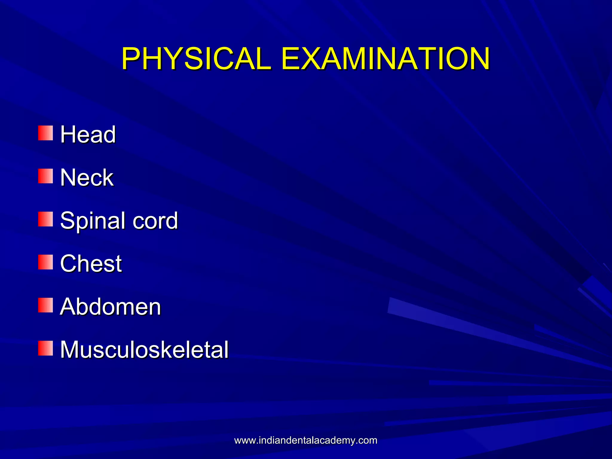
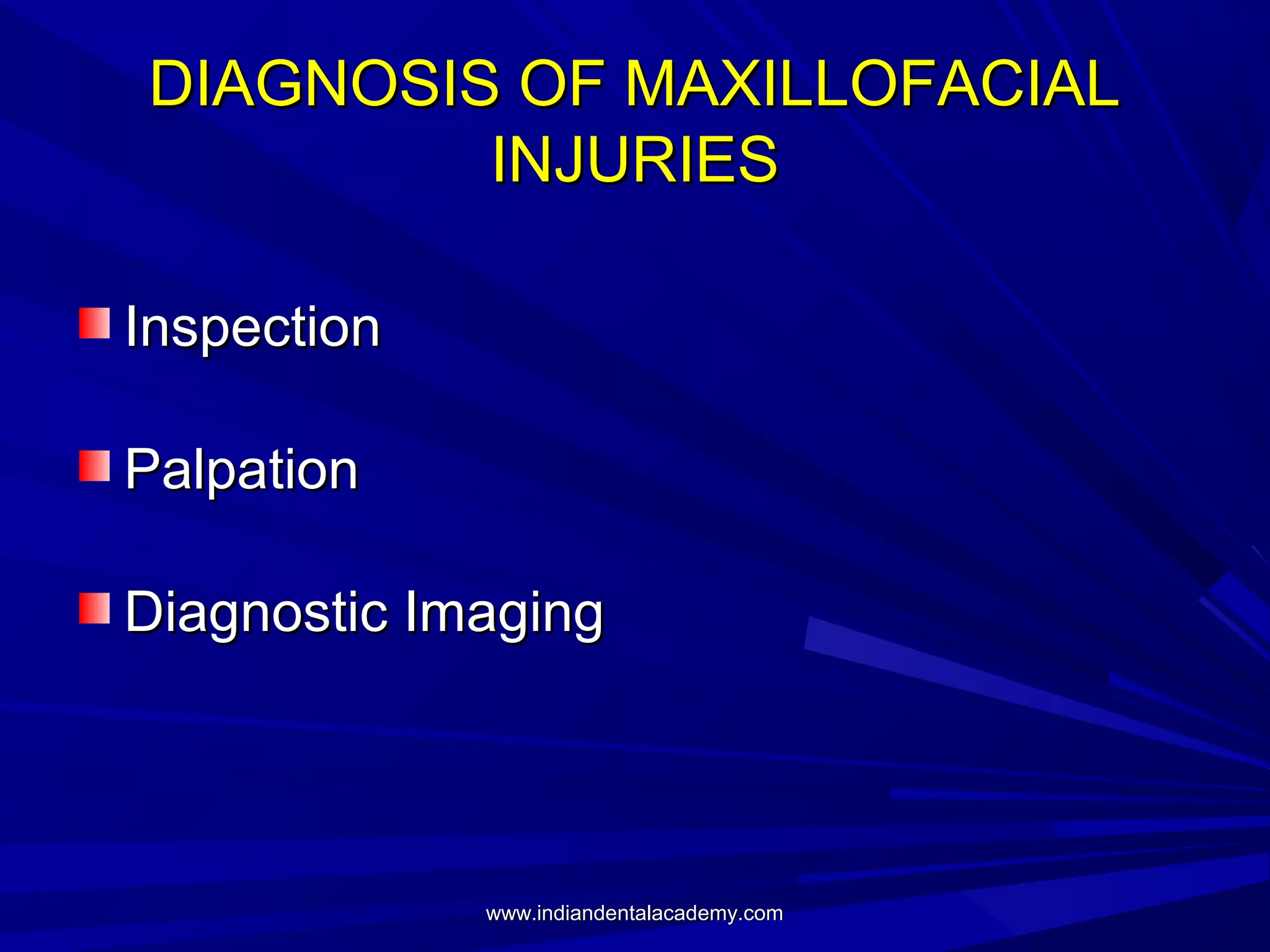
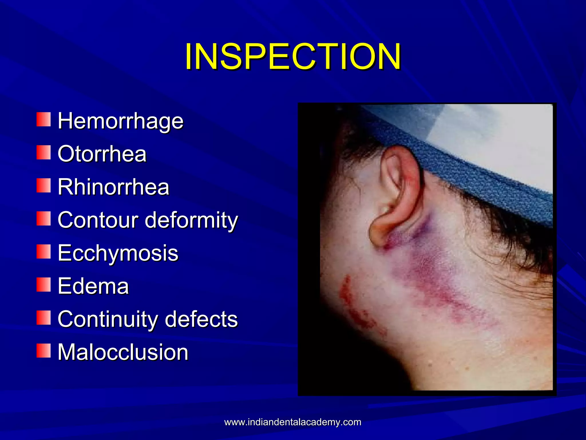
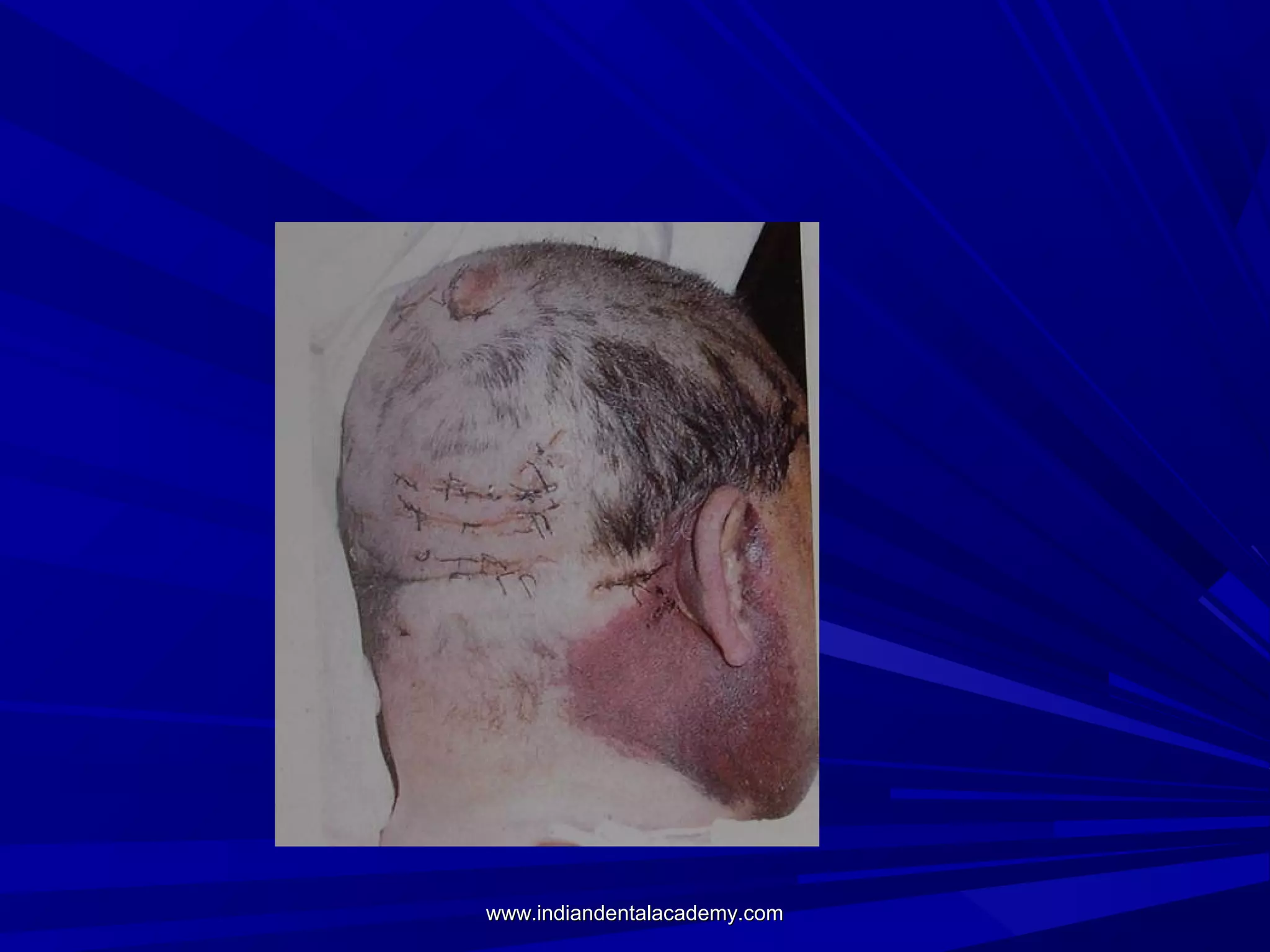
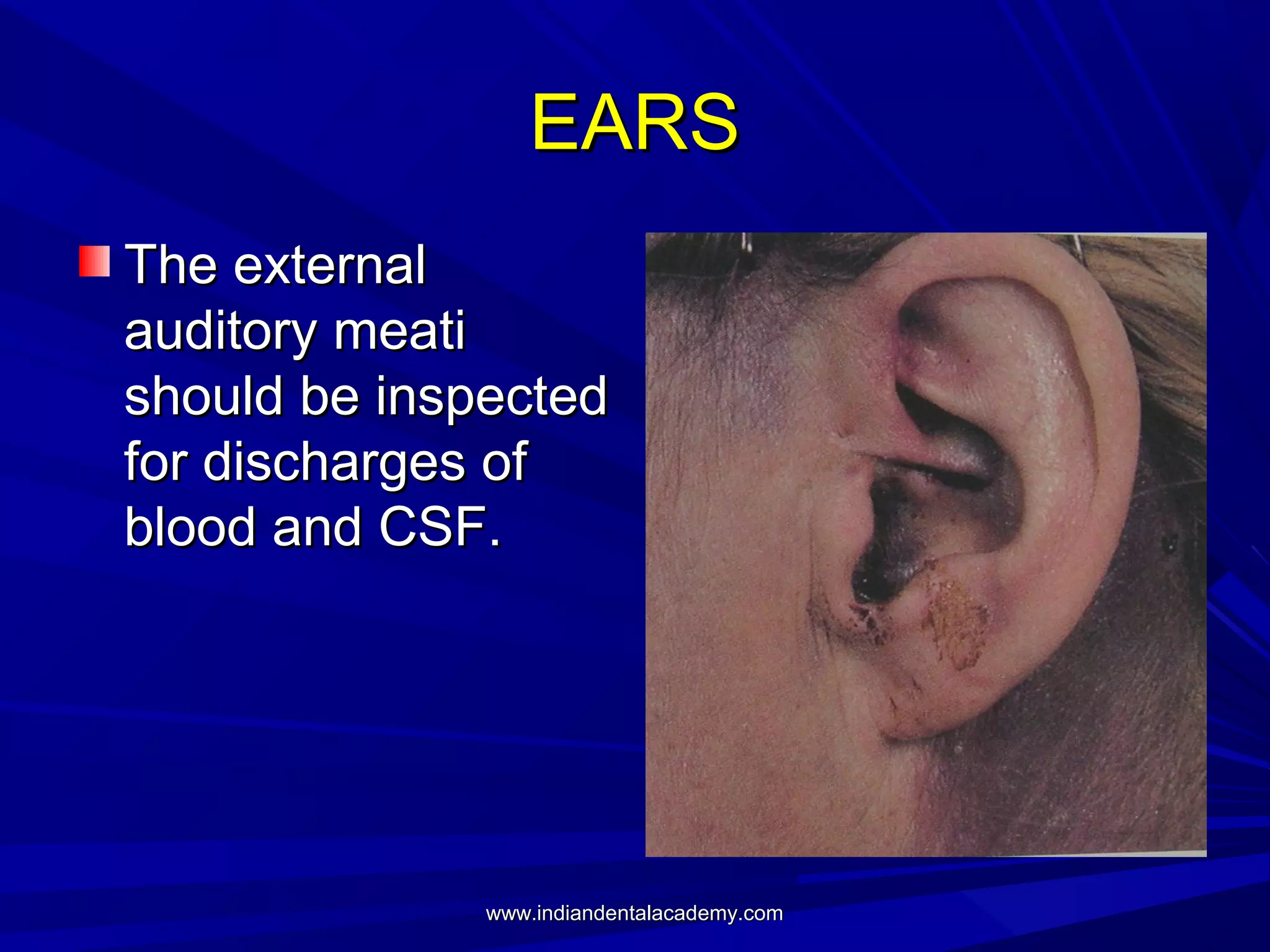
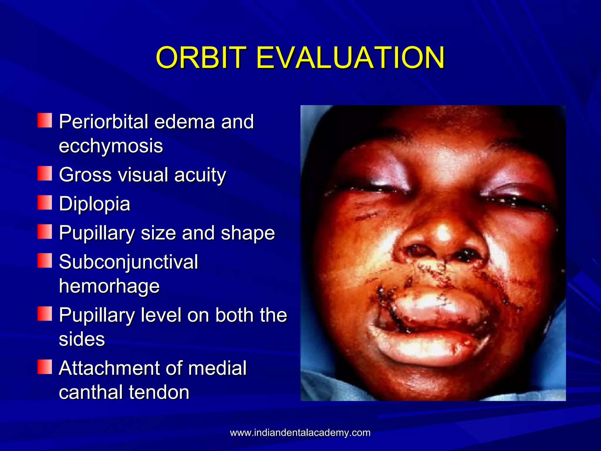
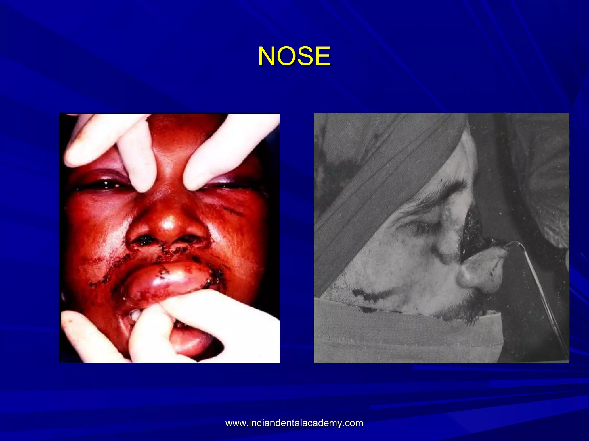
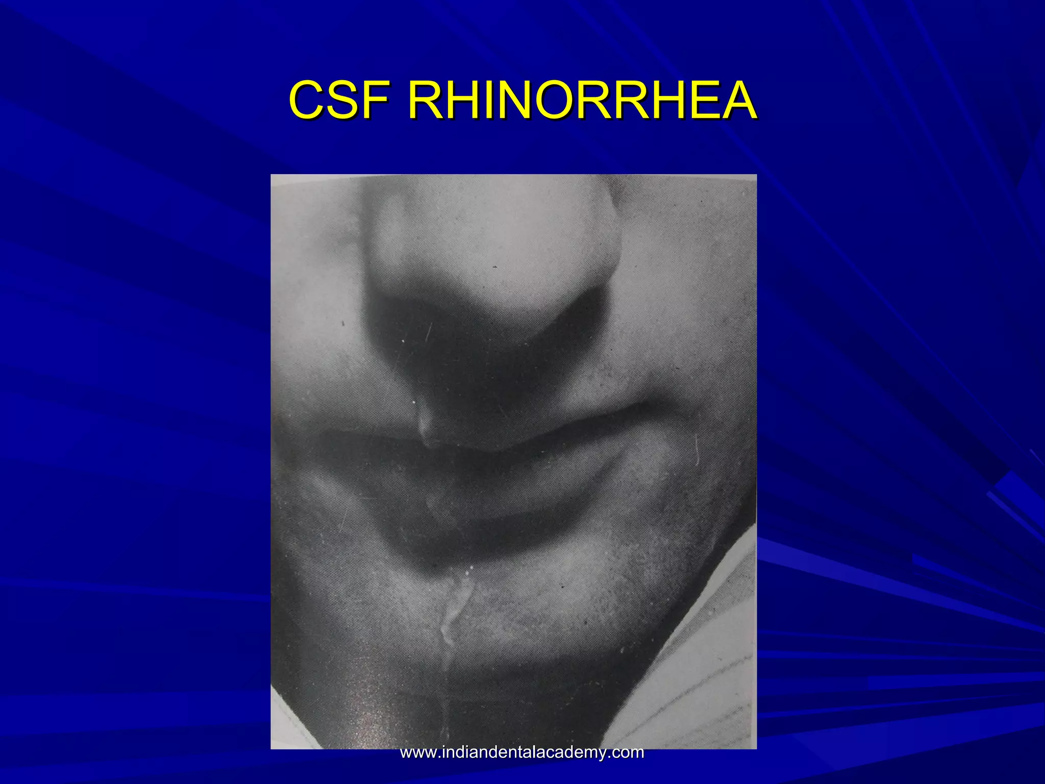
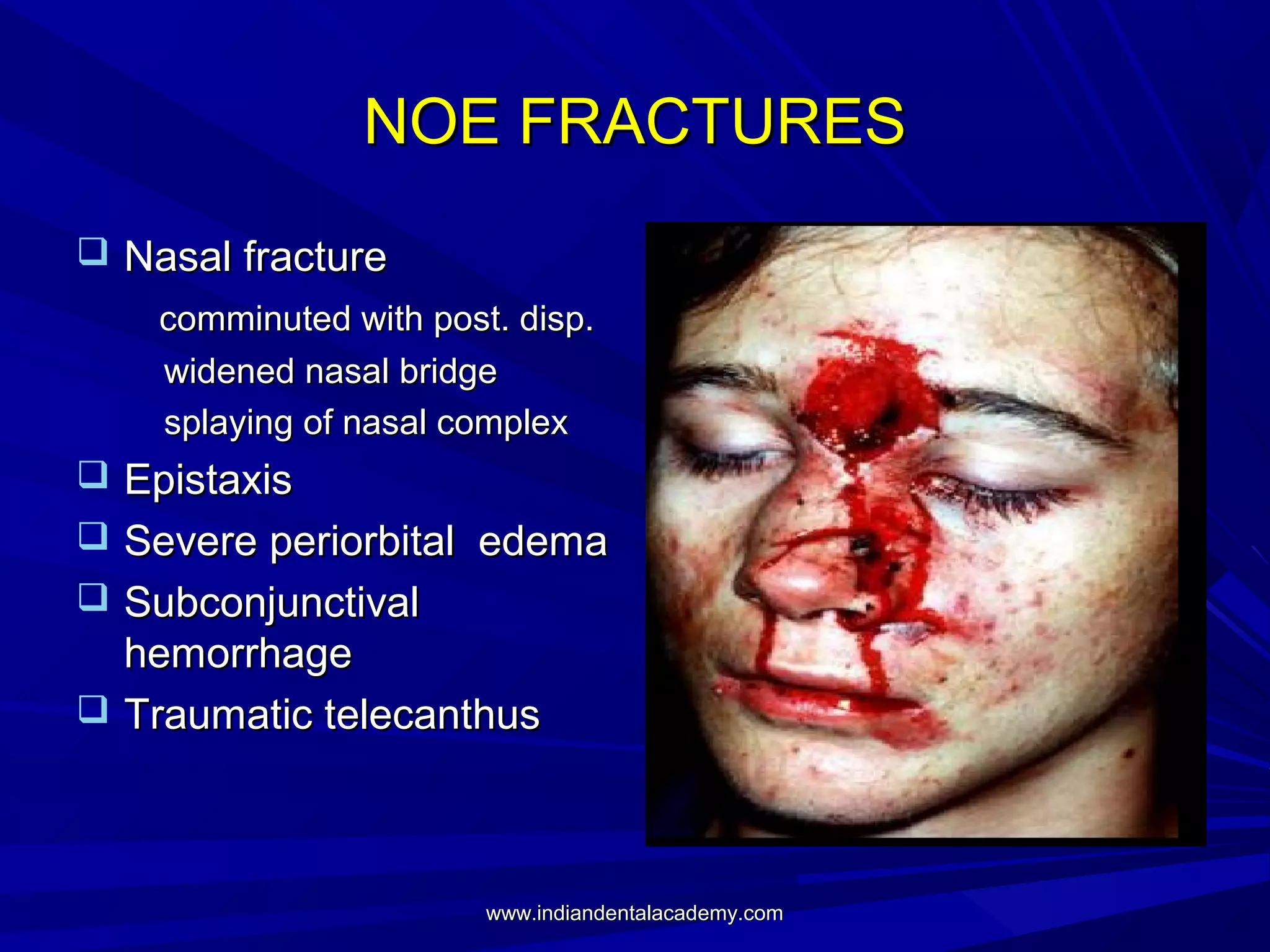
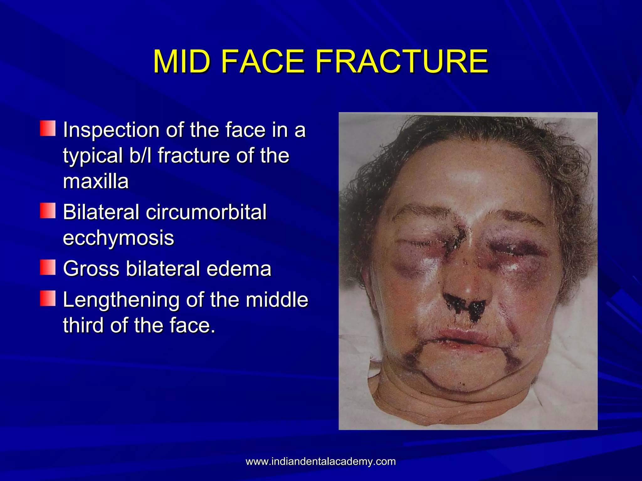
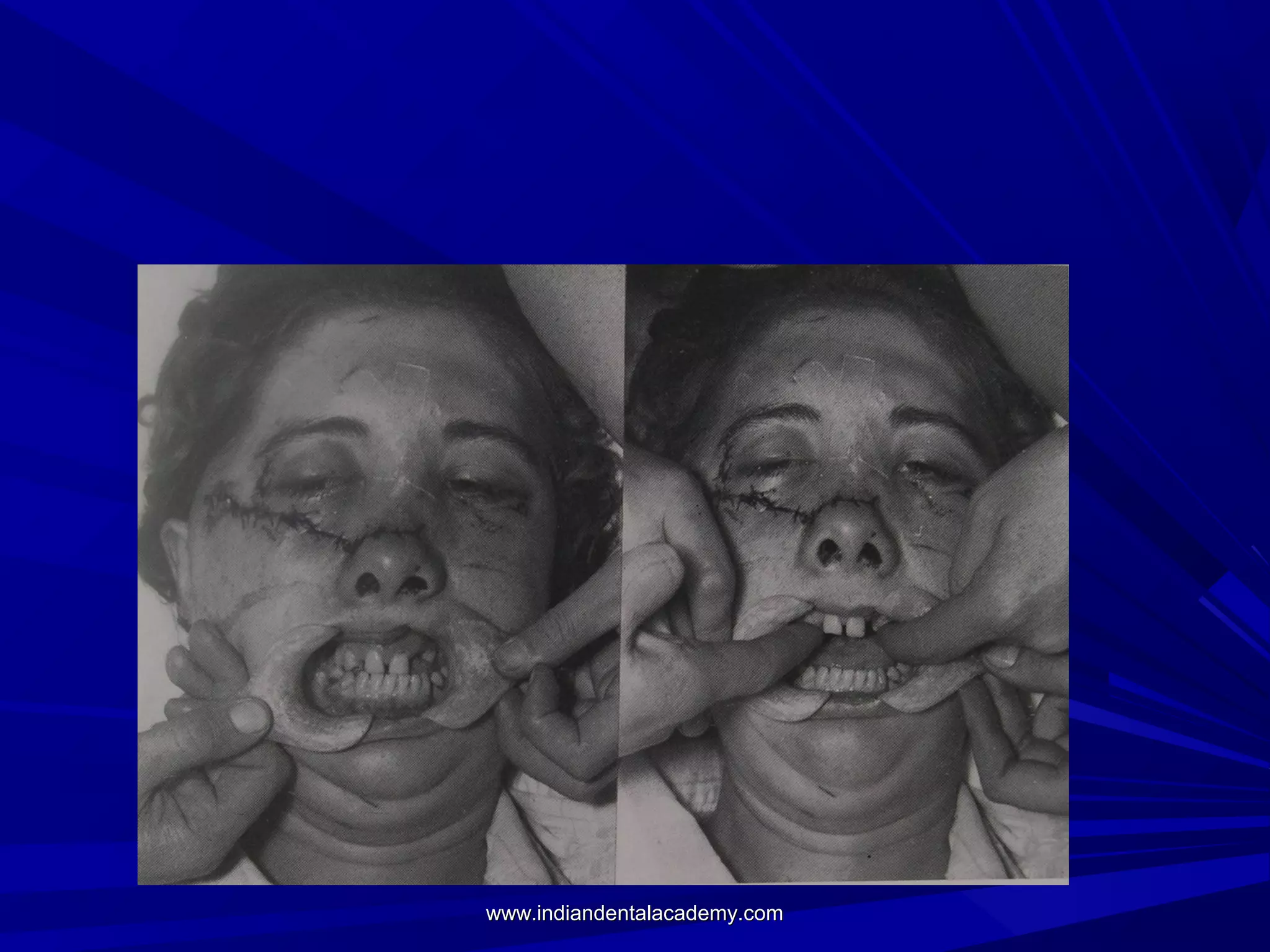
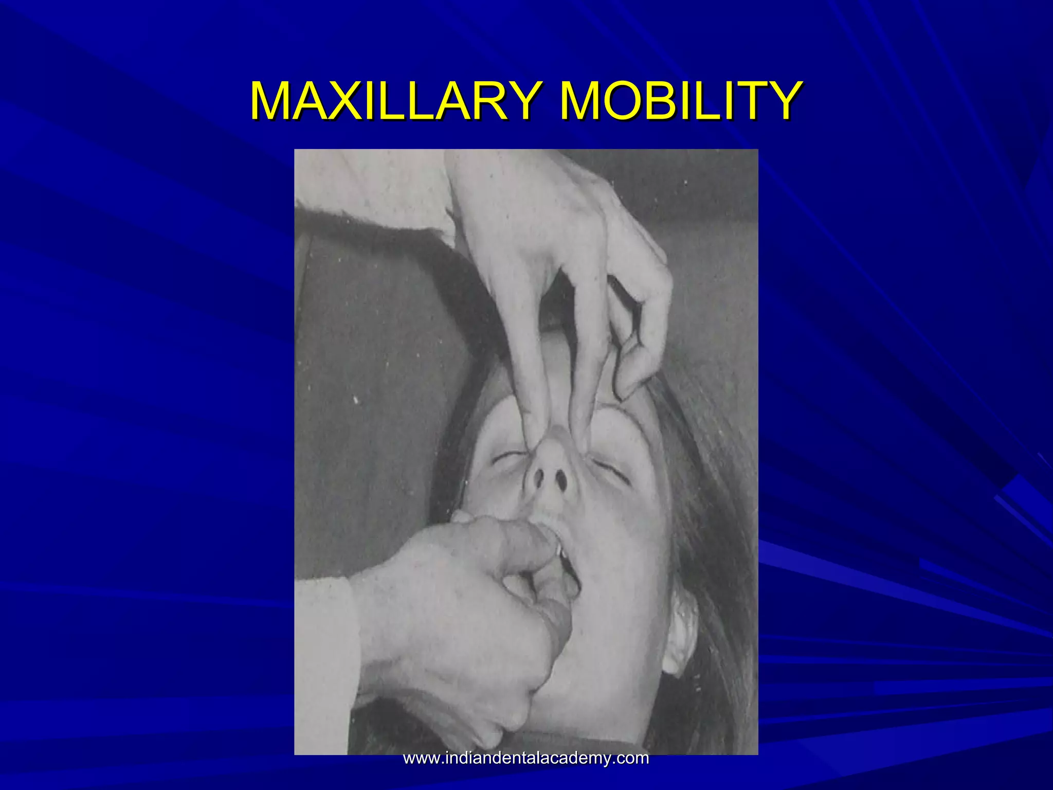
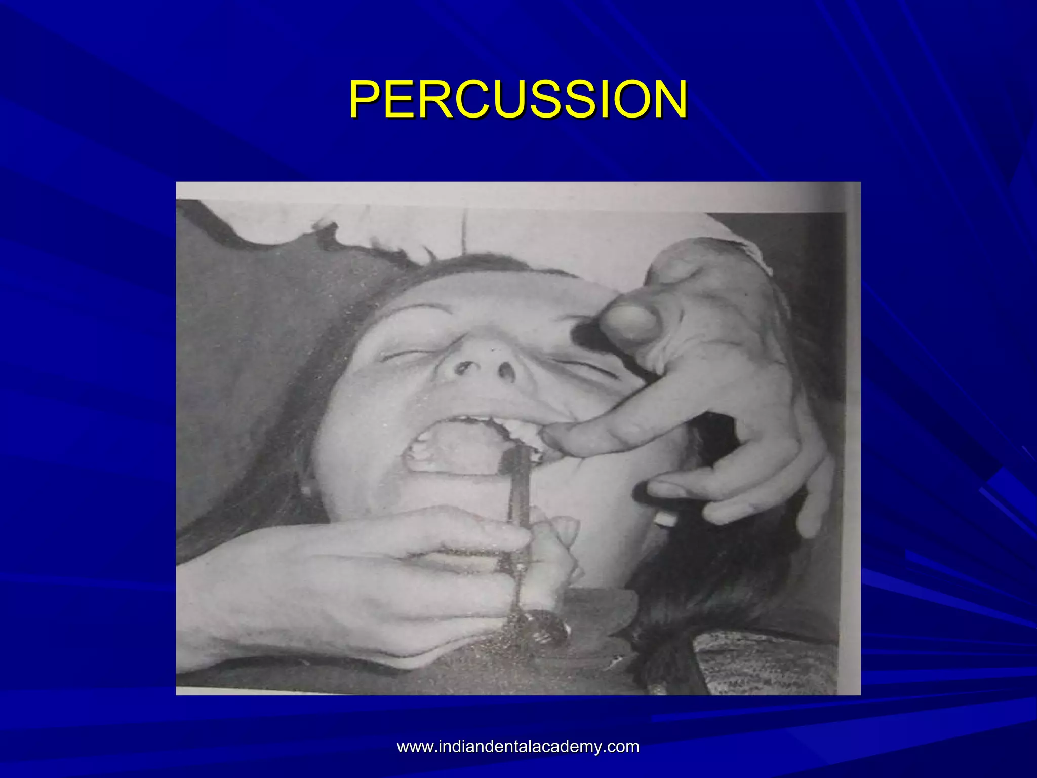
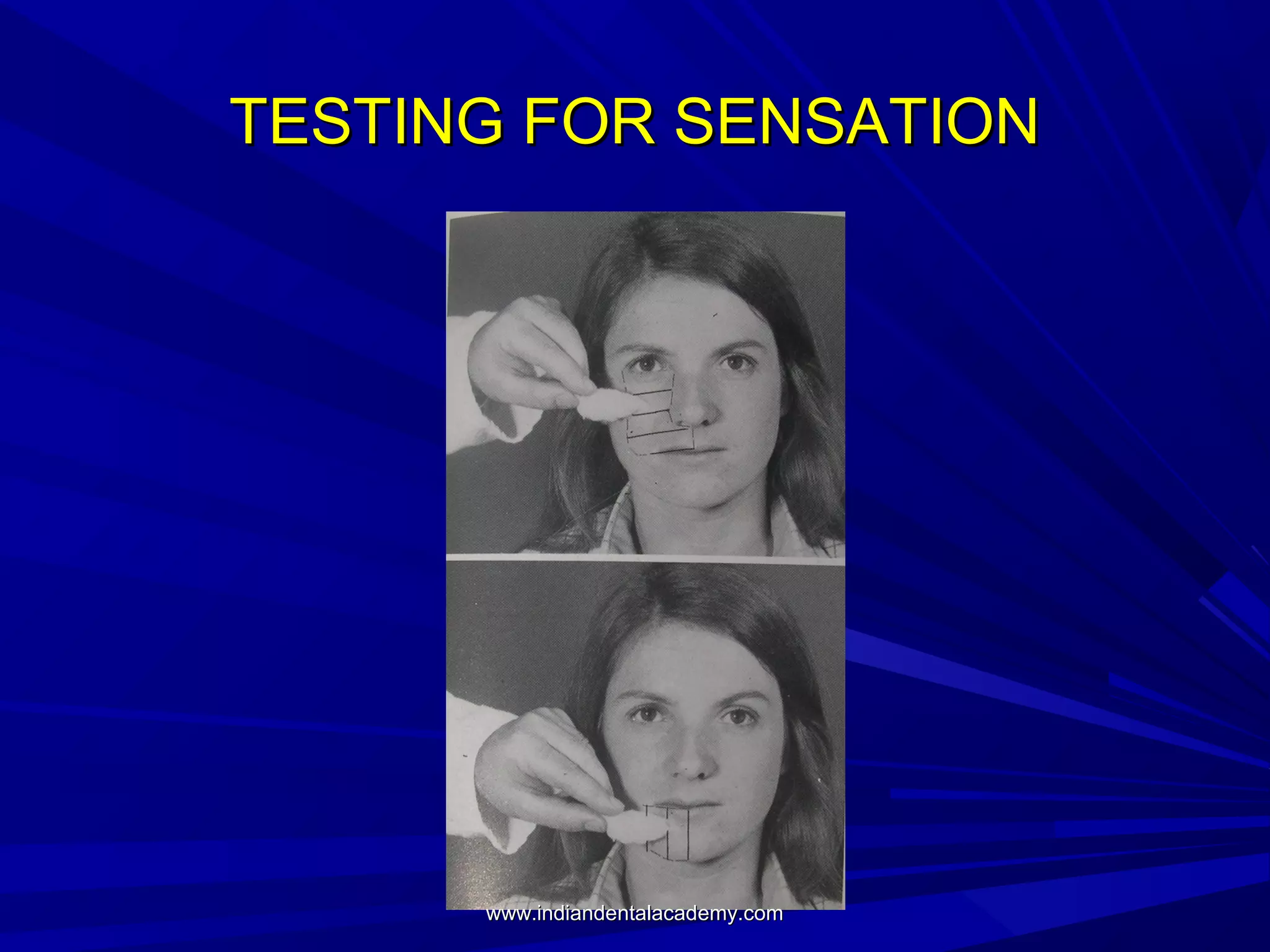
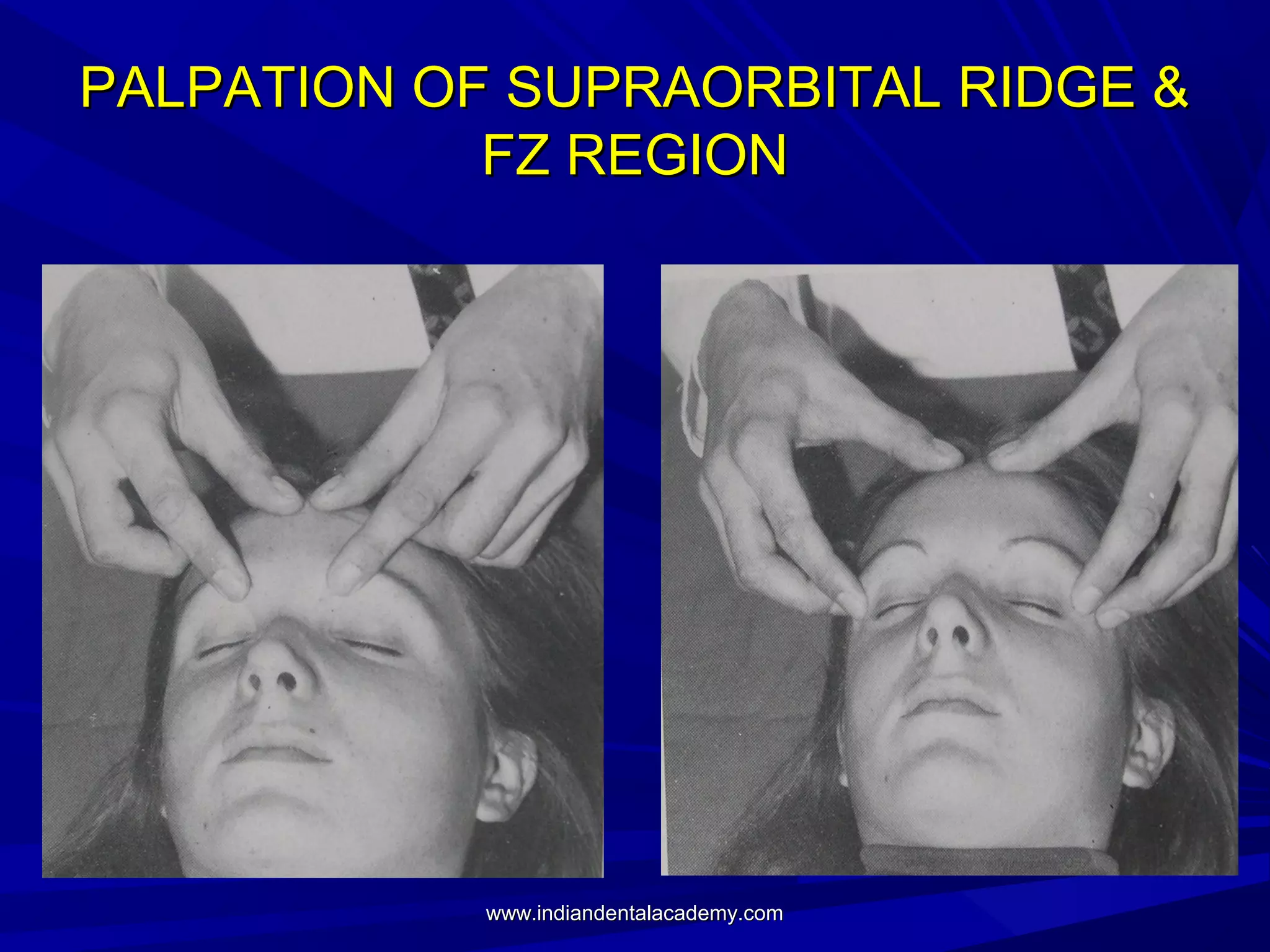
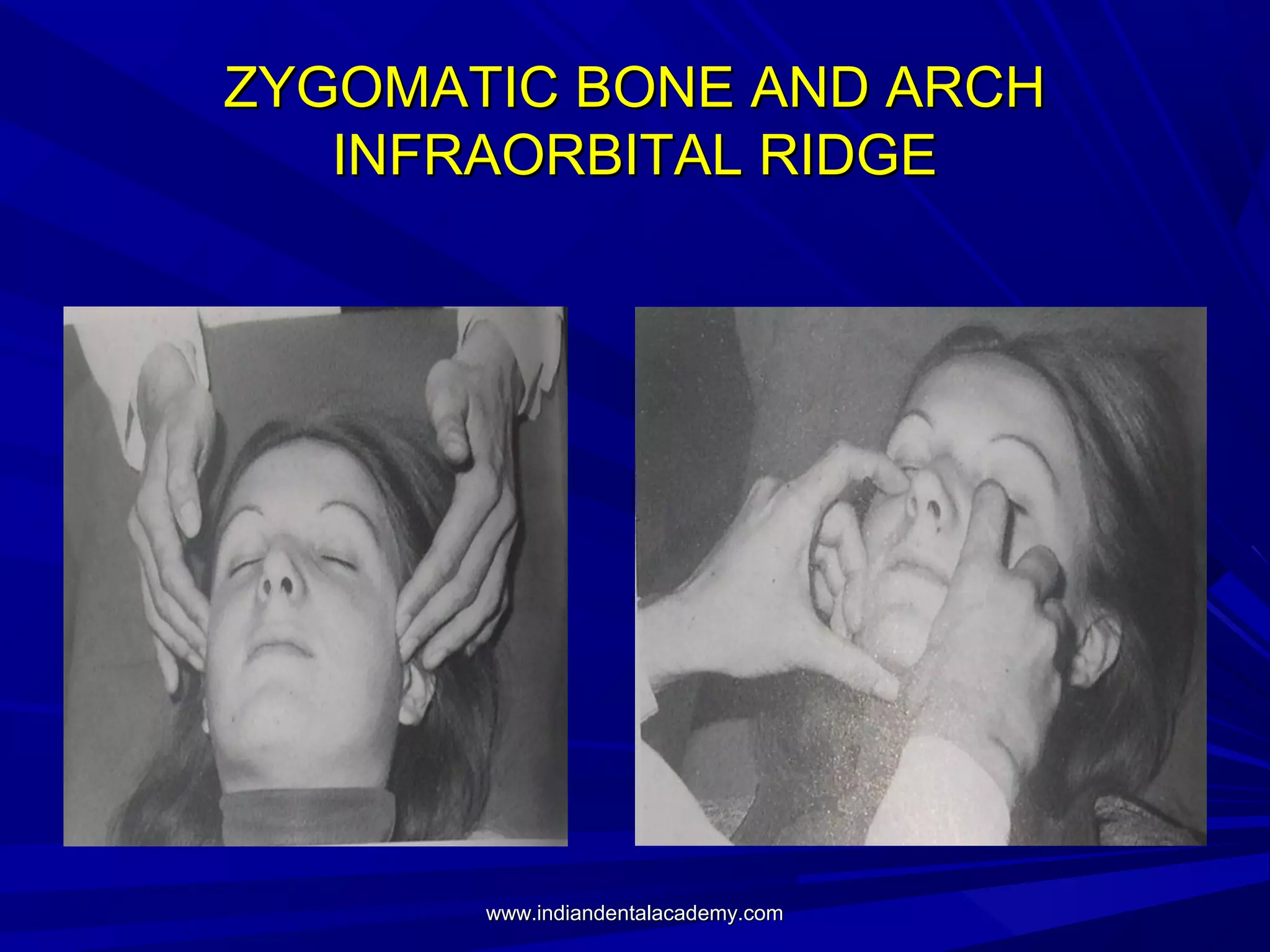
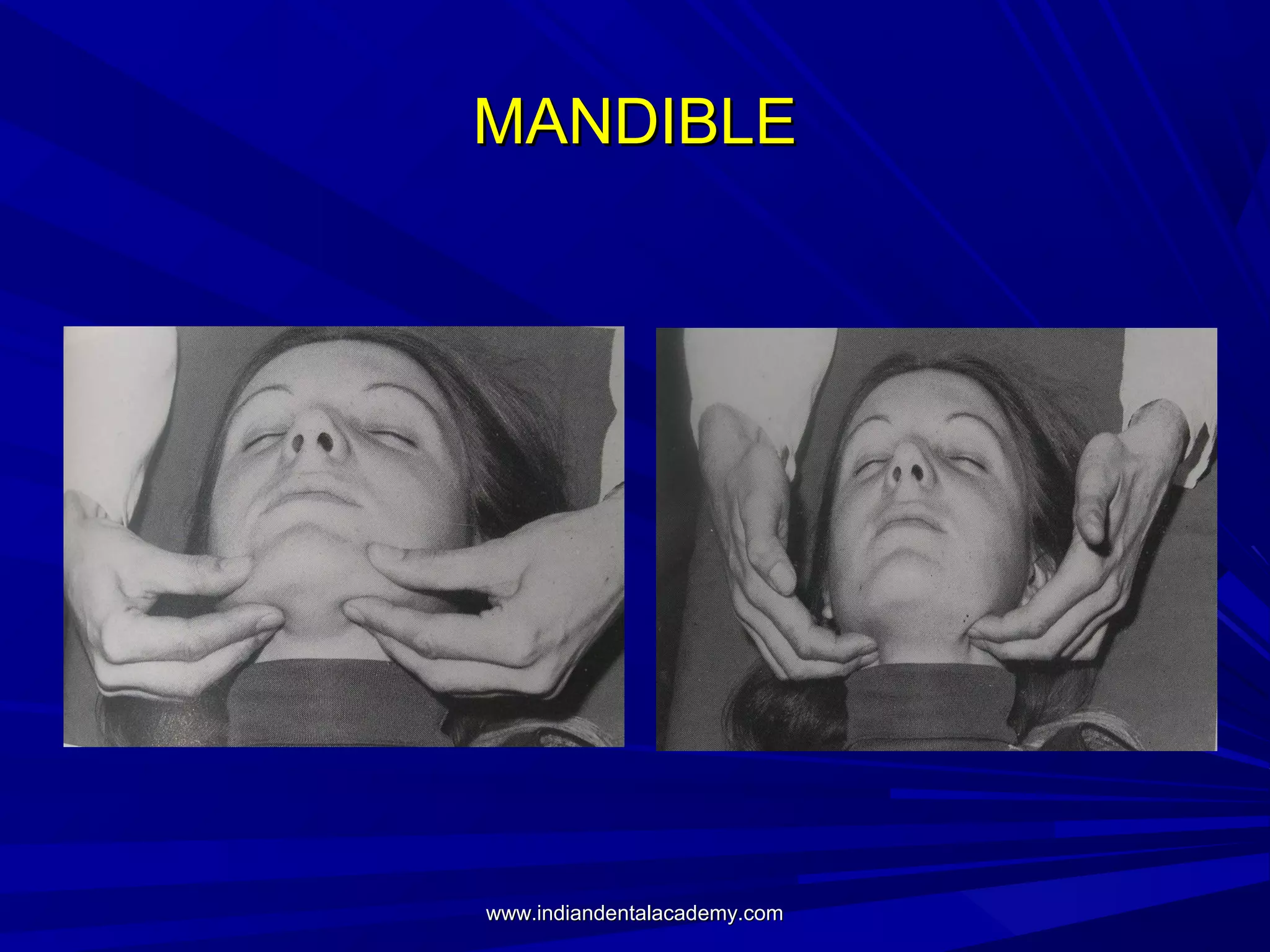
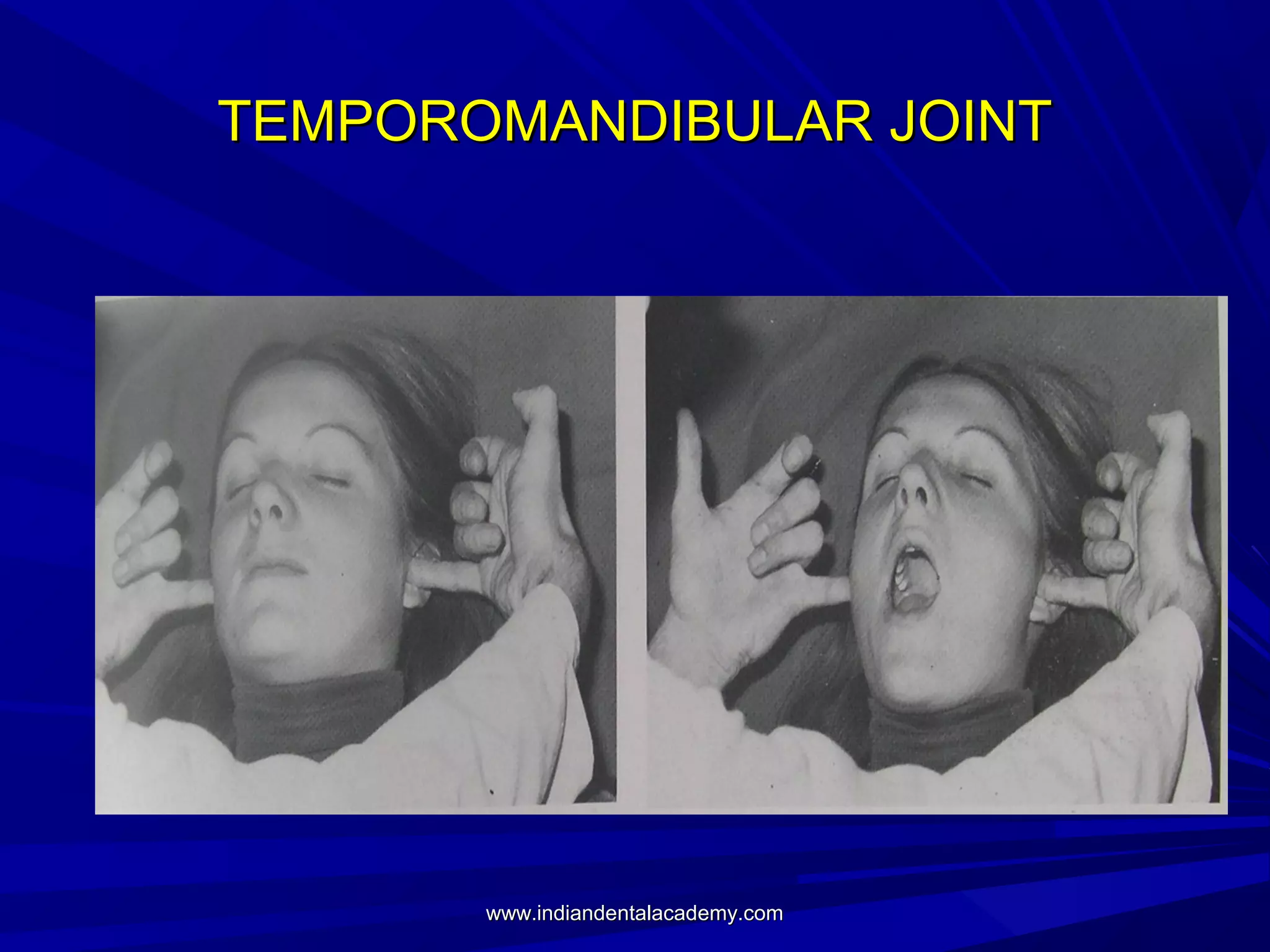
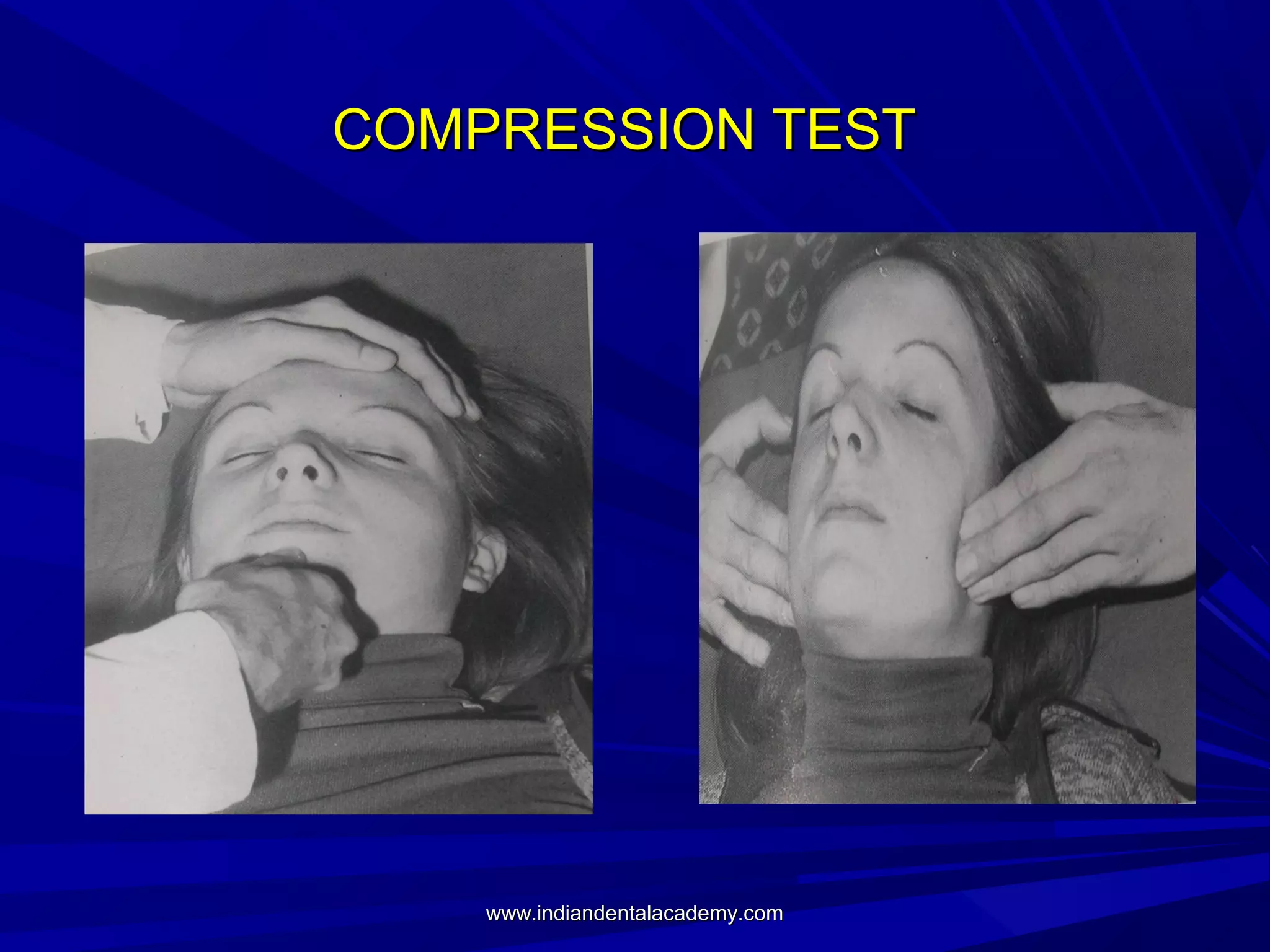
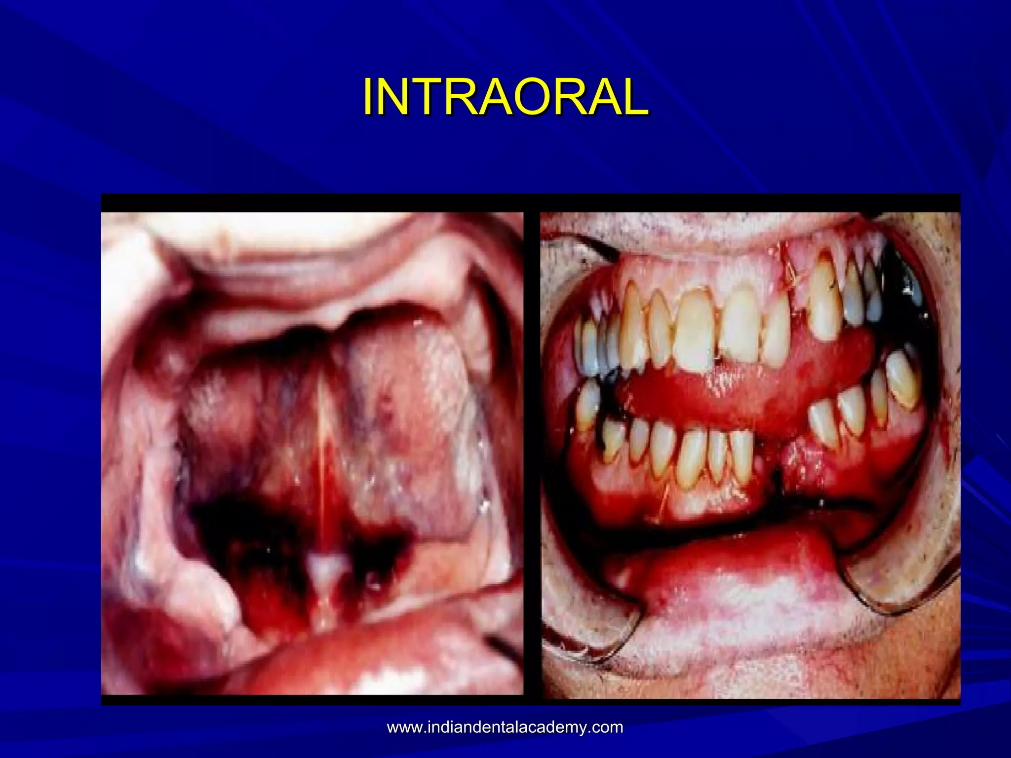
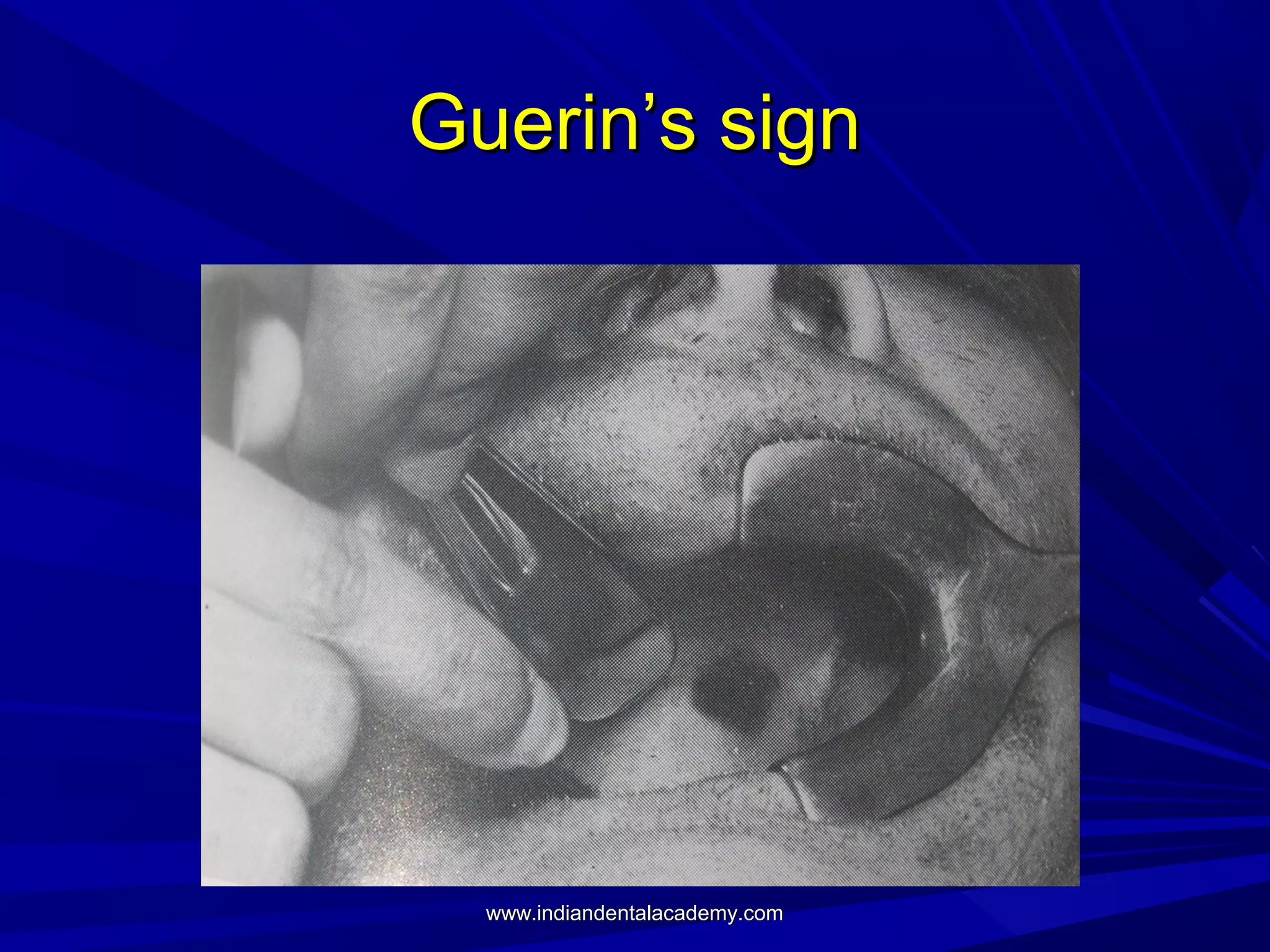
PRIMARY ASSESSMENT AND
PREOPERATIVE MANAGEMENT OF
MAXILLOFACIL AND HEAD INJURIES
INDIAN DENTAL ACADEMY
Leader in continuing dental education
www.indiandentalacademy.com

www.indiandentalacademy.com
INTRODUCTION

www.indiandentalacademy.com
DEATHS FOLLOWING RTA’S
Immmediate deaths (50 per cent)
Early deaths (30 per cent)
Late deaths (20 per cent)

www.indiandentalacademy.com
TRIMODAL DISTRIBUTION OF DEATH

www.indiandentalacademy.com
STEPS IN MANAGEMENT
Primary survey- identify what is killing the
patient.
Resuscitation- treat what is killing the
patient.
Secondary survey- proceed to identify all
other injuries.
Definitive care- develop a definitive
management plan.
www.indiandentalacademy.com
PRIMARY SURVEY AND
RESUSCITATION
Primary survey of the patient follows a strict
sequential protocol.
Airway and Cervical spine control.
Breathing and Ventilation.
Circulation and Hemmorhage control.
Disability = Neurological status.
Exposure + Environment.
www.indiandentalacademy.com
CERVICAL SPINE
There should be high index of suspicion for
cervical spine injury in the patient with
maxillofacial injuries or multisystem trauma, if
the patient has an altered level of
consciousness or if there is history of high
speed impact.
www.indiandentalacademy.com
CERVICAL SPINE
Assume injury to cervical spine if –
Blunt trauma above clavicles
Head injury
Maxillofacial trauma
Multiple trauma

www.indiandentalacademy.com
CERVICAL SPINE CONTROL
Definitive cervical spine
control requires the
application of semi rigid
cervical collar, sandbags
placed on either side of the
head and tapes over the
forehead and chin,
immobilizing the head and
neck to the trolley.

www.indiandentalacademy.com
CERVICAL SPINE CONTROL

In the restless and agitated patient, immobilizing
the head and neck while allowing the rest of the
body to move can damage the cervical spine and
here just a semirigid collar is acceptable.
www.indiandentalacademy.com
AIRWAY
Assessment must rapidly be made as to
whether the patient can maintain and
protect his own airway.
If the patient is able to respond verbally,
the airway, at least for the time being, is
patent but repeated assessment is
prudent.
www.indiandentalacademy.com
AIRWAY
Teeth, dentures, vomitus, hematoma and
other foreign bodies may block the airway
- right main bronchus - susceptible.
Earlv in the primary survey, the oral cavity
should be cleared using a finger sweep
followed by aspiration.

www.indiandentalacademy.com
AIRWAY
Hemorrhage may result from several causes
to obstruct the airway.
Bleeding from vessels in open wounds can
be controlled by pressure with gauze swabs.
Soft tissue swelling and edema resulting
from trauma to the oral cavity may
compromise the airway.
www.indiandentalacademy.com
The fractured
nose may need
to be packed
as a result of
damage to the
anterior or
posterior
ethmoidal
vessels or the
maxillary
artery.
www.indiandentalacademy.com
AIRWAY
The tongue can often fall back and
obstruct the airway in unconscious
patients and in these a simple chin lift or
jaw thrust maneuver can be used to
correct the tongue position and open the
airway.
www.indiandentalacademy.com
CHIN LIFT
Chin lift should be
performed without
hyperextending the
neck.
The mandible is gently
lifted upward using the
fingers of one hand
placed under the chin.
The thumb of the same
hand lightly depresses
the lower lip to open the
mouth.
www.indiandentalacademy.com
JAW THRUST
Jaw thrust is performed
by grasping the angles
of the mandible with
one hand on each side
and displacing the
mandible forward.

www.indiandentalacademy.com
TONGUE STAY SUTURE

www.indiandentalacademy.com
INDICATIONS FOR DEFINITIVE AIRWAY
Apnea
Inability to maintain patent airway by other
means
need to protect the lower airway from blood or
vomit
potential compromise of the airway, e.g.
following inhalational injury, facial fractures etc.
The presence of a closed head injury requiring
assisted ventilation (GCS ≤ 8)
An inability to maintain adequate oxygenation by
face mask oxygen supplementation.
www.indiandentalacademy.com
DEFINITIVE AIRWAY
This provides oxygen assisted ventilation
via a cuffed tube present in the trachea
with the cuff inflated and the tube secured
in place with a tape.
Three types
Orotracheal intubation
Nasotracheal intubation
Surgical (cricothyroidotomy &
tracheostomy)
www.indiandentalacademy.com
ALOGRITHM LEADING TO
SURGICAL AIRWAY

www.indiandentalacademy.com
CRICOTHYRIODOTOMY

www.indiandentalacademy.com
CRICOTHYRIODOTOMY

www.indiandentalacademy.com
CRICOTHYRIODOTOMY

www.indiandentalacademy.com
TRACHEOSTOMY

www.indiandentalacademy.com
TRACHEOSTOMY

www.indiandentalacademy.com
TRACHEOSTOMY

www.indiandentalacademy.com
TRACHEOSTOMY

www.indiandentalacademy.com
TRACHEOSTOMY

www.indiandentalacademy.com
SUPPLEMENTAL OXYGEN
Supplemental oxygen delivered through a
well-fitting re-breathing mask at a rate of l5
litres per minute to achieve maximum
oxygenation of the tissues, should be given
to every trauma patient.
www.indiandentalacademy.com
BREATHING AND VENTILATION
Once the airway has been secured, breathing and
ventilation must be assessed.
Direct trauma to the chest - pain with breathing
and leads to rapid shallow breathing - hypoxemia.
Intracranial injury - abnormal patterns of
breathing and compromise the adequacy of
ventilation.
Cervical spinal cord injury - diaphragmatic
breathing and interfere with the ability to meet
increased oxygen demands.
www.indiandentalacademy.com
BREATHING AND VENTILATION
Thoracic injuries that are immediately life
threatening include
Flail chest
Tension pneumothorax
Open pneumothorax
Massive hemothorax
Cardiac tamponade
These should be identified in the primary
survey.
www.indiandentalacademy.com
EXAMINATION
All the clothes covering the front and sides of the
patients chest must be removed.
The respiratory rate, effort and symmetry should
be recorded, because these are sensitive
indicators of underlying pulmonary contusion,
hemothorax, pneumothorax and fractured ribs.

www.indiandentalacademy.com
FLAIL CHEST
This occurs when a segment of the chest
wall looses bony continuity with the rest of
the thoracic cage, usually as a result
trauma associated with multiple rib
fractures.
Associated pain with chest wall movement
and underlying lung injury add to the
patient’s hypoxia.
www.indiandentalacademy.com
FLAIL CHEST
Initial treatment includes adequate
ventiliation, administration of humidified
oxygen and fluid resuscitation.
Definitive treatment - re-expand the lung,
ensure oxygenation, administer fluids and
provide analgesia to improve ventilation.
www.indiandentalacademy.com
OPEN PNEUMOTHORAX
Large defects of the chest wall which remain
open result in an open pneumothorax.
If the opening in the chest wall is approximately
two-thirds the diameter of the trachea, air
passes preferentially through the chest defect
with each respiratory effort.
Effective ventilation is thereby impaired, leading
to hypoxia

www.indiandentalacademy.com
OPEN PNEUMOTHORAX
Initial management is by promptly closing
the defect with a sterile occlusive dressing
large enough to overlap the wound's edges
that is taped securely on three sides which
provides a flutter - type valve effect.
A chest drain should be placed remote
from the site as soon as possible.
Definitive surgical closure later.
www.indiandentalacademy.com
TENSION PNEUMOTHORAX
Tension pneumothorax develops when a
one way valve air leak occurs either from
the lung or through the chest wall.
Air is forced into the thoracic cavity,
between the parietal and visceral pleura,
without any means of escape, completely
collapsing the affected lung.
www.indiandentalacademy.com
TENSION PNEUMOTHORAX
Chest pain
Air hunger
Respiratory distress
Tachycardia
Hypotension
Tracheal deviation
Unilateral absence of breath sounds
Neck vein distention and Cyanosis - late
manifestation.
www.indiandentalacademy.com
TENSION PNEUMOTHORAX
It requires immediate decompression by
inserting a large bore needle into the
second intercostal space in the
midclavicular line of the affected
hemithorax.
Definitive treatment requires the insertion
of a chest drain into the fifth intercostal
space (nipple level) between the anterior
and midaxillary lines
www.indiandentalacademy.com
MASSIVE HEMOTHORAX
This results from a rapid accumulation of more
than 1500 ml blood in the chest cavity.
It is most commonly caused by a penetrating
wound that disrupts the systemic or hilar vessels
but can also result from blunt trauma.
lt is discovered when shock is associated with the
absence of breath sounds and/or dullness to
percussion on one side of the chest.
It is initially managed by a simultaneous
restoration of blood volume and decompression
of the chest cavity.
www.indiandentalacademy.com
CARDIAC TAMPONADE
Cardiac tamponade results commonly from
penetrating injuries, but blunt injury may
also cause the pericardium to fill with blood
from the heart, great vessels or pericardial
vessels.
Pericardiocentesis, may result in immediate
improvement.
Definitive treatment - pericardiotomy
www.indiandentalacademy.com
CIRCULATION WITH HEMORRHAGE
CONTROL
Shock is defined as an abnormality of the
circulation that results in inadequate organ
perfusion and tissue oxygenation.
If unchecked this will lead to end organ
dysfunction.
Hemorrhage is the main cause of post injury
deaths that are preventable by rapid treatment in
the hospital setting.
Hypotension following injury must be considered
to be hypovolemic in origin until proved otherwise.
www.indiandentalacademy.com
Classification and signs of hypovolemic shock
(in an adult assuming a
70 kg patient with normally 5 litres of circulating volume)

www.indiandentalacademy.com
HEMORRHAGE
Hemorrhage is an acute loss of circulating
blood volume.
After airway and breathing, the initial
treatment of shock is directed toward
restoring cellular and organ perfusion with
adequately oxygenated blood.

www.indiandentalacademy.com
MANAGEMENT OF HEMORRHAGE
The priority is to control the source of
hemorrhage.
External hemorrhage - controlled by direct
manual pressure.
Occult hemorrhage - immediate surgical
intervention.
www.indiandentalacademy.com
FLUID REPLACEMENT
The aim of fluid management in hypotensive
resuscitation - restore critical organ perfusion.
Therefore in a standard adult trauma victim, 2
litres of warmed crystalloid, preferably Ringer's
lactate, should be given and then the patient
reassessed.
In reassessing the circulatory state, one of three
responses are seen :- Responder, Transient
responder and non responder
www.indiandentalacademy.com
BLOOD REPLACEMENT
Either whole blood or packed cells can he
used to resuscitate the trauma patient.
The main purpose in transfusing blood is
to restore the oxygen-carrying capacity of
the intravascular volume.
Volume resuscitation itself can be
accomplished with crystalloids.

www.indiandentalacademy.com
DISABILITY (NEUROLOGICAL
EVALUATION)
A rapid evaluation is performed at the end
of the primary survey and this establishes
the level of the patient's consciousness, as
well as pupillary size and reaction.
The Glasgow Coma Scale (GCS) is a
detailed, quick, simple and predictive of
patient outcome.
www.indiandentalacademy.com
GLASGOW COMA SCALE

www.indiandentalacademy.com
Exposure and environment
The patient should be completely undressed
to facilitate a thorough examination and
assessment.
After the assessment is completed it is
important to cover the patient with warm
blankets or an external warming device to
prevent hypothermia.
Intravenous fluids should be warmed before
infusion.
www.indiandentalacademy.com
Secondary survey
This does not begin until the primary
survey has been completed and the
patient stabilized.
It is a head to toe and front to back
evaluation of the trauma patient.
A thorough history and physical
examination, including a reassessment of
all vital signs, are carried out and each
region of the body is completely
examined.
www.indiandentalacademy.com
HISTORY
A through history should be taken from the
patient and family.
Allergies
Medications currently used
Past illness
Pregnancy
Last meal
Events/ environment relating to injury
www.indiandentalacademy.com
PHYSICAL EXAMINATION
Head
Neck
Spinal cord
Chest
Abdomen
Musculoskeletal

www.indiandentalacademy.com
DIAGNOSIS OF MAXILLOFACIAL
INJURIES
Inspection
Palpation
Diagnostic Imaging

www.indiandentalacademy.com
INSPECTION
Hemorrhage
Otorrhea
Rhinorrhea
Contour deformity
Ecchymosis
Edema
Continuity defects
Malocclusion
www.indiandentalacademy.com
SCALP AND SKULL
Examination should commence with an
inspection of the scalp for contusions and
lacerations concealed by the hair;
particular attention should be paid to the
back of the head where such injuries may
be overlooked.
www.indiandentalacademy.com
EARS
The external
auditory meati
should be inspected
for discharges of
blood and CSF.

www.indiandentalacademy.com
ORBIT EVALUATION
Periorbital edema and
ecchymosis
Gross visual acuity
Diplopia
Pupillary size and shape
Subconjunctival
hemorhage
Pupillary level on both the
sides
Attachment of medial
canthal tendon
www.indiandentalacademy.com
NOSE

www.indiandentalacademy.com
CSF RHINORRHEA

www.indiandentalacademy.com
NOE FRACTURES
 Nasal fracture
comminuted with post. disp.
widened nasal bridge
splaying of nasal complex

 Epistaxis
 Severe periorbital edema
 Subconjunctival

hemorrhage
 Traumatic telecanthus

www.indiandentalacademy.com
MID FACE FRACTURE
Inspection of the face in a
typical b/l fracture of the
maxilla
Bilateral circumorbital
ecchymosis
Gross bilateral edema
Lengthening of the middle
third of the face.

www.indiandentalacademy.com
MAXILLARY MOBILITY

www.indiandentalacademy.com
PERCUSSION

www.indiandentalacademy.com
TESTING FOR SENSATION

www.indiandentalacademy.com
PALPATION OF SUPRAORBITAL RIDGE &
FZ REGION

www.indiandentalacademy.com
ZYGOMATIC BONE AND ARCH
INFRAORBITAL RIDGE

www.indiandentalacademy.com
MANDIBLE

www.indiandentalacademy.com
TEMPOROMANDIBULAR JOINT

www.indiandentalacademy.com
COMPRESSION TEST

www.indiandentalacademy.com
INTRAORAL

www.indiandentalacademy.com
Guerin’s sign

www.indiandentalacademy.com
FURTHER MANAGEMENT

www.indiandentalacademy.com
GOD GAVE YOU EARS, EYES AND
HANDS; USE THEM ON THE PATIENT
IN THAT ORDER.
Sir William Kelsey Fry

www.indiandentalacademy.com
Thank you
For more details please visit
www.indiandentalacademy.com

www.indiandentalacademy.com

Ap (nx power lite) /certified fixed orthodontic courses by Indian dental academy

  • 1.
    PRIMARY ASSESSMENT AND PREOPERATIVEMANAGEMENT OF MAXILLOFACIL AND HEAD INJURIES INDIAN DENTAL ACADEMY Leader in continuing dental education www.indiandentalacademy.com www.indiandentalacademy.com
  • 2.
  • 3.
    DEATHS FOLLOWING RTA’S Immmediatedeaths (50 per cent) Early deaths (30 per cent) Late deaths (20 per cent) www.indiandentalacademy.com
  • 4.
    TRIMODAL DISTRIBUTION OFDEATH www.indiandentalacademy.com
  • 5.
    STEPS IN MANAGEMENT Primarysurvey- identify what is killing the patient. Resuscitation- treat what is killing the patient. Secondary survey- proceed to identify all other injuries. Definitive care- develop a definitive management plan. www.indiandentalacademy.com
  • 6.
    PRIMARY SURVEY AND RESUSCITATION Primarysurvey of the patient follows a strict sequential protocol. Airway and Cervical spine control. Breathing and Ventilation. Circulation and Hemmorhage control. Disability = Neurological status. Exposure + Environment. www.indiandentalacademy.com
  • 7.
    CERVICAL SPINE There shouldbe high index of suspicion for cervical spine injury in the patient with maxillofacial injuries or multisystem trauma, if the patient has an altered level of consciousness or if there is history of high speed impact. www.indiandentalacademy.com
  • 8.
    CERVICAL SPINE Assume injuryto cervical spine if – Blunt trauma above clavicles Head injury Maxillofacial trauma Multiple trauma www.indiandentalacademy.com
  • 9.
    CERVICAL SPINE CONTROL Definitivecervical spine control requires the application of semi rigid cervical collar, sandbags placed on either side of the head and tapes over the forehead and chin, immobilizing the head and neck to the trolley. www.indiandentalacademy.com
  • 10.
    CERVICAL SPINE CONTROL Inthe restless and agitated patient, immobilizing the head and neck while allowing the rest of the body to move can damage the cervical spine and here just a semirigid collar is acceptable. www.indiandentalacademy.com
  • 11.
    AIRWAY Assessment must rapidlybe made as to whether the patient can maintain and protect his own airway. If the patient is able to respond verbally, the airway, at least for the time being, is patent but repeated assessment is prudent. www.indiandentalacademy.com
  • 12.
    AIRWAY Teeth, dentures, vomitus,hematoma and other foreign bodies may block the airway - right main bronchus - susceptible. Earlv in the primary survey, the oral cavity should be cleared using a finger sweep followed by aspiration. www.indiandentalacademy.com
  • 13.
    AIRWAY Hemorrhage may resultfrom several causes to obstruct the airway. Bleeding from vessels in open wounds can be controlled by pressure with gauze swabs. Soft tissue swelling and edema resulting from trauma to the oral cavity may compromise the airway. www.indiandentalacademy.com
  • 14.
    The fractured nose mayneed to be packed as a result of damage to the anterior or posterior ethmoidal vessels or the maxillary artery. www.indiandentalacademy.com
  • 15.
    AIRWAY The tongue canoften fall back and obstruct the airway in unconscious patients and in these a simple chin lift or jaw thrust maneuver can be used to correct the tongue position and open the airway. www.indiandentalacademy.com
  • 16.
    CHIN LIFT Chin liftshould be performed without hyperextending the neck. The mandible is gently lifted upward using the fingers of one hand placed under the chin. The thumb of the same hand lightly depresses the lower lip to open the mouth. www.indiandentalacademy.com
  • 17.
    JAW THRUST Jaw thrustis performed by grasping the angles of the mandible with one hand on each side and displacing the mandible forward. www.indiandentalacademy.com
  • 18.
  • 19.
    INDICATIONS FOR DEFINITIVEAIRWAY Apnea Inability to maintain patent airway by other means need to protect the lower airway from blood or vomit potential compromise of the airway, e.g. following inhalational injury, facial fractures etc. The presence of a closed head injury requiring assisted ventilation (GCS ≤ 8) An inability to maintain adequate oxygenation by face mask oxygen supplementation. www.indiandentalacademy.com
  • 20.
    DEFINITIVE AIRWAY This providesoxygen assisted ventilation via a cuffed tube present in the trachea with the cuff inflated and the tube secured in place with a tape. Three types Orotracheal intubation Nasotracheal intubation Surgical (cricothyroidotomy & tracheostomy) www.indiandentalacademy.com
  • 21.
    ALOGRITHM LEADING TO SURGICALAIRWAY www.indiandentalacademy.com
  • 22.
  • 23.
  • 24.
  • 25.
  • 26.
  • 27.
  • 28.
  • 29.
  • 30.
    SUPPLEMENTAL OXYGEN Supplemental oxygendelivered through a well-fitting re-breathing mask at a rate of l5 litres per minute to achieve maximum oxygenation of the tissues, should be given to every trauma patient. www.indiandentalacademy.com
  • 31.
    BREATHING AND VENTILATION Oncethe airway has been secured, breathing and ventilation must be assessed. Direct trauma to the chest - pain with breathing and leads to rapid shallow breathing - hypoxemia. Intracranial injury - abnormal patterns of breathing and compromise the adequacy of ventilation. Cervical spinal cord injury - diaphragmatic breathing and interfere with the ability to meet increased oxygen demands. www.indiandentalacademy.com
  • 32.
    BREATHING AND VENTILATION Thoracicinjuries that are immediately life threatening include Flail chest Tension pneumothorax Open pneumothorax Massive hemothorax Cardiac tamponade These should be identified in the primary survey. www.indiandentalacademy.com
  • 33.
    EXAMINATION All the clothescovering the front and sides of the patients chest must be removed. The respiratory rate, effort and symmetry should be recorded, because these are sensitive indicators of underlying pulmonary contusion, hemothorax, pneumothorax and fractured ribs. www.indiandentalacademy.com
  • 34.
    FLAIL CHEST This occurswhen a segment of the chest wall looses bony continuity with the rest of the thoracic cage, usually as a result trauma associated with multiple rib fractures. Associated pain with chest wall movement and underlying lung injury add to the patient’s hypoxia. www.indiandentalacademy.com
  • 35.
    FLAIL CHEST Initial treatmentincludes adequate ventiliation, administration of humidified oxygen and fluid resuscitation. Definitive treatment - re-expand the lung, ensure oxygenation, administer fluids and provide analgesia to improve ventilation. www.indiandentalacademy.com
  • 36.
    OPEN PNEUMOTHORAX Large defectsof the chest wall which remain open result in an open pneumothorax. If the opening in the chest wall is approximately two-thirds the diameter of the trachea, air passes preferentially through the chest defect with each respiratory effort. Effective ventilation is thereby impaired, leading to hypoxia www.indiandentalacademy.com
  • 37.
    OPEN PNEUMOTHORAX Initial managementis by promptly closing the defect with a sterile occlusive dressing large enough to overlap the wound's edges that is taped securely on three sides which provides a flutter - type valve effect. A chest drain should be placed remote from the site as soon as possible. Definitive surgical closure later. www.indiandentalacademy.com
  • 38.
    TENSION PNEUMOTHORAX Tension pneumothoraxdevelops when a one way valve air leak occurs either from the lung or through the chest wall. Air is forced into the thoracic cavity, between the parietal and visceral pleura, without any means of escape, completely collapsing the affected lung. www.indiandentalacademy.com
  • 39.
    TENSION PNEUMOTHORAX Chest pain Airhunger Respiratory distress Tachycardia Hypotension Tracheal deviation Unilateral absence of breath sounds Neck vein distention and Cyanosis - late manifestation. www.indiandentalacademy.com
  • 40.
    TENSION PNEUMOTHORAX It requiresimmediate decompression by inserting a large bore needle into the second intercostal space in the midclavicular line of the affected hemithorax. Definitive treatment requires the insertion of a chest drain into the fifth intercostal space (nipple level) between the anterior and midaxillary lines www.indiandentalacademy.com
  • 41.
    MASSIVE HEMOTHORAX This resultsfrom a rapid accumulation of more than 1500 ml blood in the chest cavity. It is most commonly caused by a penetrating wound that disrupts the systemic or hilar vessels but can also result from blunt trauma. lt is discovered when shock is associated with the absence of breath sounds and/or dullness to percussion on one side of the chest. It is initially managed by a simultaneous restoration of blood volume and decompression of the chest cavity. www.indiandentalacademy.com
  • 42.
    CARDIAC TAMPONADE Cardiac tamponaderesults commonly from penetrating injuries, but blunt injury may also cause the pericardium to fill with blood from the heart, great vessels or pericardial vessels. Pericardiocentesis, may result in immediate improvement. Definitive treatment - pericardiotomy www.indiandentalacademy.com
  • 43.
    CIRCULATION WITH HEMORRHAGE CONTROL Shockis defined as an abnormality of the circulation that results in inadequate organ perfusion and tissue oxygenation. If unchecked this will lead to end organ dysfunction. Hemorrhage is the main cause of post injury deaths that are preventable by rapid treatment in the hospital setting. Hypotension following injury must be considered to be hypovolemic in origin until proved otherwise. www.indiandentalacademy.com
  • 44.
    Classification and signsof hypovolemic shock (in an adult assuming a 70 kg patient with normally 5 litres of circulating volume) www.indiandentalacademy.com
  • 45.
    HEMORRHAGE Hemorrhage is anacute loss of circulating blood volume. After airway and breathing, the initial treatment of shock is directed toward restoring cellular and organ perfusion with adequately oxygenated blood. www.indiandentalacademy.com
  • 46.
    MANAGEMENT OF HEMORRHAGE Thepriority is to control the source of hemorrhage. External hemorrhage - controlled by direct manual pressure. Occult hemorrhage - immediate surgical intervention. www.indiandentalacademy.com
  • 47.
    FLUID REPLACEMENT The aimof fluid management in hypotensive resuscitation - restore critical organ perfusion. Therefore in a standard adult trauma victim, 2 litres of warmed crystalloid, preferably Ringer's lactate, should be given and then the patient reassessed. In reassessing the circulatory state, one of three responses are seen :- Responder, Transient responder and non responder www.indiandentalacademy.com
  • 48.
    BLOOD REPLACEMENT Either wholeblood or packed cells can he used to resuscitate the trauma patient. The main purpose in transfusing blood is to restore the oxygen-carrying capacity of the intravascular volume. Volume resuscitation itself can be accomplished with crystalloids. www.indiandentalacademy.com
  • 49.
    DISABILITY (NEUROLOGICAL EVALUATION) A rapidevaluation is performed at the end of the primary survey and this establishes the level of the patient's consciousness, as well as pupillary size and reaction. The Glasgow Coma Scale (GCS) is a detailed, quick, simple and predictive of patient outcome. www.indiandentalacademy.com
  • 50.
  • 51.
    Exposure and environment Thepatient should be completely undressed to facilitate a thorough examination and assessment. After the assessment is completed it is important to cover the patient with warm blankets or an external warming device to prevent hypothermia. Intravenous fluids should be warmed before infusion. www.indiandentalacademy.com
  • 52.
    Secondary survey This doesnot begin until the primary survey has been completed and the patient stabilized. It is a head to toe and front to back evaluation of the trauma patient. A thorough history and physical examination, including a reassessment of all vital signs, are carried out and each region of the body is completely examined. www.indiandentalacademy.com
  • 53.
    HISTORY A through historyshould be taken from the patient and family. Allergies Medications currently used Past illness Pregnancy Last meal Events/ environment relating to injury www.indiandentalacademy.com
  • 54.
  • 55.
  • 56.
  • 57.
    SCALP AND SKULL Examinationshould commence with an inspection of the scalp for contusions and lacerations concealed by the hair; particular attention should be paid to the back of the head where such injuries may be overlooked. www.indiandentalacademy.com
  • 58.
  • 59.
    EARS The external auditory meati shouldbe inspected for discharges of blood and CSF. www.indiandentalacademy.com
  • 60.
    ORBIT EVALUATION Periorbital edemaand ecchymosis Gross visual acuity Diplopia Pupillary size and shape Subconjunctival hemorhage Pupillary level on both the sides Attachment of medial canthal tendon www.indiandentalacademy.com
  • 61.
  • 62.
  • 63.
    NOE FRACTURES  Nasalfracture comminuted with post. disp. widened nasal bridge splaying of nasal complex  Epistaxis  Severe periorbital edema  Subconjunctival hemorrhage  Traumatic telecanthus www.indiandentalacademy.com
  • 64.
    MID FACE FRACTURE Inspectionof the face in a typical b/l fracture of the maxilla Bilateral circumorbital ecchymosis Gross bilateral edema Lengthening of the middle third of the face. www.indiandentalacademy.com
  • 65.
  • 66.
  • 67.
  • 68.
  • 69.
    PALPATION OF SUPRAORBITALRIDGE & FZ REGION www.indiandentalacademy.com
  • 70.
    ZYGOMATIC BONE ANDARCH INFRAORBITAL RIDGE www.indiandentalacademy.com
  • 71.
  • 72.
  • 73.
  • 74.
  • 75.
  • 76.
  • 77.
    GOD GAVE YOUEARS, EYES AND HANDS; USE THEM ON THE PATIENT IN THAT ORDER. Sir William Kelsey Fry www.indiandentalacademy.com
  • 78.
    Thank you For moredetails please visit www.indiandentalacademy.com www.indiandentalacademy.com