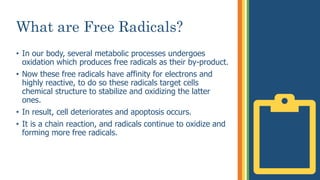
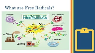
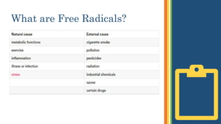
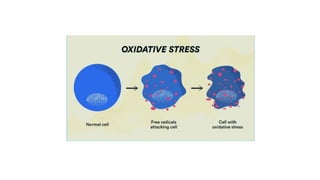
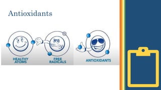
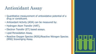
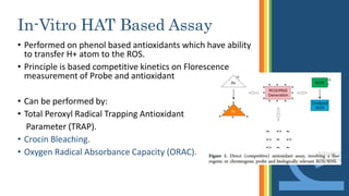
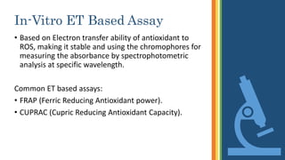
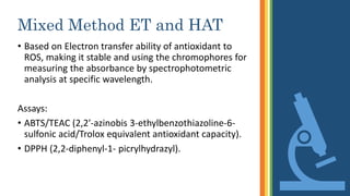
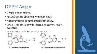
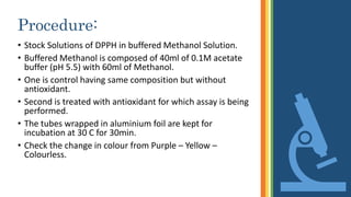
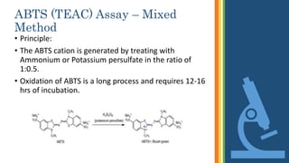
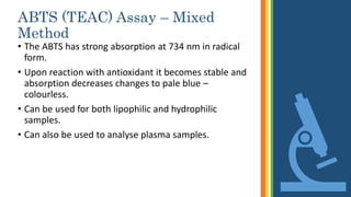
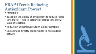
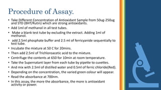
Anti Oxidant
Assays
Dr. MAHESH KUMAR,
M.Phil
SHU
What are Free Radicals?
• In our body, several metabolic processes undergoes
oxidation which produces free radicals as their by-product.
• Now these free radicals have affinity for electrons and
highly reactive, to do so these radicals target cells
chemical structure to stabilize and oxidizing the latter
ones.
• In result, cell deteriorates and apoptosis occurs.
• It is a chain reaction, and radicals continue to oxidize and
forming more free radicals.
What are Free Radicals?
Antioxidants
• Coumpounds that inhibit Oxidation or slows down the
process.
• Terminate these free radical chain reactions.
• Antioxidants stabilize free radicals and can be acquired
through a nutritious and balanced diet.
• Vitamin C, E, selenium, carotenoids are examples of
antioxidants.
• Indigenous Antioxidants include: Catalase, Glutathion
Synthetase (GSH) and Superoxide Dismutase (SOD).
• They are electron donors.
Antioxidants
Scavenging
• Clearance or sweeping the free radicals out of the body
after converting them into stable molecule is called
“Scavenging”
• The Antioxidant activity is measuring the scavenging
ability.
Antioxidant Assay
• Quantitative measurement of antioxidative potential of a
drug or constituent.
• Antioxidant Activity (AOA) can be measured by:
• Hydrogen Atom Transfer (HAT),
• Electron Transfer (ET) based assays.
• Lipid Peroxidation Assay.
• Reactive Oxygen Species (ROS)/Reactive Nitrogen Species
(RNS) Scavenging Assay.
In-Vitro HAT Based Assay
• Performed on phenol based antioxidants which have ability
to transfer H+ atom to the ROS.
• Principle is based competitive kinetics on Florescence
measurement of Probe and antioxidant
• Can be performed by:
• Total Peroxyl Radical Trapping Antioxidant
Parameter (TRAP).
• Crocin Bleaching.
• Oxygen Radical Absorbance Capacity (ORAC).
In-Vitro ET Based Assay
• Based on Electron transfer ability of antioxidant to
ROS, making it stable and using the chromophores for
measuring the absorbance by spectrophotometric
analysis at specific wavelength.
Common ET based assays:
• FRAP (Ferric Reducing Antioxidant power).
• CUPRAC (Cupric Reducing Antioxidant Capacity).
Mixed Method ET and HAT
• Based on Electron transfer ability of antioxidant to
ROS, making it stable and using the chromophores for
measuring the absorbance by spectrophotometric
analysis at specific wavelength.
Assays:
• ABTS/TEAC (2,2′-azinobis 3-ethylbenzothiazoline-6-
sulfonic acid/Trolox equivalent antioxidant capacity).
• DPPH (2,2-diphenyl-1- picrylhydrazyl).
Enzymatic Assay
• Based on absorption parameter, the absorbance of
H2O2 decreases on oxidation by antioxidant.
Assays:
• Hydrogen Peroxide Scavenging Activity (HPSA).
DPPH Assay
• Simple and sensitive.
• Results can be obtained within an hour.
• Non-enzymatic natural antioxidant assay.
• DPPH is stable in powder form and commercially
available.
• Can only be used for organic media.
DPPH Assay
• DPPH has very purple color and shows maximum
absorption at 517nm (purple band).
• Upon reaction with antioxidant, the hydrazine
derivative becomes yellow to colourless.
• Antioxidant activity is inversely proportional to
absorbance. The higher the activity, the lesser is
absorbance and can be easily evaluated by
spectrophotometry taking absorbance as measuring
parameter.
Procedure:
• Stock Solutions of DPPH in buffered Methanol Solution.
• Buffered Methanol is composed of 40ml of 0.1M acetate
buffer (pH 5.5) with 60ml of Methanol.
• One is control having same composition but without
antioxidant.
• Second is treated with antioxidant for which assay is being
performed.
• The tubes wrapped in aluminium foil are kept for
incubation at 30 C for 30min.
• Check the change in colour from Purple – Yellow –
Colourless.
Procedure:
Procedure:
• Calculate the Scavenging Activity
• Scavenging Activity = (Absorbance of Control-Absorbance of Sample) × 100
Absorbance of Control
Procedure:
• Stock Solutions of DPPH in buffered Methanol Solution.
• Buffered Methanol is composed of 40ml of 0.1M acetate
buffer (pH 5.5) with 60ml of Methanol.
• One is control having same composition but without
antioxidant.
• Second is treated with antioxidant for which assay is being
performed.
• The tubes wrapped in aluminium foil are kept for
incubation at 30 C for 30min.
• Check the change in colour from Purple – Yellow –
Colourless.
ABTS (TEAC) Assay – Mixed
Method
• Principle:
• The ABTS cation is generated by treating with
Ammonium or Potassium persulfate in the ratio of
1:0.5.
• Oxidation of ABTS is a long process and requires 12-16
hrs of incubation.
ABTS (TEAC) Assay – Mixed
Method
• The ABTS has strong absorption at 734 nm in radical
form.
• Upon reaction with antioxidant it becomes stable and
absorption decreases changes to pale blue –
colourless.
• Can be used for both lipophilic and hydrophilic
samples.
• Can also be used to analyse plasma samples.
FRAP (Ferric Reducing
Antioxidant Power)
• Principle :
• Based on the ability of antioxidant to reduce Ferric
Ions (Fe+3) – Red in colour to Ferrous Ions (Fe+2) –
Gain of Electron.
• Reduction will produce Green Colour complex.
• Colouring is directly proportional to Antioxidant
activity.
Procedure of Assay.
• Take Different Concentration of Antioxidant Sample from 50ug-250ug
and STD (BHT/Rutin) which are strong antioxidants.
• Add 1ml of methanol in all test tubes.
• Make a blank test tube by excluding the extract. Adding 1ml of
methanol.
• add 2.5ml phosphate buffer and 2.5 ml of ferricyanide sequentially to
test tube.
• Incubate the mixture at 50 C for 20mins.
• Then add 2.5ml of Trichloroacetic acid to the mixture.
• Centrifuge the contents at 650 for 10min at room temperature.
• Take the Supernatant layer from each tube by pipette to cuvettes.
• And mix with 2.5ml of distilled water and 0.5ml of ferric chloride(Red).
• Depending on the concentration, the varied green colour will appear.
• Read the absorbance at 700nm.
• In this assay, the more the absorbance, the more is antioxidant
activity or power.
Catalase Assay
• Catalase (CAT) activity was estimated by the UV
method of Aebi (1983).
• The reaction mixture contained 50 µL of enzyme
extract.
• Add 500 µL of 10 mM H2O2 (pale blue) and 2450 µL of
50 mM potassium phosphate buffer.
• The decrease in absorbance recorded at 240 nm for 60
seconds.
Superoxide Dismutase
• A reaction mixture containing 200 µl of 50 mM potassium phosphate buffer,
300 µl of 12 mM methionine, 300 µl of 75 µM NBT (Nitro Blue Tetrazolium)
and 1mL of 50 mM Na2CO3 was used as control.
• Take 300 µl of enzyme extract to another same mixture.
• At the end, 30 µl of 1 µM riboflavin was added to each mixture.
• The tubes were shaken and placed 30 cm far from light source consisting of a
40 W fluorescent lamps.
• The reaction was allowed to run for 15 min and then stopped by switching
off the light.
• The tubes were immediately covered with black cloth.
• The absorbance was recorded at 560 nm.
• A non-irradiated reaction mixture which did not develop a color, served as
the control.
• However, in the presence of SOD, the reaction was inhibited and the amount
of inhibition was used to quantify the enzyme.
• The absorbance of enzyme extract volume corresponding to 50 percent
inhibition of the photochemical reaction was obtained and considered as
one enzyme unit.
Remember…
Safety First!

Antiioxidant Assay.pptx

  • 1.
  • 2.
    What are FreeRadicals? • In our body, several metabolic processes undergoes oxidation which produces free radicals as their by-product. • Now these free radicals have affinity for electrons and highly reactive, to do so these radicals target cells chemical structure to stabilize and oxidizing the latter ones. • In result, cell deteriorates and apoptosis occurs. • It is a chain reaction, and radicals continue to oxidize and forming more free radicals.
  • 3.
    What are FreeRadicals?
  • 4.
    What are FreeRadicals?
  • 6.
    Antioxidants • Coumpounds thatinhibit Oxidation or slows down the process. • Terminate these free radical chain reactions. • Antioxidants stabilize free radicals and can be acquired through a nutritious and balanced diet. • Vitamin C, E, selenium, carotenoids are examples of antioxidants. • Indigenous Antioxidants include: Catalase, Glutathion Synthetase (GSH) and Superoxide Dismutase (SOD). • They are electron donors.
  • 7.
  • 8.
    Scavenging • Clearance orsweeping the free radicals out of the body after converting them into stable molecule is called “Scavenging” • The Antioxidant activity is measuring the scavenging ability.
  • 9.
    Antioxidant Assay • Quantitativemeasurement of antioxidative potential of a drug or constituent. • Antioxidant Activity (AOA) can be measured by: • Hydrogen Atom Transfer (HAT), • Electron Transfer (ET) based assays. • Lipid Peroxidation Assay. • Reactive Oxygen Species (ROS)/Reactive Nitrogen Species (RNS) Scavenging Assay.
  • 10.
    In-Vitro HAT BasedAssay • Performed on phenol based antioxidants which have ability to transfer H+ atom to the ROS. • Principle is based competitive kinetics on Florescence measurement of Probe and antioxidant • Can be performed by: • Total Peroxyl Radical Trapping Antioxidant Parameter (TRAP). • Crocin Bleaching. • Oxygen Radical Absorbance Capacity (ORAC).
  • 11.
    In-Vitro ET BasedAssay • Based on Electron transfer ability of antioxidant to ROS, making it stable and using the chromophores for measuring the absorbance by spectrophotometric analysis at specific wavelength. Common ET based assays: • FRAP (Ferric Reducing Antioxidant power). • CUPRAC (Cupric Reducing Antioxidant Capacity).
  • 12.
    Mixed Method ETand HAT • Based on Electron transfer ability of antioxidant to ROS, making it stable and using the chromophores for measuring the absorbance by spectrophotometric analysis at specific wavelength. Assays: • ABTS/TEAC (2,2′-azinobis 3-ethylbenzothiazoline-6- sulfonic acid/Trolox equivalent antioxidant capacity). • DPPH (2,2-diphenyl-1- picrylhydrazyl).
  • 13.
    Enzymatic Assay • Basedon absorption parameter, the absorbance of H2O2 decreases on oxidation by antioxidant. Assays: • Hydrogen Peroxide Scavenging Activity (HPSA).
  • 14.
    DPPH Assay • Simpleand sensitive. • Results can be obtained within an hour. • Non-enzymatic natural antioxidant assay. • DPPH is stable in powder form and commercially available. • Can only be used for organic media.
  • 15.
    DPPH Assay • DPPHhas very purple color and shows maximum absorption at 517nm (purple band). • Upon reaction with antioxidant, the hydrazine derivative becomes yellow to colourless. • Antioxidant activity is inversely proportional to absorbance. The higher the activity, the lesser is absorbance and can be easily evaluated by spectrophotometry taking absorbance as measuring parameter.
  • 16.
    Procedure: • Stock Solutionsof DPPH in buffered Methanol Solution. • Buffered Methanol is composed of 40ml of 0.1M acetate buffer (pH 5.5) with 60ml of Methanol. • One is control having same composition but without antioxidant. • Second is treated with antioxidant for which assay is being performed. • The tubes wrapped in aluminium foil are kept for incubation at 30 C for 30min. • Check the change in colour from Purple – Yellow – Colourless.
  • 17.
  • 18.
    Procedure: • Calculate theScavenging Activity • Scavenging Activity = (Absorbance of Control-Absorbance of Sample) × 100 Absorbance of Control
  • 19.
    Procedure: • Stock Solutionsof DPPH in buffered Methanol Solution. • Buffered Methanol is composed of 40ml of 0.1M acetate buffer (pH 5.5) with 60ml of Methanol. • One is control having same composition but without antioxidant. • Second is treated with antioxidant for which assay is being performed. • The tubes wrapped in aluminium foil are kept for incubation at 30 C for 30min. • Check the change in colour from Purple – Yellow – Colourless.
  • 20.
    ABTS (TEAC) Assay– Mixed Method • Principle: • The ABTS cation is generated by treating with Ammonium or Potassium persulfate in the ratio of 1:0.5. • Oxidation of ABTS is a long process and requires 12-16 hrs of incubation.
  • 21.
    ABTS (TEAC) Assay– Mixed Method • The ABTS has strong absorption at 734 nm in radical form. • Upon reaction with antioxidant it becomes stable and absorption decreases changes to pale blue – colourless. • Can be used for both lipophilic and hydrophilic samples. • Can also be used to analyse plasma samples.
  • 22.
    FRAP (Ferric Reducing AntioxidantPower) • Principle : • Based on the ability of antioxidant to reduce Ferric Ions (Fe+3) – Red in colour to Ferrous Ions (Fe+2) – Gain of Electron. • Reduction will produce Green Colour complex. • Colouring is directly proportional to Antioxidant activity.
  • 23.
    Procedure of Assay. •Take Different Concentration of Antioxidant Sample from 50ug-250ug and STD (BHT/Rutin) which are strong antioxidants. • Add 1ml of methanol in all test tubes. • Make a blank test tube by excluding the extract. Adding 1ml of methanol. • add 2.5ml phosphate buffer and 2.5 ml of ferricyanide sequentially to test tube. • Incubate the mixture at 50 C for 20mins. • Then add 2.5ml of Trichloroacetic acid to the mixture. • Centrifuge the contents at 650 for 10min at room temperature. • Take the Supernatant layer from each tube by pipette to cuvettes. • And mix with 2.5ml of distilled water and 0.5ml of ferric chloride(Red). • Depending on the concentration, the varied green colour will appear. • Read the absorbance at 700nm. • In this assay, the more the absorbance, the more is antioxidant activity or power.
  • 24.
    Catalase Assay • Catalase(CAT) activity was estimated by the UV method of Aebi (1983). • The reaction mixture contained 50 µL of enzyme extract. • Add 500 µL of 10 mM H2O2 (pale blue) and 2450 µL of 50 mM potassium phosphate buffer. • The decrease in absorbance recorded at 240 nm for 60 seconds.
  • 25.
    Superoxide Dismutase • Areaction mixture containing 200 µl of 50 mM potassium phosphate buffer, 300 µl of 12 mM methionine, 300 µl of 75 µM NBT (Nitro Blue Tetrazolium) and 1mL of 50 mM Na2CO3 was used as control. • Take 300 µl of enzyme extract to another same mixture. • At the end, 30 µl of 1 µM riboflavin was added to each mixture. • The tubes were shaken and placed 30 cm far from light source consisting of a 40 W fluorescent lamps. • The reaction was allowed to run for 15 min and then stopped by switching off the light. • The tubes were immediately covered with black cloth. • The absorbance was recorded at 560 nm. • A non-irradiated reaction mixture which did not develop a color, served as the control. • However, in the presence of SOD, the reaction was inhibited and the amount of inhibition was used to quantify the enzyme. • The absorbance of enzyme extract volume corresponding to 50 percent inhibition of the photochemical reaction was obtained and considered as one enzyme unit.
  • 26.