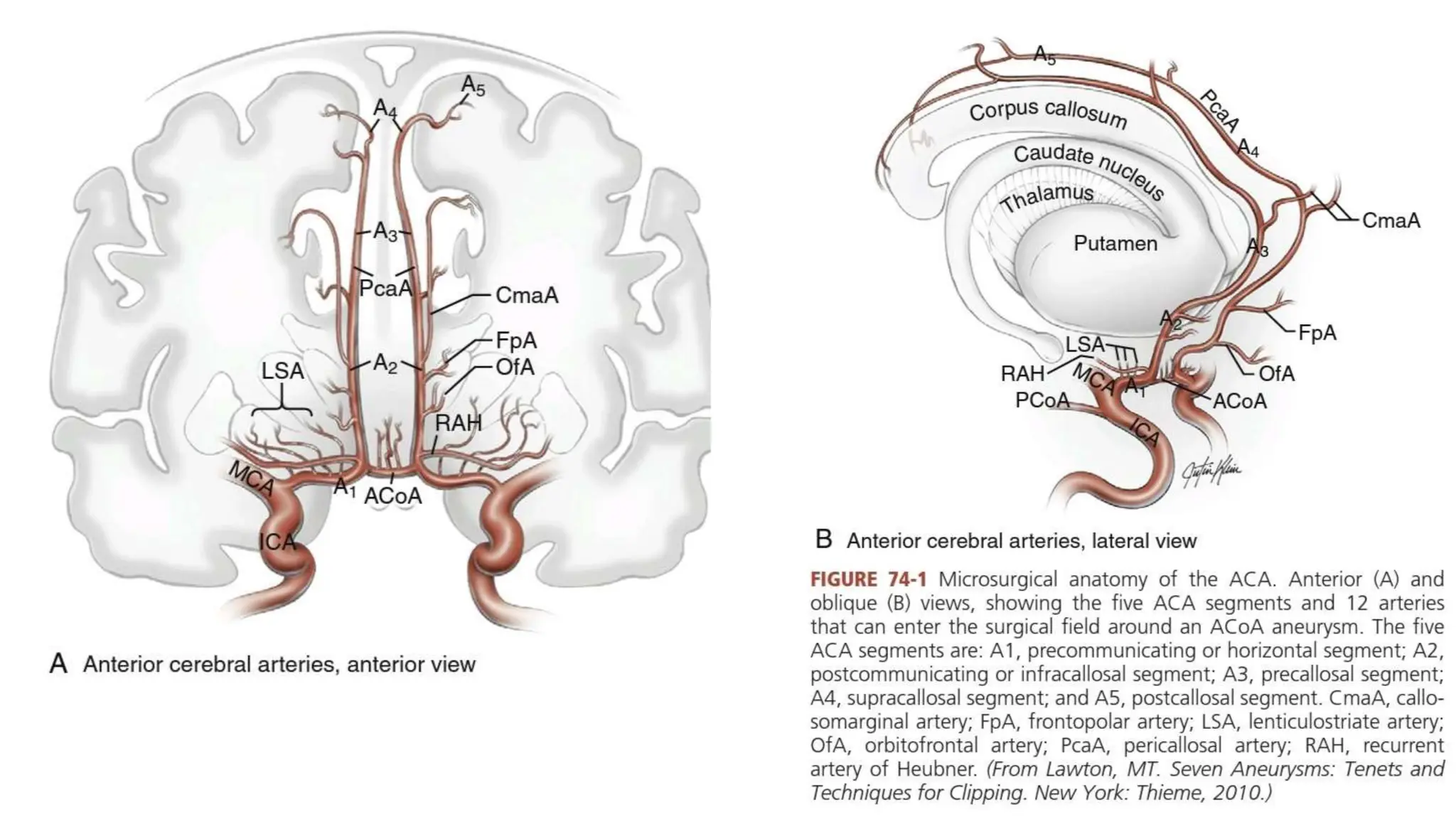
ANTERIOR COMMUNICATING
ARTERY ANEURYSMS
MCH RESIDENT
DEPARTMENT OF NEUROSURGERY
INTRODUCTION
• Most common site
• Bleed in young
• Requires surgery
• Clipping better than coiling
• high risk in surgery since close to hypothalamus,optic
apparatus,emotional control areas
CIRCLE OF WILLIS
ACA SEGMENTS
A1.PRECOMMUNICATING-ICA TO ACOM
A2.POST COMMUNICATING-ACOM TO GENU
A3.GENU TO BODY
A4.AFTER BODY INFRONT OF THE PLANE OF CORONAL SUTURE
A5.BEYOND THE PLANE OF CORONAL SUTURE
BRANCHES OF DIFFERENT SEGMENTS
• A1-MEDIAL LENTICULO STRIATE A
• A2-1.RECURRENT BRANCH OF HEUBNER along the lateral wall
2.ORBITOFRONTAL A
• A3-1.PERICALLOSAL A
2.CALLOSOMARGINAL A
RECURRENT ARTERY OF HEUBNER
• Important land mark to identifyA1
• diameter 1mm
present in 98% cases
seen by retracting frontal lobe even before A1
• Variations-
1.from dista A1-14%
2.at the level of Acomm-8%
3.Duplication-2%
4.superior to A1-60%
5.Anterior to A1-40%
supplies CN,GP,AL of Internal capsule
C/L face,arm paresis with expressive aphasia
ANTERIOR COMMUNICATING ARTERY
• always present
• Incease in diameter as caliber if A1 decreases
• duplicaton in a third
• triplicate in 10%
• Unvisualised in angio in certain cases
• do carotid cross compression test
• anteriro perforators-54
• posterior perforatrs-36
• VARIATIONS-1.accessory Acom
2.duplicates should be preserved
3.only 18 % are transversely oriented.
A1 VARIATIONS
• hypoplastic<1.5 mm
• Asymmetry in 10%
• <1mm in 2 %
• APPROACH THE ANEURYSM FROM THE SIDE OF THE DOMINANT A1
BECAUSE THE ANEURYSM PROJECTS TO THE OPPOSITE SIDE.
OTHER ARTERIES
ORBITO FRONTAL A
5mm from Acom
supplies gyrus rectus
leads to neck of anurysm
FRONTO POLAR A
14mm from Acom
PERICALLOSAL AND CALLOSO MARGINAL
arise from near Genu
supply cingulate
EFFERENT VARIATIONS
BAPTISTA CLASSIFICATION
• TYPE 1-AZYGOS/UNPAIRED ACA-0.3 TO 2%
• TYPE 2-BIHEMISPHERIC A2 ACA-12%
• TYPE 3-ACCESSORY ACA FROM ACOM
MACC
ANTERIORLY PROJECTING ANEURYSM
• Favourable for NEUROSURGEONS
1.90 degree away from A2
2.posterior perforators are away
3.ruptured dome is away
4.dome is usually sealed inside frontal lobe allwing addditional
maneuverability
5.tilted away from dominat A1
6.Easy visualisation of C/L A1
SUPERIORLY PROJECTING ANEURYSM
• Less favorable for access
1.interposed between C/L A2 AND NEUROSURGEON
2.often adherent to A2-need dissection
3.Displaces perforators posterolaterally
4.requires mobilisation of ipsilateral A2 anteriorly to visualise C/L A2
5.May require fenestrated clip for ipsilateral A2,
POSTERIORLY PROJECTING
• most challenging
• Difficult to preserve perforators
• Difficult to apply clip at neck
• Difficult to dissect parent arteries
INFERIORLY PROJECTING ANEURYSMS
• DANGEROUS
• Even before proximal control is achieved it may rupture on retractinof
frontal lobes itself. since it may be attached to the optic apparatus
beneath.
SURGERY
• Identify 11 arteries
2 ACA
ACOM
2 RHA
2OFA
2FPA
2CMA
CONTRIBUTIONS
• YASARGIL-PTERIONAL
• DANDY-LATERAL SUBFRONTAL
• KEMPES-SPHENOID REMOVAL
• NORLEN-GYRUS RECTUS RESECTION
• TANNIS/PPOL-INTERHEMISPHERIC APPROACH
PROCEDURE
THANK YOU

ANTERIOR COMMUNICATING ARTERY ANEURYSMS.pptx

  • 1.
    ANTERIOR COMMUNICATING ARTERY ANEURYSMS MCHRESIDENT DEPARTMENT OF NEUROSURGERY
  • 2.
    INTRODUCTION • Most commonsite • Bleed in young • Requires surgery • Clipping better than coiling • high risk in surgery since close to hypothalamus,optic apparatus,emotional control areas
  • 3.
  • 4.
    ACA SEGMENTS A1.PRECOMMUNICATING-ICA TOACOM A2.POST COMMUNICATING-ACOM TO GENU A3.GENU TO BODY A4.AFTER BODY INFRONT OF THE PLANE OF CORONAL SUTURE A5.BEYOND THE PLANE OF CORONAL SUTURE
  • 6.
    BRANCHES OF DIFFERENTSEGMENTS • A1-MEDIAL LENTICULO STRIATE A • A2-1.RECURRENT BRANCH OF HEUBNER along the lateral wall 2.ORBITOFRONTAL A • A3-1.PERICALLOSAL A 2.CALLOSOMARGINAL A
  • 7.
    RECURRENT ARTERY OFHEUBNER • Important land mark to identifyA1 • diameter 1mm present in 98% cases seen by retracting frontal lobe even before A1 • Variations- 1.from dista A1-14% 2.at the level of Acomm-8% 3.Duplication-2% 4.superior to A1-60% 5.Anterior to A1-40% supplies CN,GP,AL of Internal capsule C/L face,arm paresis with expressive aphasia
  • 8.
    ANTERIOR COMMUNICATING ARTERY •always present • Incease in diameter as caliber if A1 decreases • duplicaton in a third • triplicate in 10% • Unvisualised in angio in certain cases • do carotid cross compression test • anteriro perforators-54 • posterior perforatrs-36 • VARIATIONS-1.accessory Acom 2.duplicates should be preserved 3.only 18 % are transversely oriented.
  • 9.
    A1 VARIATIONS • hypoplastic<1.5mm • Asymmetry in 10% • <1mm in 2 % • APPROACH THE ANEURYSM FROM THE SIDE OF THE DOMINANT A1 BECAUSE THE ANEURYSM PROJECTS TO THE OPPOSITE SIDE.
  • 10.
    OTHER ARTERIES ORBITO FRONTALA 5mm from Acom supplies gyrus rectus leads to neck of anurysm FRONTO POLAR A 14mm from Acom PERICALLOSAL AND CALLOSO MARGINAL arise from near Genu supply cingulate
  • 11.
    EFFERENT VARIATIONS BAPTISTA CLASSIFICATION •TYPE 1-AZYGOS/UNPAIRED ACA-0.3 TO 2% • TYPE 2-BIHEMISPHERIC A2 ACA-12% • TYPE 3-ACCESSORY ACA FROM ACOM MACC
  • 12.
    ANTERIORLY PROJECTING ANEURYSM •Favourable for NEUROSURGEONS 1.90 degree away from A2 2.posterior perforators are away 3.ruptured dome is away 4.dome is usually sealed inside frontal lobe allwing addditional maneuverability 5.tilted away from dominat A1 6.Easy visualisation of C/L A1
  • 14.
    SUPERIORLY PROJECTING ANEURYSM •Less favorable for access 1.interposed between C/L A2 AND NEUROSURGEON 2.often adherent to A2-need dissection 3.Displaces perforators posterolaterally 4.requires mobilisation of ipsilateral A2 anteriorly to visualise C/L A2 5.May require fenestrated clip for ipsilateral A2,
  • 15.
    POSTERIORLY PROJECTING • mostchallenging • Difficult to preserve perforators • Difficult to apply clip at neck • Difficult to dissect parent arteries
  • 16.
    INFERIORLY PROJECTING ANEURYSMS •DANGEROUS • Even before proximal control is achieved it may rupture on retractinof frontal lobes itself. since it may be attached to the optic apparatus beneath.
  • 17.
    SURGERY • Identify 11arteries 2 ACA ACOM 2 RHA 2OFA 2FPA 2CMA
  • 18.
    CONTRIBUTIONS • YASARGIL-PTERIONAL • DANDY-LATERALSUBFRONTAL • KEMPES-SPHENOID REMOVAL • NORLEN-GYRUS RECTUS RESECTION • TANNIS/PPOL-INTERHEMISPHERIC APPROACH
  • 19.
  • 32.