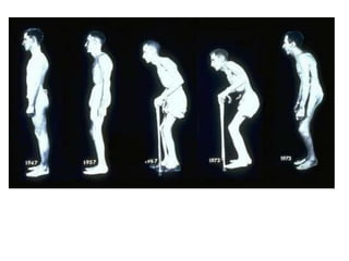
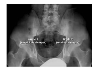
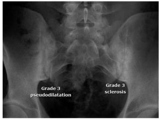
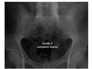
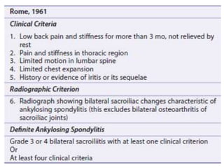
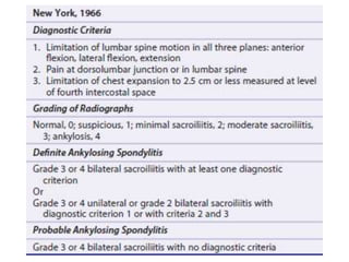
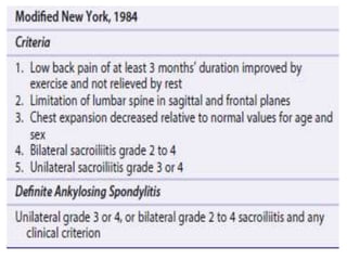
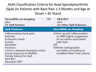
Ankylosing spondylitis
• Ankylosis-stiffening of a joint, spondylos-
vertebra
• Inflammatory disorder associated with fibrous
or bony bridging of joints in the spine,
including the bridging of intervertebral discs.
Pathogenesis
• Predominantly axial disease
• The pathologic changes include inflammation,
structural damage, and excessive new bone
formation.
• Targeted biologic agents- tumor necrosis
factor (TNF)-alpha and interleukin (IL)-17,play
a major role in mediating disease activity.
• HLA-B27
• IL-23/IL-17 pathway
Central role of articulations of axial
skeleton
• Sites
– sacroiliac joints
– Spine
– Hips
– Shoulders
– knees and heels.
• Inflammation, structural destruction, and new bone
formation.
• Focused at locations where ligaments, tendons, joint
capsules, fibrocartilage or other cartilaginous structures,
and bone are adjacent to one another.
• Historically, many of these anatomical sites have been
termed entheses
• Bone destruction and new bone formation seen
in X ray,CT or MRI.
• Involvement of the sacroiliac joints is the most
characteristic.
• Bone destruction can be visualized on imaging as
erosions, and new bone formation appears as
sclerosis and may result in fusion of the joint.
• Vertebral involvement is important in the
development of disability.
• At the vertebrae, bone destruction is usually
observed first at the vertebral corners.
• New bone formation at the vertebral corners
results in the formation of syndesmophytes.
• Bone destruction and new bone formation
also take place at the zygapophyseal joints of
the spine.
• Bridging of syndesmophytes across all the
vertebrae, together with calcification of the
longitudinal ligaments, leads to the
radiographic appearance described as the
classic bamboo spine appearance.
• New bone formation can also be seen at the
heels in the form of bone spurs
• Histomorphological and histomorphometric
analysis of the zygapophyseal joints in AS
suggest that the process
– begins with degeneration of the cartilage and
bone plate
– followed by fusion of the cartilage across the
joints
– replacement of the bone marrow by fibrous
tissues
Epidemiology
• Male-to-female ratio is about 2- 3:1
• Among relatives of an affected patient:
– monozygotic twins – 63 percent
– first-degree relatives – 8.2 percent
– second-degree relatives – 1.0 percent
– third-degree relatives – 0.7 percent
– parent-child – 7.9 percent
• Among siblings, AS occurred almost exclusively
among HLA-B27-positive individuals
Musculoskeletal signs and symptoms
• Common sites
– Spinal and SI involvement
– Hip and shoulder ("root" joint) involvement
– Costovertebral, manubriosternal, sternoclavicular,
and costochondral inflammation
– Inflammation of extraspinal entheses
– Dactylitis
– Peripheral arthritis
Postural abnormalities
• Postural abnormalities(esp hyperkyphosis),
within the first 10 years of disease in some
patients.
• Typical stooped posture of a patient with
advanced disease is caused by flexion
deformity of the neck, thoracic kyphosis, loss
of normal lumbar lordosis, and flexion
deformities of the hips.
Limited spinal mobility and chest
expansion
• Cervical spine
• Forward stooping of the thoracic and cervical
spine.
• The degree of flexion deformity is measured
by asking the patient to stand erect with heels
and buttocks against a wall and to extend his
or her neck, while keeping the chin (mandible)
horizontal in an attempt to touch the wall.
• Almost all normal individuals can touch the
wall with the occiput.
• The distance between the occiput and the
wall in a patient with AS reflects the
degree of cervical deformity.
Thoracic spine
• The range of motion of the costovertebral joints
is measured by the degree of chest expansion.
• Chest expansion is measured at the level of the
xiphoid.
• The patients are instructed to raise their arms
above their heads and to exert a maximal forced
expiration followed by a maximal inspiration.
• Normal expansion is usually >2 cm
Lower spine
• Range of motion of the lower spine is tested in
the sagittal plane by the Schober test, and in
the coronal plane by the extent of lateral
spinal flexion.
Schober test
• The patient stands erect
• A mark is placed at the midpoint of a line
joining the posterior superior iliac spines
(dimples of Venus)
• Another mark is made 10 cm above it in the
midline.
• The patient then bends forward maximally
without bending the knees and the distance is
measured again.
• In normal individuals, the difference between the
two measurements should exceed 2 cm
• Lateral spinal flexion –patient standing erect with
heel and back against a wall and knees and hands
extended, the distance between the tip of the
middle finger and the floor is measured.
• The patient is then instructed to bend sideways
without bending the knees or lifting the heels.
• A second measurement is made and the
difference between the two is recorded.
• The final result is the averaged measurements
of right and left flexion. Normal is greater than
10 cm.
• SI joint tenderness.
• Sausage digit (dactylitis) – The number of
digits that have sausage appearance should be
determined
Achilles tendinitis
• The most common and readily evident site of
enthesitis on physical examination is the heel,
where tenderness and/or swelling may involve
affected patients at the insertions of the
plantar fascii and/or Achilles tendon into the
calcaneus
• Skin- Psoriasis
• Eye- Uveitis.
Laboratory testing
• HLA-B27
• Useful in patients without plain radiographic
evidence of sacroiliitis, where its presence may
aid in making the diagnosis.
• Should be obtained when the diagnosis or
exclusion remains uncertain despite the findings
of the history, physical examination, and plain
radiographic imaging of the pelvis and sacroiliac
(SI) joints, and thus plays an important role in
making the diagnosis.
• The prevalence of AS in the HLA-B27-positive
population is only about 5 percent.
• Thus, a positive test for HLA-B27 alone is not
diagnostic.
• Elevated CRP levels are observed in almost 40
percent of patients.
• However, a normal CRP does not exclude the
diagnosis or the presence of disease activity
Imaging
• X ray
• AP View
– Erosions
– Sclerosis
– Changes in joint width
– Ankylosis
• Strongly supports the diagnosis of AS.
• The SI joint abnormalities are typically graded
to identify the nature and severity of
involvement.
• Grade 0 : Normal.
• Grade 1: Suspicious(but not definite) changes
• Grade 2: minimal abnormality- small areas of
erosions or sclerosis without alteration of joint
width.
• Grade 3: unequivocal abnormality – moderate
or advanced sacroilitis with one or more of
the following: erosions, sclerosis, joint space
widening, narrowing, or partial ankylosis.
• Grade 4: total ankylosis of joints
Approach to diagnosis
• chronic back pain (at least three months),
• Onset before age 45,
• Pelvis with both hips AP.
• A diagnosis of AS can be made if the image
meets criteria for sacroiliitis (at least grade 2
bilaterally or grade 3 unilaterally).
• Those not positive for sacroiliitis by plain
radiography of the pelvis,
• The presence or history of each of 11 features
of AS should be ascertained.
• Patient with at least 4 of the 11 SpA features
can usually be diagnosed.
• Inflammatory back pain
• Heel pain (enthesitis)
• Dactylitis
• Uveitis
• Positive family history for SpA
• Inflammatory bowel disease
• Alternating buttock pain
• Psoriasis
• Asymmetric arthritis
• Positive response to nonsteroidal
antiinflammatory drugs (NSAIDs)
• Elevated acute phase reactants (ESR or CRP)
ASAS Classification Criteria for Axial Spondyloarthritis
(SpA) (in Patients with Back Pain ≥ 3 Months and Age at
Onset < 45 Years)
Treatment
• Include exercise programmes to maintain
posture and range of motion.
• NSAIDs- first line of treatment.
• Continuous high dose NSAID therapy slows
progression.
• COX-2 Inhibitors can be used in patients at risk
of acid peptic disease.
• Failure to respond to more than 2 drugs
should lead to other alternatives.
Second line drugs
• The primary indication for the use of
sulfasalazine in routine practice is a patient
who has concomitant peripheral arthritis and
has had an inadequate response to NSAIDs
and physical modalities.
• The efficacy of methotrexate is yet to be
proved.
• Corticosteroids may be effective for local intra-
articular treatment in AS including the SI joints
Biologic therapies
• Finding of TNF expression in SI joint biopsies
of AS patients, overexpression of TNF leads to
sacroiliitis in animal models and clinical trials.
• Four anti-TNF agents of proven benefit.
– infliximab,
– etanercept,
– adalimumab,
– golimumab.
• Infliximab is an IgG1 chimeric monoclonal
antibody with the Fab portion derived from
the mouse.
• Given in a dose of 3 to 5 mg/kg every 6 to 8
weeks after loading at 0, 2, and 6 weeks.
• Etanercept is a recombinant 75-kD TNF
receptor IgG1 fusion protein that is self-
administered by subcutaneous injection either
once (50 mg) or twice (25 mg) weekly
• Adalimumab and golimumab are human
monoclonal antibodies that are self-
administered by subcutaneous injection on
alternate weeks (40 mg) or monthly (50 mg),
respectively.
Surgeries
• Involvement of the hip joint may cause serious
disability..
• Ectopic bone formation may occur, but the
outcome of total hip replacement is generally
favorable.
• Vertebral osteotomy may be required in selected
cases to correct marked flexion deformity when
forward vision is severely impaired.
• Diaphragmatic herniation may result from the
procedure.
Diffuse Idiopathic skeletal
hyperostosis
Introduction
• Calcification of ligaments and soft tissues-
mainly ligaments and entheses.
• Described by Forestier and Rotes-Querol.
• Axial skeleton- thoracic spine.
• Production of coarse flowing osteophytes,
involving in particular the right side of thoracic
spine.
• Intervertebral disc space is preserved.
• Calcification and ossification of posterior
longitudnal ligament.
• Males more commonly affected.
Suggested diagnostic criteria
• Flowing calcification and ossification along the
anterolateral aspect of atleast four contiguous
vertebral bodies.
• Preservation of the intervertebral disc height
in the involved vertebral segment and absence
of extensive radiographic changes of degn disc
disease.
• Absence of apophyseal joint bony ankylosis
and sacroilliac joint erosion, sclerosis or
intraarticular osseous fusion.
• Resnick and Niwayama.
Associated conditions
• NIDDM
• Obesity
• High waist
circumference ratio
• Dyslipidemia
• Hypertension
• Hyperuricemia
• Hyperinsulinemia
• Elevated IGF-1
• Elevated growth
hormone
• Use of retinoids
• Genetic predisposition.
Pathogenesis
• Higher likelihood to be affected by metabolic
syndrome and higher coronary artery risk.
• Insulin promotes mesenchymal cell
differentiation into chondrocytes and
enchondral ossification in ligaments.
• BMP-2 promotes cell proliferation, ALP activity
and collagen synthesis
• Differentiation of mesenchymal cells into
osteoblasts and chondroblasts.
• Growth hormone promotes IGF-1
• Vitamin A is implicated owing to their ability
to promote new bone formation.
Clinical manifestations
• Morning stiffness
• Dorsolumbar pain
• Diffuse range of motion
• Extermity pain-
peripheral large and
small joints
• Peripheral enthesis
involvement
– Heel
– Achilles tendon
– Shoulder
– Patella
– Olecranon
• Spinal stenosis and stiffening increase the
chances of fractures
• Accentuated kyphosis
• Reduced mobility of spine
• Enthesopathy
• High incidence of Heterotrophic ossification
after surgery.
Suspect DISH in-
• Patients with OA in atypical locations(elbow)
• Hypertrophic osteoarthritis
• Large enthesopathies and entrapment
neuropathies of uncertain origin.
• Chest X rays- useful screening tool with 77%
sensitivity and 97% specificity.
Treatment goals
• Symptomatic relief of pain and stiffness
• Prevent, retard or arrest progression
• Treatment of associated metabolic disorder.
• Prevent spontaneous complications
• Prevent traumatic complications
• Prevent complications that might emerge
during diagnostic or therapeutic procedures.
Difference from OA
• OA
• Lower cervical and
lumbar spine
• Reduced intervertebral
height
• Primary target-
Cartilage in discs and
facet joints
• DISH
• Thoracic spine
• Preserved intervertebral
height
• Primary target- spinal
ligaments and enthesis
• Involvement of MCP
joints, elbows and
shoulders.
• Light exercises
• Heat
• Analgesics and NSAIDs
• Local soft applications- insoles for plantar
spurs or protective bandages at other sites.
• Reduce cardiovascular risk factors- Weight
reduction, physical activity, low fat, low
carbohydrate diet.
• Oral hypoglycemics- Decrease insulin
resistance
• ACE inhibitors, CCBs and alpha blocker in
concomintant hypertensives
Complications-
• Prevent aspiration pneumonia
• Minimise trauma to soft tissues during
intubations or upper GI scopy.
• HO- NSAIDs, irradiation.
• Common measures to prevent fall and
trauma.

Ank spond and dish

  • 1.
  • 2.
    • Ankylosis-stiffening ofa joint, spondylos- vertebra • Inflammatory disorder associated with fibrous or bony bridging of joints in the spine, including the bridging of intervertebral discs.
  • 3.
    Pathogenesis • Predominantly axialdisease • The pathologic changes include inflammation, structural damage, and excessive new bone formation. • Targeted biologic agents- tumor necrosis factor (TNF)-alpha and interleukin (IL)-17,play a major role in mediating disease activity. • HLA-B27
  • 4.
  • 5.
    Central role ofarticulations of axial skeleton • Sites – sacroiliac joints – Spine – Hips – Shoulders – knees and heels. • Inflammation, structural destruction, and new bone formation. • Focused at locations where ligaments, tendons, joint capsules, fibrocartilage or other cartilaginous structures, and bone are adjacent to one another. • Historically, many of these anatomical sites have been termed entheses
  • 6.
    • Bone destructionand new bone formation seen in X ray,CT or MRI. • Involvement of the sacroiliac joints is the most characteristic. • Bone destruction can be visualized on imaging as erosions, and new bone formation appears as sclerosis and may result in fusion of the joint. • Vertebral involvement is important in the development of disability.
  • 7.
    • At thevertebrae, bone destruction is usually observed first at the vertebral corners. • New bone formation at the vertebral corners results in the formation of syndesmophytes. • Bone destruction and new bone formation also take place at the zygapophyseal joints of the spine.
  • 8.
    • Bridging ofsyndesmophytes across all the vertebrae, together with calcification of the longitudinal ligaments, leads to the radiographic appearance described as the classic bamboo spine appearance. • New bone formation can also be seen at the heels in the form of bone spurs
  • 9.
    • Histomorphological andhistomorphometric analysis of the zygapophyseal joints in AS suggest that the process – begins with degeneration of the cartilage and bone plate – followed by fusion of the cartilage across the joints – replacement of the bone marrow by fibrous tissues
  • 10.
    Epidemiology • Male-to-female ratiois about 2- 3:1 • Among relatives of an affected patient: – monozygotic twins – 63 percent – first-degree relatives – 8.2 percent – second-degree relatives – 1.0 percent – third-degree relatives – 0.7 percent – parent-child – 7.9 percent • Among siblings, AS occurred almost exclusively among HLA-B27-positive individuals
  • 11.
    Musculoskeletal signs andsymptoms • Common sites – Spinal and SI involvement – Hip and shoulder ("root" joint) involvement – Costovertebral, manubriosternal, sternoclavicular, and costochondral inflammation – Inflammation of extraspinal entheses – Dactylitis – Peripheral arthritis
  • 12.
    Postural abnormalities • Posturalabnormalities(esp hyperkyphosis), within the first 10 years of disease in some patients. • Typical stooped posture of a patient with advanced disease is caused by flexion deformity of the neck, thoracic kyphosis, loss of normal lumbar lordosis, and flexion deformities of the hips.
  • 14.
    Limited spinal mobilityand chest expansion • Cervical spine • Forward stooping of the thoracic and cervical spine. • The degree of flexion deformity is measured by asking the patient to stand erect with heels and buttocks against a wall and to extend his or her neck, while keeping the chin (mandible) horizontal in an attempt to touch the wall.
  • 15.
    • Almost allnormal individuals can touch the wall with the occiput. • The distance between the occiput and the wall in a patient with AS reflects the degree of cervical deformity.
  • 16.
    Thoracic spine • Therange of motion of the costovertebral joints is measured by the degree of chest expansion. • Chest expansion is measured at the level of the xiphoid. • The patients are instructed to raise their arms above their heads and to exert a maximal forced expiration followed by a maximal inspiration. • Normal expansion is usually >2 cm
  • 17.
    Lower spine • Rangeof motion of the lower spine is tested in the sagittal plane by the Schober test, and in the coronal plane by the extent of lateral spinal flexion.
  • 18.
  • 19.
    • The patientstands erect • A mark is placed at the midpoint of a line joining the posterior superior iliac spines (dimples of Venus) • Another mark is made 10 cm above it in the midline. • The patient then bends forward maximally without bending the knees and the distance is measured again.
  • 20.
    • In normalindividuals, the difference between the two measurements should exceed 2 cm • Lateral spinal flexion –patient standing erect with heel and back against a wall and knees and hands extended, the distance between the tip of the middle finger and the floor is measured. • The patient is then instructed to bend sideways without bending the knees or lifting the heels.
  • 21.
    • A secondmeasurement is made and the difference between the two is recorded. • The final result is the averaged measurements of right and left flexion. Normal is greater than 10 cm. • SI joint tenderness. • Sausage digit (dactylitis) – The number of digits that have sausage appearance should be determined
  • 22.
    Achilles tendinitis • Themost common and readily evident site of enthesitis on physical examination is the heel, where tenderness and/or swelling may involve affected patients at the insertions of the plantar fascii and/or Achilles tendon into the calcaneus
  • 24.
  • 25.
    Laboratory testing • HLA-B27 •Useful in patients without plain radiographic evidence of sacroiliitis, where its presence may aid in making the diagnosis. • Should be obtained when the diagnosis or exclusion remains uncertain despite the findings of the history, physical examination, and plain radiographic imaging of the pelvis and sacroiliac (SI) joints, and thus plays an important role in making the diagnosis.
  • 26.
    • The prevalenceof AS in the HLA-B27-positive population is only about 5 percent. • Thus, a positive test for HLA-B27 alone is not diagnostic. • Elevated CRP levels are observed in almost 40 percent of patients. • However, a normal CRP does not exclude the diagnosis or the presence of disease activity
  • 27.
    Imaging • X ray •AP View – Erosions – Sclerosis – Changes in joint width – Ankylosis • Strongly supports the diagnosis of AS.
  • 28.
    • The SIjoint abnormalities are typically graded to identify the nature and severity of involvement. • Grade 0 : Normal. • Grade 1: Suspicious(but not definite) changes • Grade 2: minimal abnormality- small areas of erosions or sclerosis without alteration of joint width.
  • 30.
    • Grade 3:unequivocal abnormality – moderate or advanced sacroilitis with one or more of the following: erosions, sclerosis, joint space widening, narrowing, or partial ankylosis. • Grade 4: total ankylosis of joints
  • 33.
    Approach to diagnosis •chronic back pain (at least three months), • Onset before age 45, • Pelvis with both hips AP. • A diagnosis of AS can be made if the image meets criteria for sacroiliitis (at least grade 2 bilaterally or grade 3 unilaterally).
  • 34.
    • Those notpositive for sacroiliitis by plain radiography of the pelvis, • The presence or history of each of 11 features of AS should be ascertained. • Patient with at least 4 of the 11 SpA features can usually be diagnosed.
  • 35.
    • Inflammatory backpain • Heel pain (enthesitis) • Dactylitis • Uveitis • Positive family history for SpA
  • 36.
    • Inflammatory boweldisease • Alternating buttock pain • Psoriasis • Asymmetric arthritis • Positive response to nonsteroidal antiinflammatory drugs (NSAIDs) • Elevated acute phase reactants (ESR or CRP)
  • 40.
    ASAS Classification Criteriafor Axial Spondyloarthritis (SpA) (in Patients with Back Pain ≥ 3 Months and Age at Onset < 45 Years)
  • 41.
    Treatment • Include exerciseprogrammes to maintain posture and range of motion. • NSAIDs- first line of treatment. • Continuous high dose NSAID therapy slows progression. • COX-2 Inhibitors can be used in patients at risk of acid peptic disease. • Failure to respond to more than 2 drugs should lead to other alternatives.
  • 42.
    Second line drugs •The primary indication for the use of sulfasalazine in routine practice is a patient who has concomitant peripheral arthritis and has had an inadequate response to NSAIDs and physical modalities. • The efficacy of methotrexate is yet to be proved. • Corticosteroids may be effective for local intra- articular treatment in AS including the SI joints
  • 43.
    Biologic therapies • Findingof TNF expression in SI joint biopsies of AS patients, overexpression of TNF leads to sacroiliitis in animal models and clinical trials. • Four anti-TNF agents of proven benefit. – infliximab, – etanercept, – adalimumab, – golimumab.
  • 44.
    • Infliximab isan IgG1 chimeric monoclonal antibody with the Fab portion derived from the mouse. • Given in a dose of 3 to 5 mg/kg every 6 to 8 weeks after loading at 0, 2, and 6 weeks. • Etanercept is a recombinant 75-kD TNF receptor IgG1 fusion protein that is self- administered by subcutaneous injection either once (50 mg) or twice (25 mg) weekly
  • 45.
    • Adalimumab andgolimumab are human monoclonal antibodies that are self- administered by subcutaneous injection on alternate weeks (40 mg) or monthly (50 mg), respectively.
  • 46.
    Surgeries • Involvement ofthe hip joint may cause serious disability.. • Ectopic bone formation may occur, but the outcome of total hip replacement is generally favorable. • Vertebral osteotomy may be required in selected cases to correct marked flexion deformity when forward vision is severely impaired. • Diaphragmatic herniation may result from the procedure.
  • 47.
  • 48.
    Introduction • Calcification ofligaments and soft tissues- mainly ligaments and entheses. • Described by Forestier and Rotes-Querol. • Axial skeleton- thoracic spine. • Production of coarse flowing osteophytes, involving in particular the right side of thoracic spine. • Intervertebral disc space is preserved.
  • 49.
    • Calcification andossification of posterior longitudnal ligament. • Males more commonly affected.
  • 50.
    Suggested diagnostic criteria •Flowing calcification and ossification along the anterolateral aspect of atleast four contiguous vertebral bodies. • Preservation of the intervertebral disc height in the involved vertebral segment and absence of extensive radiographic changes of degn disc disease.
  • 51.
    • Absence ofapophyseal joint bony ankylosis and sacroilliac joint erosion, sclerosis or intraarticular osseous fusion. • Resnick and Niwayama.
  • 52.
    Associated conditions • NIDDM •Obesity • High waist circumference ratio • Dyslipidemia • Hypertension • Hyperuricemia • Hyperinsulinemia • Elevated IGF-1 • Elevated growth hormone • Use of retinoids • Genetic predisposition.
  • 53.
    Pathogenesis • Higher likelihoodto be affected by metabolic syndrome and higher coronary artery risk. • Insulin promotes mesenchymal cell differentiation into chondrocytes and enchondral ossification in ligaments. • BMP-2 promotes cell proliferation, ALP activity and collagen synthesis • Differentiation of mesenchymal cells into osteoblasts and chondroblasts.
  • 54.
    • Growth hormonepromotes IGF-1 • Vitamin A is implicated owing to their ability to promote new bone formation.
  • 55.
    Clinical manifestations • Morningstiffness • Dorsolumbar pain • Diffuse range of motion • Extermity pain- peripheral large and small joints • Peripheral enthesis involvement – Heel – Achilles tendon – Shoulder – Patella – Olecranon
  • 56.
    • Spinal stenosisand stiffening increase the chances of fractures • Accentuated kyphosis • Reduced mobility of spine • Enthesopathy • High incidence of Heterotrophic ossification after surgery.
  • 57.
    Suspect DISH in- •Patients with OA in atypical locations(elbow) • Hypertrophic osteoarthritis • Large enthesopathies and entrapment neuropathies of uncertain origin. • Chest X rays- useful screening tool with 77% sensitivity and 97% specificity.
  • 59.
    Treatment goals • Symptomaticrelief of pain and stiffness • Prevent, retard or arrest progression • Treatment of associated metabolic disorder. • Prevent spontaneous complications • Prevent traumatic complications • Prevent complications that might emerge during diagnostic or therapeutic procedures.
  • 60.
    Difference from OA •OA • Lower cervical and lumbar spine • Reduced intervertebral height • Primary target- Cartilage in discs and facet joints • DISH • Thoracic spine • Preserved intervertebral height • Primary target- spinal ligaments and enthesis • Involvement of MCP joints, elbows and shoulders.
  • 61.
    • Light exercises •Heat • Analgesics and NSAIDs • Local soft applications- insoles for plantar spurs or protective bandages at other sites. • Reduce cardiovascular risk factors- Weight reduction, physical activity, low fat, low carbohydrate diet.
  • 62.
    • Oral hypoglycemics-Decrease insulin resistance • ACE inhibitors, CCBs and alpha blocker in concomintant hypertensives
  • 63.
    Complications- • Prevent aspirationpneumonia • Minimise trauma to soft tissues during intubations or upper GI scopy. • HO- NSAIDs, irradiation. • Common measures to prevent fall and trauma.