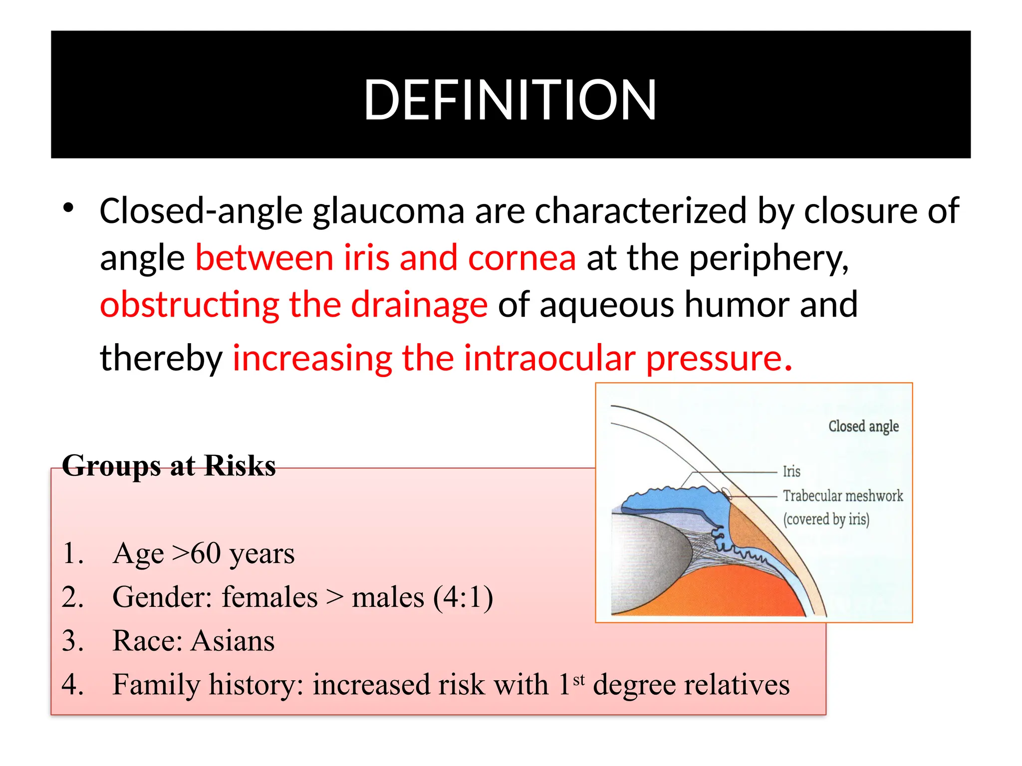
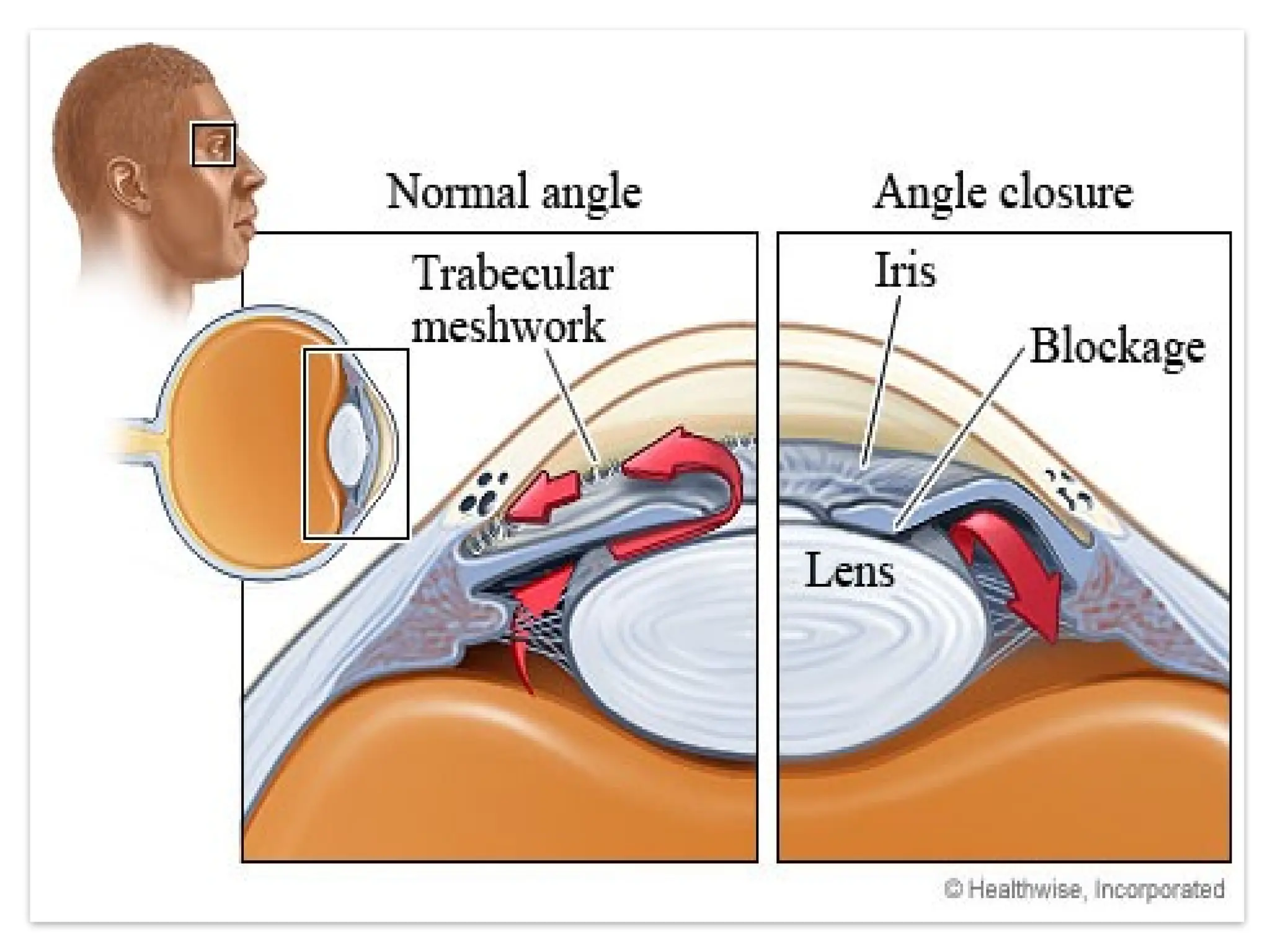
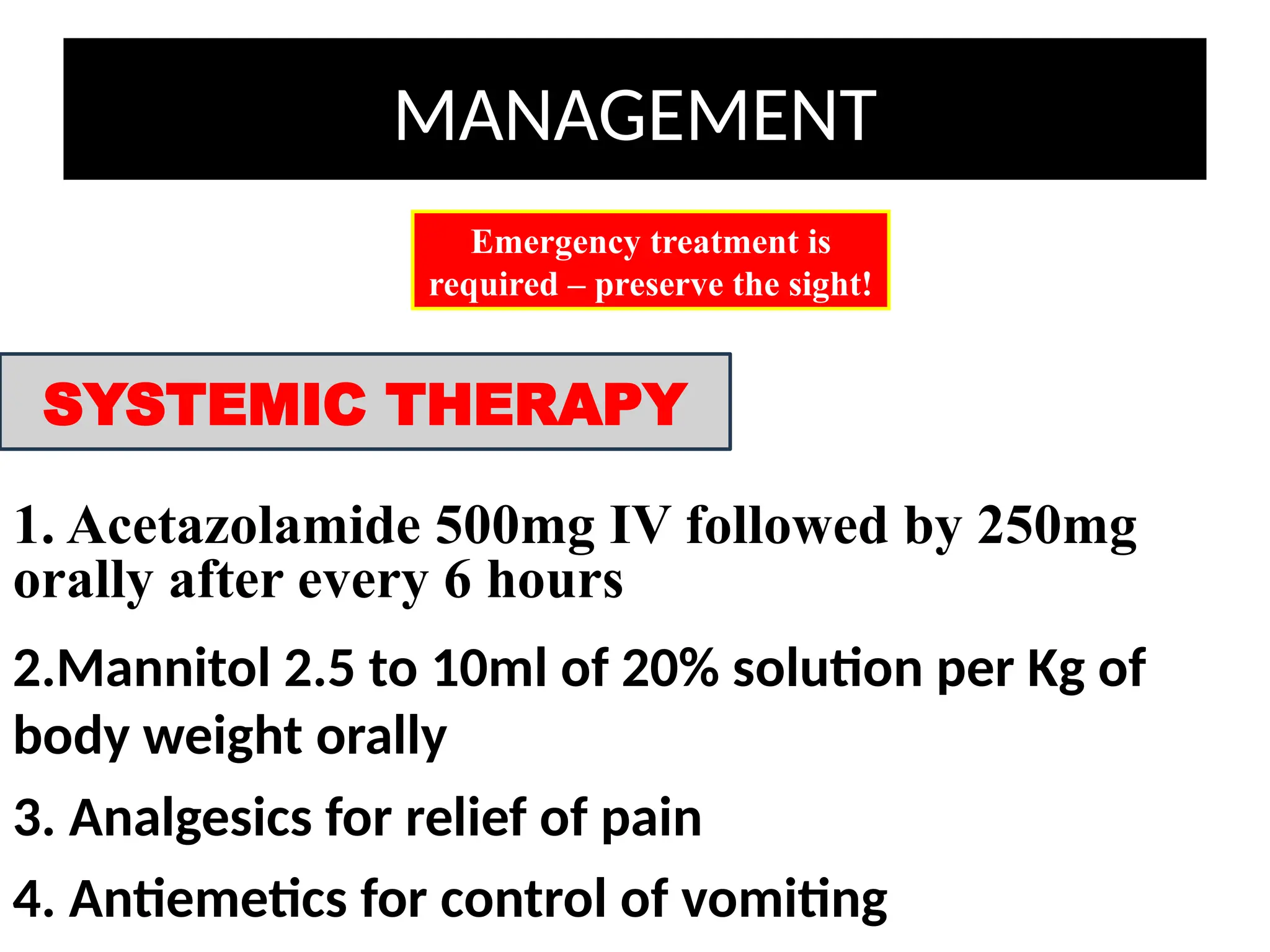
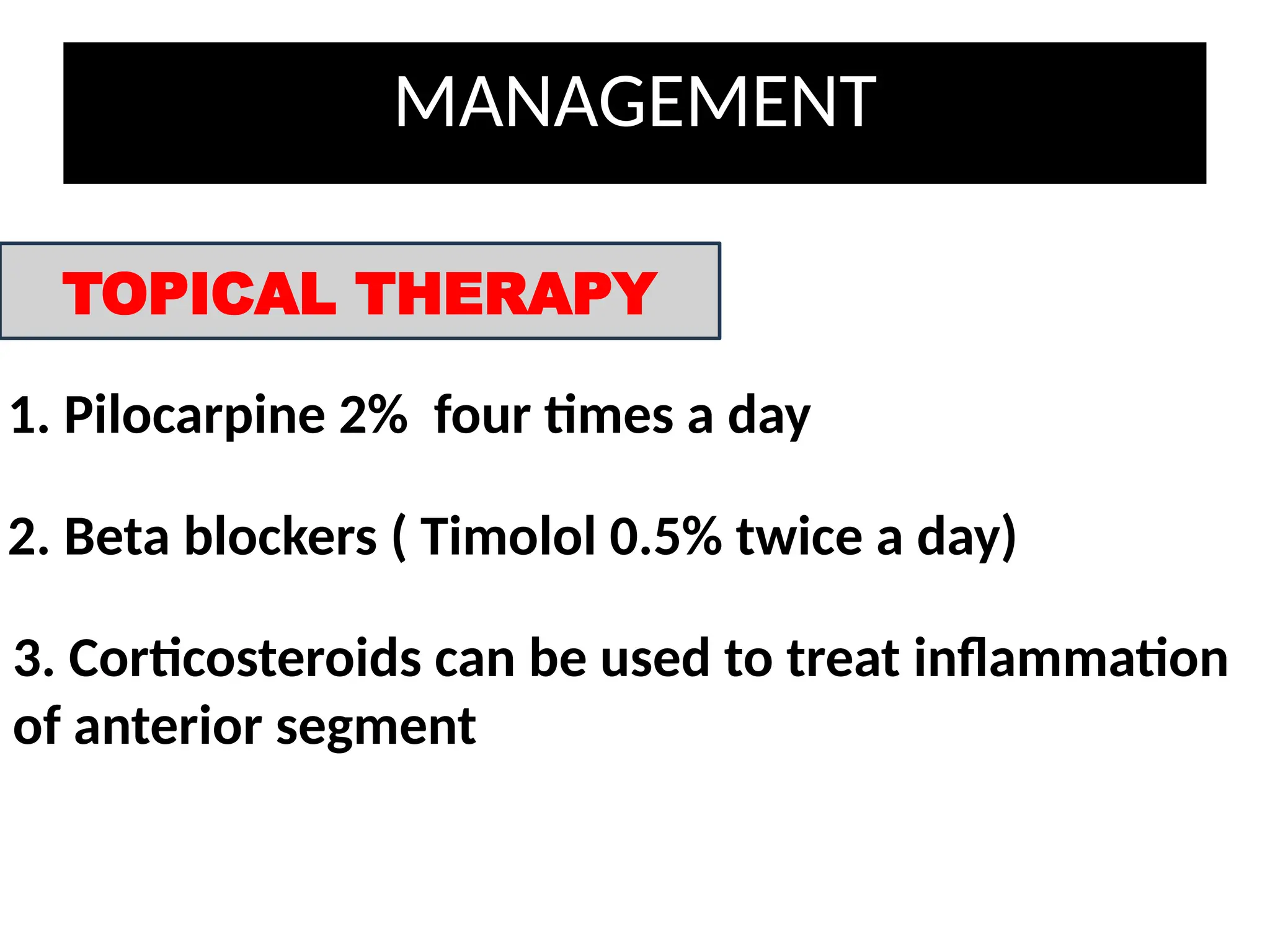
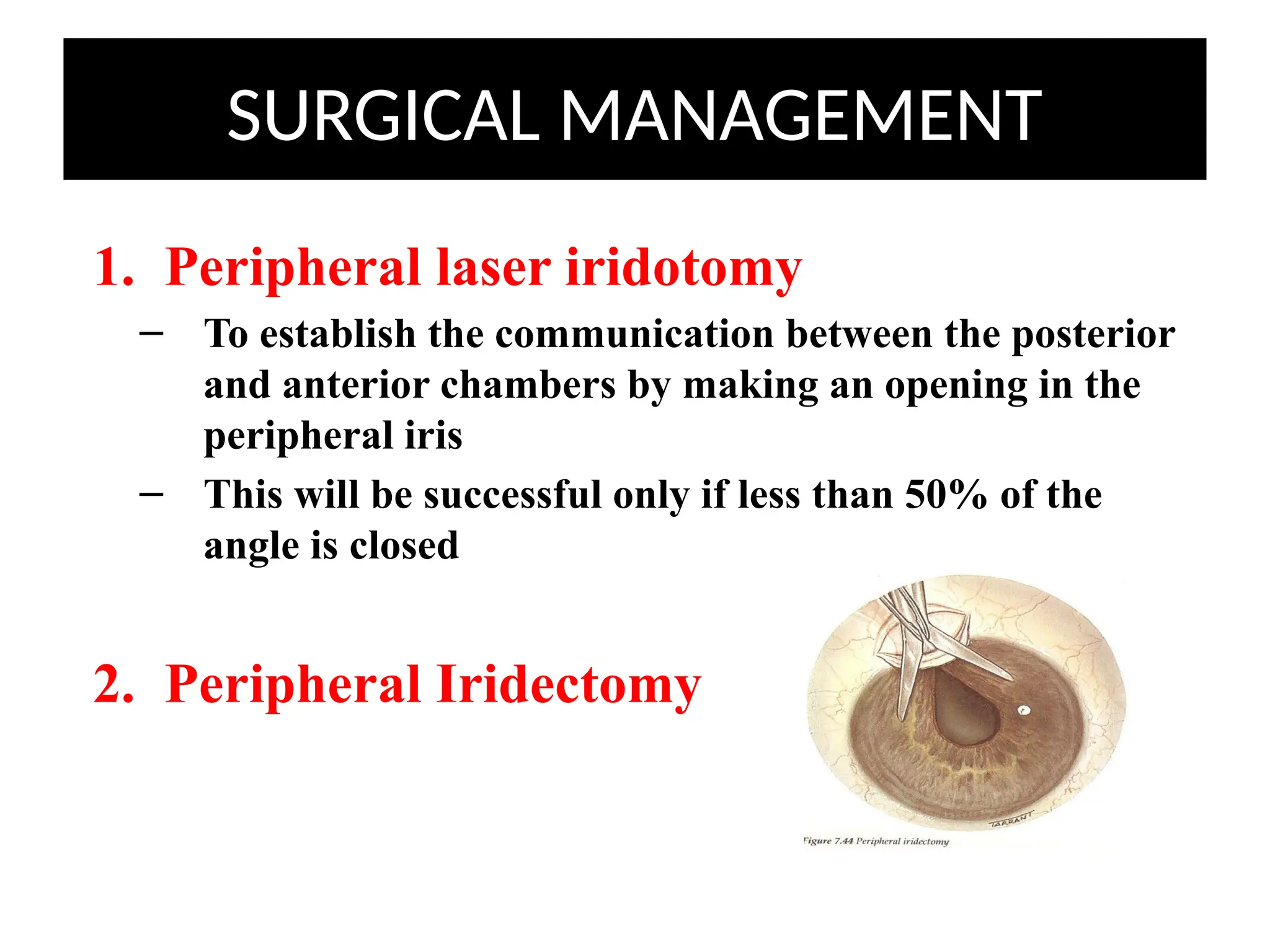
Angle Closure
Glaucoma
ABUBAKAR AMEER
ROLL NO. 140
LEARNING OBJECTIVES
Define Angle Closure Glaucoma (ACG)
Signs of ACG
Symptoms of ACG
Treatment of ACG
DEFINITION
• Closed-angle glaucoma are characterized by closure of
angle between iris and cornea at the periphery,
obstructing the drainage of aqueous humor and
thereby increasing the intraocular pressure.
Groups at Risks
1. Age >60 years
2. Gender: females > males (4:1)
3. Race: Asians
4. Family history: increased risk with 1st
degree relatives
CLASSIFICATION
Angle-Closure
Glaucoma
Primary
Acute (AACG) Chronic (CACG)
Secondary
Lens abnormalities
(thick cataract)
Lens dislocation
PREDISPOSING FACTORS
• Relative anterior position of iris-lens
diaphragm
• Shallow anterior chamber
• Narrow entrance to angle
Anatomical
• Physiological pupillary block
Physiological
PHYSIOLOGICAL PUPILLARY BLOCK
1. Iris has large arc of
contact with anterior
surface of lens
2. Resistance to
aqueous flow from
posterior to anterior
chamber (relative
pupil block)
3. Pupil dilates,
peripheral iris
becomes more
flaccid and pushed
anteriorly
4. Iris lies against
trabecular
meshwork 
impede aqueous
humor drainage 
↑ IOP
SYMPTOMS
1. Rapidly progressive impairment of
vision
2. Painful eye
3. Red eye
4. Nausea, vomiting
5. Photophobia
6. Haloes, transient blurring – indicate previous
intermittent attacks
7. Hx of similar attacks in the past
** CACG: usually asymptomatic due to slow onset of
disease
SIGNS
1. Reduced visual acuity
2. Cornea cloudy and oedematous
3. Pupil oval, fixed and moderately dilated
4. Ciliary injection
5. Eye feels hard on palpation
6. Elevated IOP (50-100 mmHg)
7. Narrow chamber angle with peripheral
iridocorneal contact
8. Gonioscopy – complete peripheral
iridocorneal contact
Severe
edematous
cornea.
Dilated,
unreactive,
vertically oval
pupil
Shallow
anterior
chamber
Complete
angle closure
MANAGEMENT
1. Acetazolamide 500mg IV followed by 250mg
orally after every 6 hours
Emergency treatment is
required – preserve the sight!
SYSTEMIC THERAPY
2.Mannitol 2.5 to 10ml of 20% solution per Kg of
body weight orally
3. Analgesics for relief of pain
4. Antiemetics for control of vomiting
MANAGEMENT
TOPICAL THERAPY
1. Pilocarpine 2% four times a day
2. Beta blockers ( Timolol 0.5% twice a day)
3. Corticosteroids can be used to treat inflammation
of anterior segment
SURGICAL MANAGEMENT
1. Peripheral laser iridotomy
– To establish the communication between the posterior
and anterior chambers by making an opening in the
peripheral iris
– This will be successful only if less than 50% of the
angle is closed
2. Peripheral Iridectomy
SECONDARY ANGLE CLOSURE GLAUCOMA
• Angle-closure secondary to a variety of
ocular disorders
– Lens abnormalities (thick cataract)
– Lens dislocation
– Inflammation (uveitis, scleritis, extensive retinal
photocoagulation)
• Signs and symptoms
– Same as PACG
TAKE HOME MESSAGE…
THANK YOU!
…

ANGLE CLOSURE GLAUCOMA SIMPLE REVIEW.pptx

  • 1.
  • 2.
    LEARNING OBJECTIVES Define AngleClosure Glaucoma (ACG) Signs of ACG Symptoms of ACG Treatment of ACG
  • 3.
    DEFINITION • Closed-angle glaucomaare characterized by closure of angle between iris and cornea at the periphery, obstructing the drainage of aqueous humor and thereby increasing the intraocular pressure. Groups at Risks 1. Age >60 years 2. Gender: females > males (4:1) 3. Race: Asians 4. Family history: increased risk with 1st degree relatives
  • 5.
    CLASSIFICATION Angle-Closure Glaucoma Primary Acute (AACG) Chronic(CACG) Secondary Lens abnormalities (thick cataract) Lens dislocation
  • 6.
    PREDISPOSING FACTORS • Relativeanterior position of iris-lens diaphragm • Shallow anterior chamber • Narrow entrance to angle Anatomical • Physiological pupillary block Physiological
  • 7.
    PHYSIOLOGICAL PUPILLARY BLOCK 1.Iris has large arc of contact with anterior surface of lens 2. Resistance to aqueous flow from posterior to anterior chamber (relative pupil block) 3. Pupil dilates, peripheral iris becomes more flaccid and pushed anteriorly 4. Iris lies against trabecular meshwork  impede aqueous humor drainage  ↑ IOP
  • 8.
    SYMPTOMS 1. Rapidly progressiveimpairment of vision 2. Painful eye 3. Red eye 4. Nausea, vomiting 5. Photophobia 6. Haloes, transient blurring – indicate previous intermittent attacks 7. Hx of similar attacks in the past ** CACG: usually asymptomatic due to slow onset of disease
  • 9.
    SIGNS 1. Reduced visualacuity 2. Cornea cloudy and oedematous 3. Pupil oval, fixed and moderately dilated 4. Ciliary injection 5. Eye feels hard on palpation 6. Elevated IOP (50-100 mmHg) 7. Narrow chamber angle with peripheral iridocorneal contact 8. Gonioscopy – complete peripheral iridocorneal contact
  • 10.
  • 11.
    MANAGEMENT 1. Acetazolamide 500mgIV followed by 250mg orally after every 6 hours Emergency treatment is required – preserve the sight! SYSTEMIC THERAPY 2.Mannitol 2.5 to 10ml of 20% solution per Kg of body weight orally 3. Analgesics for relief of pain 4. Antiemetics for control of vomiting
  • 12.
    MANAGEMENT TOPICAL THERAPY 1. Pilocarpine2% four times a day 2. Beta blockers ( Timolol 0.5% twice a day) 3. Corticosteroids can be used to treat inflammation of anterior segment
  • 13.
    SURGICAL MANAGEMENT 1. Peripherallaser iridotomy – To establish the communication between the posterior and anterior chambers by making an opening in the peripheral iris – This will be successful only if less than 50% of the angle is closed 2. Peripheral Iridectomy
  • 14.
    SECONDARY ANGLE CLOSUREGLAUCOMA • Angle-closure secondary to a variety of ocular disorders – Lens abnormalities (thick cataract) – Lens dislocation – Inflammation (uveitis, scleritis, extensive retinal photocoagulation) • Signs and symptoms – Same as PACG
  • 15.
  • 16.