
More Related Content
Similar to anatomyandphsiologyqqqqqqqqqqqqqqqqqqqqqqq
Similar to anatomyandphsiologyqqqqqqqqqqqqqqqqqqqqqqq (20)
Recently uploaded
Recently uploaded (20)
anatomyandphsiologyqqqqqqqqqqqqqqqqqqqqqqq
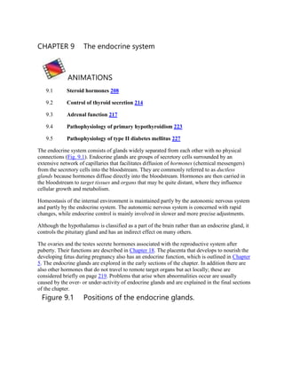
- 1. CHAPTER 9 The endocrine system ANIMATIONS 9.1 Steroid hormones 208 9.2 Control of thyroid secretion 214 9.3 Adrenal function 217 9.4 Pathophysiology of primary hypothyroidism 223 9.5 Pathophysiology of type II diabetes mellitus 227 The endocrine system consists of glands widely separated from each other with no physical connections (Fig. 9.1). Endocrine glands are groups of secretory cells surrounded by an extensive network of capillaries that facilitates diffusion of hormones (chemical messengers) from the secretory cells into the bloodstream. They are commonly referred to as ductless glands because hormones diffuse directly into the bloodstream. Hormones are then carried in the bloodstream to target tissues and organs that may be quite distant, where they influence cellular growth and metabolism. Homeostasis of the internal environment is maintained partly by the autonomic nervous system and partly by the endocrine system. The autonomic nervous system is concerned with rapid changes, while endocrine control is mainly involved in slower and more precise adjustments. Although the hypothalamus is classified as a part of the brain rather than an endocrine gland, it controls the pituitary gland and has an indirect effect on many others. The ovaries and the testes secrete hormones associated with the reproductive system after puberty. Their functions are described in Chapter 18. The placenta that develops to nourish the developing fetus during pregnancy also has an endocrine function, which is outlined in Chapter 5. The endocrine glands are explored in the early sections of the chapter. In addition there are also other hormones that do not travel to remote target organs but act locally; these are considered briefly on page 219. Problems that arise when abnormalities occur are usually caused by the over- or under-activity of endocrine glands and are explained in the final sections of the chapter. Figure 9.1 Positions of the endocrine glands.
- 3. Box 9.1 Examples of lipid-based and peptide hormones Lipid-based hormones Peptide hormones Steroids e.g. glucocorticoids, mineralocorticoids Adrenaline (epinephrine), noradrenaline (norepinephrine) Thyroid hormones Insulin Glucagon Overview of hormone action When a hormone arrives at its target cell, it binds to a specific receptor, where it acts as a switch influencing chemical or metabolic reactions inside the cell. The receptors for peptide hormones are situated on the cell membrane and those for lipid-based hormones are inside the cell. Examples are shown in Box 9.1. www.rossandwilson.com The level of a hormone in the blood is variable and self-regulating within its normal range. A hormone is released in response to a specific stimulus and usually its action reverses or negates the stimulus through a negative feedback mechanism (see p. 6). This may be controlled either indirectly through the release of hormones by the hypothalamus and the anterior pituitary gland, e.g. steroid and thyroid hormones, or directly by blood levels of the stimulus, e.g. insulin and glucagon. The effect of a positive feedback mechanism is amplification of the stimulus and increasing release of the hormone until a particular process is complete and the stimulus ceases, e.g. release of oxytocin during labour (p. 7). Pituitary gland and hypothalamus Learning outcomes After studying this section you should be able to: ▪ describe the structure of the hypothalamus and the pituitary gland ▪ explain the influence of the hypothalamus on the lobes of the pituitary gland
- 4. ▪ outline the actions of the hormones secreted by the anterior and posterior lobes of the pituitary gland. Figure 9.2 The position of the pituitary gland and its associated structures.
- 5. The pituitary gland and the hypothalamus act as a unit, regulating the activity of most of the other endocrine glands. The pituitary gland lies in the hypophyseal fossa of the sphenoid bone below the hypothalamus, to which it is attached by a stalk (Fig. 9.2). It is the size of a pea, weighs about 500 mg and consists of three distinct parts that originate from different
- 6. types of cells. The anterior pituitary (adenohypophysis) is an upgrowth of glandular epithelium from the pharynx and the posterior pituitary (neurohypophysis) is a downgrowth of nervous tissue from the brain. There is a network of nerve fibres between the hypothalamus and the posterior pituitary. Between these lobes is a thin strip of tissue called the intermediate lobe and its function in humans is not known. Blood supply Arterial blood This is supplied by branches from the internal carotid artery. The anterior lobe is supplied indirectly by blood that has already passed through a capillary bed in the hypothalamus but the posterior lobe is supplied directly. Venous drainage This comes from both lobes, containing hormones, and leaves the gland in short veins that enter the venous sinuses between the layers of dura mater. The influence of the hypothalamus on the pituitary gland The influence of the hypothalamus on the release of hormones is different in the anterior and posterior lobes of the pituitary gland. The anterior pituitary This is supplied indirectly with arterial blood that has already passed through a capillary bed in the hypothalamus (Fig. 9.3A). This network of blood vessels forms part of the pituitary portal system, which transports blood from the hypothalamus to the anterior pituitary where it enters thin-walled sinusoids that are in close contact with the secretory cells. As well as providing oxygen and nutrients, this blood transports releasing and inhibiting hormones secreted by the hypothalamus. These hormones influence secretion and release of other hormones formed in the anterior pituitary. The releasing and inhibiting hormones that stimulate and inhibit secretion of specific anterior pituitary hormones are shown in Table 9.1. The posterior pituitary This is formed from nervous tissue and consists of nerve cells surrounded by supporting cells called pituicytes. These neurones have their cell bodies in the supraoptic and paraventricular nuclei of the hypothalamus and their axons form a bundle known as the hypothalamohypophyseal tract (Fig. 9.3A). Posterior pituitary hormones are synthesised in the nerve cell bodies, transported along the axons and stored in vesicles within the
- 7. axon terminals in the posterior pituitary (Fig. 9.3B). Their release by exocytosis is triggered by nerve impulses from the hypothalamus. Anterior pituitary Some of the hormones secreted by the anterior lobe stimulate or inhibit secretion by other endocrine glands (target glands) while others have a direct effect on target tissues. Table 9.1 summarises the main relationships between the hormones of the hypothalamus, the anterior pituitary and target glands or tissues. The release of an anterior pituitary hormone follows stimulation of the gland by a specific releasing hormone produced by the hypothalamus and carried to the gland through the pituitary portal system of blood vessels. The whole system is controlled by a negative feedback mechanism. That is, when there is a low level of a hormone in the blood supplying the hypothalamus it produces the appropriate releasing hormone that stimulates release of a trophic hormone by the anterior pituitary. This in turn stimulates the target gland to produce and release its hormone. As a result the blood level of that hormone rises and inhibits the secretion of releasing factor by the hypothalamus (Fig. 9.4). Figure 9.3 The pituitary gland. A. The lobes of the pituitary gland and their relationship with the hypothalamus. B. Synthesis and storage of antidiuretic hormone and oxytocin.
- 8. Growth hormone (GH) This is the most abundant hormone synthesised by the anterior pituitary. It stimulates growth and division of most body cells but especially those in the bones and skeletal muscles. Body growth in response to the secretion of GH is evident during childhood and adolescence, and thereafter secretion of GH maintains the mass of bones and skeletal muscles. It also regulates aspects of metabolism in many organs, e.g. liver, intestines and pancreas; stimulates protein synthesis, especially tissue growth and repair; promotes breakdown of fats; and increases blood glucose levels (see Ch. 12). Its release is stimulated by growth hormone releasing hormone (GHRH) and suppressed by growth hormone release inhibiting hormone (GHRIH), also known as somatostatin, both of which are secreted by the hypothalamus. Secretion of GH is greater at night during sleep and is also stimulated by hypoglycaemia, exercise and anxiety. The daily amount secreted peaks in adolescence and then declines with age. Inhibition of GH secretion occurs by a negative feedback mechanism when the blood level rises and also when GHRIH is released by the hypothalamus. GHRIH also suppresses secretion of TSH and gastrointestinal secretions, e.g. gastric juice, gastrin and cholecystokinin (see Ch. 12). Thyroid stimulating hormone (TSH)
- 9. This hormone is synthesised by the anterior pituitary and its release is stimulated by thyrotrophin releasing hormone (TRH) from the hypothalamus. It stimulates growth and activity of the thyroid gland, which secretes the hormones thyroxine (T4) and tri- iodothyronine (T3). Release is lowest in the early evening and highest during the night. Secretion is regulated by a negative feedback mechanism (Fig. 9.4). When the blood level of thyroid hormones is high, secretion of TSH is reduced, and vice versa. Adrenocorticotrophic hormone (ACTH, corticotrophin) Corticotrophin releasing hormone (CRH) from the hypothalamus promotes the synthesis and release of ACTH by the anterior pituitary. This increases the concentration of cholesterol and steroids within the adrenal cortex and the output of steroid hormones, especially cortisol. Table 9.1 Hormones of the hypothalamus, anterior pituitary and their target tissues Hypothalamus Anterior pituitary Target gland or tissue GHRH GH Most tissues Many organs GHRIH GH inhibition Thyroid gland TSH inhibition Pancreatic islets Most tissues TRH TSH
- 10. Thyroid gland CRH ACTH Adrenal cortex PRH PRL Breast PIH PRL inhibition Breast LHRH or FSH Ovaries and testes GnRH LH Ovaries and testes GHRH = growth hormone releasing hormone GH = growth hormone (somatotrophin) GHRIH = growth hormone release inhibiting hormone (somatostatin) TRH = thyrotrophin releasing hormone TSH = thyroid stimulating hormone CRH = corticotrophin releasing hormone ACTH = adrenocorticotrophic hormone PRH = prolactin releasing hormone PRL = prolactin (lactogenic hormone)
- 11. PIH = prolactin inhibiting hormone (dopamine) LHRH = luteinising hormone releasing hormone GnRH = gonadotrophin releasing hormone FSH = follicle stimulating hormone LH = luteinising hormone ACTH levels are highest at about 8 a.m. and fall to their lowest about midnight, although high levels sometimes occur at midday and 6 p.m. This circadian rhythm is maintained throughout life. It is associated with the sleep pattern and adjustment to changes takes several days, following, e.g., changing work shifts, travelling to a different time zone (jet lag). Secretion is also regulated by a negative feedback mechanism, being suppressed when the blood level of ACTH rises (Fig. 9.4). Other factors that stimulate secretion include hypoglycaemia, exercise and other stressors, e.g. emotional states and fever. Prolactin This hormone is secreted during pregnancy to prepare the breasts for lactation (milk production) after childbirth. The blood level of prolactin is stimulated by prolactin releasing hormone (PRH) released from the hypothalamus and it is lowered by prolactin inhibiting hormone (PIH, dopamine) and by an increased blood level of prolactin. Immediately after birth, suckling stimulates prolactin secretion and lactation. The resultant high blood level is a factor in reducing the incidence of conception during lactation. Figure 9.4 Negative feedback regulation of secretion of hormones by the anterior lobe of the pituitary gland.
- 13. Prolactin, together with oestrogens, corticosteroids, insulin and thyroxine, is involved in initiating and maintaining lactation. Prolactin secretion is related to sleep, i.e. it is raised during any period of sleep, night or day. Gonadotrophins Just before puberty two gonadotrophins (sex hormones) are secreted in gradually increasing amounts by the anterior pituitary in response to luteinising hormone releasing hormone (LHRH), also known as gonadotrophin releasing hormone (GnRH). At puberty secretion increases, further enabling normal adult functioning of the reproductive organs; in both males and females the hormones responsible are: • follicle stimulating hormone (FSH) • luteinising hormone (LH). In both sexes FSH stimulates production of gametes (ova or spermatozoa) by the gonads. In females LH and FSH are involved in secretion of the hormones oestrogen and progesterone during the menstrual cycle (see Figs 18.9 and 18.10, p. 444). As the levels of oestrogen and progesterone rise, secretion of LH and FSH is suppressed. Table 9.2 Summary of the hormones secreted by the anterior pituitary gland and their functions Hormone Function Growth hormone (GH) Regulates metabolism, promotes tissue growth especially of bones and muscles Thyroid stimulating hormone (TSH) Stimulates growth and activity of thyroid gland and secretion of T3 and T4 Adrenocorticotrophic hormone (ACTH) Stimulates the adrenal cortex to secrete glucocorticoids
- 14. Prolactin (PRL) Stimulates milk production in the breasts Follicle stimulating hormone (FSH) Stimulates production of sperm in the testes, stimulates secretion of oestrogen by the ovaries, maturation of ovarian follicles, ovulation Luteinising hormone (LH) Stimulates secretion of testosterone by the testes, stimulates secretion of progesterone by the corpus luteum In males LH, also called interstitial cell stimulating hormone (ICSH) stimulates the interstitial cells of the testes to secrete the hormone testosterone (see Ch. 18). Table 9.2 summarises the hormonal secretions of the anterior pituitary. Posterior pituitary The structure of the posterior pituitary gland and its relationship with the hypothalamus is explained on page 209. Oxytocin and antidiuretic hormone (ADH or vasopressin) are the hormones synthesised in nerve cell bodies in the hypothalamus and then stored in the axon terminals within the posterior pituitary gland (Fig. 9.3B). These hormones act directly on non-endocrine tissue and their release from synaptic vesicles by exocytosis is stimulated by nerve impulses from the hypothalamus. Oxytocin Oxytocin stimulates two target tissues during and after childbirth (parturition): uterine smooth muscle and the muscle cells of the lactating breast. During childbirth increasing amounts of oxytocin are released by the posterior pituitary into the bloodstream in response to increasing distension of sensory stretch receptors in the uterine cervix by the baby's head. Sensory impulses are generated and travel to the control centre in the hypothalamus, stimulating the posterior pituitary to release more oxytocin. In turn this stimulates more forceful uterine contractions and greater stretching of the uterine cervix as the baby's head is forced further downwards. This is an example of a positive feedback mechanism which stops soon after the baby is delivered and distension of the uterine cervix is greatly reduced (Fig. 9.5). The process of milk ejection also involves a positive feedback mechanism. Suckling generates sensory impulses that are transmitted from the breast to the hypothalamus. The
- 15. impulses trigger the release of oxytocin from the posterior pituitary and oxytocin stimulates Figure 9.5 Regulation of secretion of oxytocin through a positive feedback mechanism.
- 17. contraction of the myoepithelial cells around the glandular cells and ducts of the lactating breast to contract, ejecting milk. Suckling also inhibits the release of prolactin inhibiting hormone (PIH), prolonging prolactin secretion and lactation. The role of this hormone in males and non-lactating females remains unclear. Antidiuretic hormone (ADH, vasopressin) The main effect of antidiuretic hormone is to reduce urine output (diuresis is the production of a large volume of urine). ADH acts on the distal convoluted and collecting ducts of the nephrons of the kidneys (Ch. 13), increasing their permeability to water. As a result, the reabsorption of water from the glomerular filtrate is increased. The amount of ADH secreted is determined by the osmotic pressure of the blood circulating to the osmoreceptors in the hypothalamus. As the osmotic pressure rises, for example as a result of dehydration or following haemorrhage, the secretion of ADH increases. More water is therefore reabsorbed and the urine output is reduced. This means that the body retains more water and the rise in osmotic pressure is reversed. Conversely, when the osmotic pressure of the blood is low, for example after a large fluid intake, secretion of ADH is reduced, less water is reabsorbed and more urine is produced (Fig. 9.11). At high concentrations, for example after severe blood loss, ADH causes smooth muscle contraction, especially vasoconstriction in small arteries. This has a pressor effect, raising systemic blood pressure; the alternative name of this hormone, vasopressin, reflects this effect. Thyroid gland (Fig. 9.6) Learning outcomes After studying this section you should be able to: ▪ describe the position of the thyroid gland and its related structures ▪ describe the microscopic structure of the thyroid gland ▪ outline the actions of the thyroid hormones ▪ explain how blood levels of the thyroid hormones T3 and T4 are regulated. The thyroid gland is situated in the neck in front of the larynx and trachea at the level of the 5th, 6th and 7th cervical and 1st thoracic vertebrae. It is a highly vascular gland that weighs about 25 g and is surrounded by a fibrous capsule. It resembles a butterfly in shape, consisting of two lobes, one on either side of the thyroid cartilage and upper cartilaginous rings of the trachea. The lobes are joined by a narrow isthmus, lying in front of the trachea. The lobes are roughly cone shaped, about 5 cm long and 3 cm wide.
- 18. The arterial blood supply to the gland is through the superior and inferior thyroid arteries. The superior thyroid artery is a branch of the external carotid artery Figure 9.6 The position of the thyroid gland and its associated structures. Anterior view. and the inferior thyroid artery is a branch of the subclavian artery. The venous return is by the thyroid veins, which drain into the internal jugular veins. Two parathyroid glands lie against the posterior surface of each lobe and are sometimes embedded in thyroid tissue. The recurrent laryngeal nerve passes upwards close to the lobes of the gland and on the right side it lies near the inferior thyroid artery (see Fig. 9.9).
- 19. The gland is composed of cuboidal epithelium that forms spherical follicles. These secrete and store colloid, a thick sticky protein material (Fig. 9.7). Between the follicles there are other cells found singly or in small groups: parafollicular cells, also called C-cells, which secrete the hormone calcitonin. Thyroxine and tri-iodothyronine Iodine is essential for the formation of the thyroid hormones, thyroxine (T4) and tri- iodothyronine (T3), so numbered as these molecules contain four and three atoms of the element iodine respectively. The body's main dietary sources of iodine are seafood, vegetables grown in iodine-rich soil and iodinated table salt. The thyroid gland selectively takes up iodine from the blood, a process called iodine trapping. Figure 9.7 The microscopic structure of the thyroid gland. The thyroid hormones are synthesised as large precursor molecules called thyroglobulin, the major constituent of colloid. The release of T3 and T4 into the blood is stimulated by thyroid stimulating hormone (TSH) from the anterior pituitary. Secretion of TSH is stimulated by thyrotrophin releasing hormone (TRH) from the hypothalamus and secretion of TRH is stimulated by exercise, stress, malnutrition, low plasma glucose levels and sleep. The level of secretion of TSH depends on the plasma levels of T3 and T4 because these hormones affect the sensitivity of the anterior pituitary to TRH. Through the negative feedback mechanism, increased levels of T3 and T4 decrease TSH secretion and vice versa (Fig. 9.8). When the supply
- 20. Figure 9.8 Negative feedback regulation of the secretion of thyroxine (T4) and tri-iodothyronine (T3).
- 22. of iodine is deficient, excess TSH is secreted and there is proliferation of thyroid gland cells and enlargement of the gland (goitre, see Fig. 9.16). Secretion of T3 and T4 begins about the third month of fetal life and is increased at puberty and in women during the reproductive years, especially during pregnancy. Otherwise, it remains fairly constant throughout life. www.rossandwilson.com Thyroid hormones enter the cell nucleus and regulate gene expression, i.e. they increase or decrease the synthesis of some proteins including enzymes. They enhance the effects of other hormones, e.g. adrenaline (epinephrine) and noradrenaline (norepinephrine). T3 and T4 affect most cells of the body by: • increasing the basal metabolic rate and heat production • regulating metabolism of carbohydrates, proteins and fats. T3 and T4 are essential for normal growth and development, especially of the skeleton and nervous system. Most other organs and systems are also influenced by thyroid hormones – physiological effects of T3 and T4 on the heart, skeletal muscles, skin, digestive and reproductive systems are more evident when there is underactivity or overactivity of the thyroid gland. These changes are listed in Table 9.3 and the effects, especially in childhood, can be profound. Calcitonin This hormone is secreted by the parafollicular or C-cells in the thyroid gland (Fig. 9.7). It acts on bone cells and the kidneys to reduce blood calcium (Ca2+ ) levels when they are raised. It promotes storage of calcium in bones and inhibits reabsorption of calcium by the renal tubules. Its effect is opposite to that of parathyroid hormone, the hormone secreted by the parathyroid glands. Release of calcitonin is stimulated by an increase in the blood calcium levels. This hormone is important during childhood when bones undergo considerable changes in size and shape. Parathyroid glands Learning outcomes After studying this section you should be able to: ▪ describe the position and gross structure of the parathyroid glands ▪ outline the functions of parathyroid hormone and calcitonin
- 23. ▪ explain how blood levels of parathyroid hormone and calcitonin are regulated. Table 9.3 Common effects of abnormal secretion of thyroid hormones Hyperthyroidism: increased T3 and T4 secretion Hypothyroidism: decreased T3 and T4 secretion Increased basal metabolic rate Decreased basal metabolic rate Weight loss, good appetite Weight gain, anorexia Anxiety, physical restlessness, mental excitability Depression, psychosis, mental slowness, lethargy Hair loss Dry skin, brittle hair Tachycardia, palpitations, atrial fibrillation Bradycardia Warm sweaty skin, heat intolerance Dry cold skin, prone to hypothermia Diarrhoea Constipation Exophthalmos in Graves' disease (Fig. 9.15) Figure 9.9 The positions of the parathyroid glands and their related structures, viewed from behind.
- 24. There are four small parathyroid glands, two embedded in the posterior surface of each lobe of the thyroid gland (Fig. 9.9). They are surrounded by fine connective tissue capsules. The cells forming the glands are spherical in shape and are arranged in columns with sinusoids containing blood between them. Function The parathyroid glands secrete parathyroid hormone (PTH, parathormone). Secretion is regulated by blood calcium levels. When they fall, secretion of PTH is increased and vice versa. The main function of PTH is to increase the blood calcium level when it is low. This is achieved by indirectly increasing the amount of calcium absorbed from the small intestine and reabsorbed from the renal tubules. If these sources provide inadequate supplies then PTH stimulates osteoclasts (bone-destroying cells) and calcium is released from bones into the blood. Parathormone and calcitonin from the thyroid gland act in a complementary manner to maintain blood calcium levels within the normal range. This is needed for: • muscle contraction
- 25. • nerve transmission • blood clotting • normal action of many enzymes. Adrenal glands Learning outcomes After studying this section you should be able to: ▪ describe the structure of the adrenal glands ▪ describe the actions of each of the three groups of adrenocorticoid hormones ▪ explain how blood levels of glucocorticoids are regulated ▪ describe the actions of adrenaline (epinephrine) and noradrenaline (norepinephrine) ▪ outline how the adrenal glands respond to stress. The two adrenal (suprarenal) glands are situated on the upper pole of each kidney (Fig. 9.1) enclosed within the renal fascia. They are about 4 cm long and 3 cm thick. The arterial blood supply to the glands is by branches from the abdominal aorta and renal arteries. The venous return is by suprarenal veins. The right gland drains into the inferior vena cava and the left into the left renal vein. The glands are composed of two parts which have different structures and functions. The outer part is the cortex and the inner part the medulla. The adrenal cortex is essential to life but the medulla is not. Adrenal cortex The adrenal cortex produces three groups of steroid hormones from cholesterol. They are collectively called adrenocorticocoids (corticosteroids). They are: • glucocorticoids • mineralocorticoids • sex hormones (androgens).
- 26. The hormones in each group have different characteristic actions but due to their structural similarity their actions may overlap. Glucocorticoids Cortisol (hydrocortisone) is the main glucocorticoid but small amounts of corticosterone and cortisone are also produced. They are essential for life, regulating metabolism and responses to stress. Secretion is controlled through a negative feedback system involving the hypothalamus and anterior pituitary. It is stimulated by ACTH from Figure 9.10 Negative feedback regulation of glucocorticoid secretion.
- 28. the anterior pituitary and by stress (Fig. 9.10). In normal conditions, secretion shows marked circadian variations. The highest level of hormone secretion occurs between 4 a.m. and 8 a.m., and the lowest between midnight and 3 a.m. When the sleeping and waking pattern is changed it takes several days for adjustment of the ACTH/cortisol secretion to take place (see p. 211). Glucocorticoids have widespread metabolic effects generally concerned with catabolism (breakdown) of protein and fat that makes glucose and other substances available for use. These include: • gluconeogenesis (formation of new sugar from, for example, protein) and hyperglycaemia (raised blood glucose levels) • lipolysis (breakdown of triglycerides into fatty acids and glycerol for energy production) • stimulating breakdown of protein, releasing amino acids, which can be used for synthesis of other proteins, e.g. enzymes, or for energy (ATP) production (p. 24) • promoting absorption of sodium and water from renal tubules (a weak mineralocorticoid effect). In pathological and pharmacological quantities glucocorticoids (commonly referred to as ‘steroids’) also have other effects including: • anti-inflammatory actions • suppression of immune responses • delayed wound healing. Mineralocorticoids (aldosterone) Aldosterone is the main mineralocorticoid. Its functions are associated with the maintenance of water and electrolyte balance in the body. Through a negative feedback system it stimulates the reabsorption of sodium (Na+ ) by the renal tubules and excretion of potassium (K+ ) in the urine. Sodium reabsorption is also accompanied by retention of water and therefore aldosterone is involved in the regulation of blood volume and blood pressure too. The blood potassium level regulates the amount of aldosterone produced by the adrenal cortex. When blood potassium levels rise, more aldosterone is secreted (Fig. 9.11). Low blood potassium has the opposite effect. Angiotensin (see below) also stimulates the release of aldosterone. Renin–angiotensin–aldosterone system
- 29. When renal blood flow is reduced or blood sodium levels fall, the enzyme renin is secreted by kidney cells. Renin converts the plasma protein angiotensinogen, produced by the liver, to angiotensin 1. Angiotensin converting enzyme (ACE), formed in small quantities in the lungs, proximal kidney tubules and other tissues, converts angiotensin 1 to angiotensin 2, which stimulates secretion of aldosterone (Fig. 9.11). It also causes vasoconstriction and increases blood pressure. Figure 9.11 Negative feedback regulation of aldosterone secretion.
- 31. Sex hormones Sex hormones secreted by the adrenal cortex are mainly androgens (male sex hormones) and the amounts produced are insignificant compared with those secreted by the testes and ovaries in late puberty and adulthood (see Ch. 18). Adrenal medulla The medulla is completely surrounded by the adrenal cortex. It develops from nervous tissue in the embryo and is part of the sympathetic division of the autonomic nervous system (Ch. 7). It is stimulated by its extensive sympathetic nerve supply to produce the hormones adrenaline (epinephrine) and noradrenaline (norepinephrine). Adrenaline (epinephrine) and noradrenaline (norepinephrine) Noradrenaline is the postganglionic neurotransmitter of the sympathetic division of the autonomic nervous system (see Fig. 7.46, p. 168). Adrenaline and some noradrenaline are released into the blood from the adrenal medulla during stimulation of the sympathetic nervous system (see Fig. 7.47, p. 169). The action of these hormones prolongs and augments stimulation of the sympathetic nervous system. They are structurally very similar and this explains their similar effects. Together they potentiate the fight or flight response by: • increasing heart rate • increasing blood pressure • diverting blood to essential organs, including the heart, brain and skeletal muscles, by dilating their blood vessels and constricting those of less essential organs, such as the skin • increasing metabolic rate • dilating the pupils. Adrenaline has a greater effect on the heart and metabolic processes whereas noradrenaline has more influence on blood vessels. www.rossandwilson.com Response to stress When the body is under stress homeostasis is disturbed. To restore it and, in some cases, to maintain life there are immediate and, if necessary, longer-term responses. Stressors
- 32. include exercise, fasting, fright, temperature changes, infection, disease and emotional situations. The immediate response is sometimes described as preparing for ‘fight or flight’. This is mediated by the sympathetic part of the autonomic nervous system and the principal effects are shown in Figure 9.12. In the longer term, ACTH from the anterior pituitary stimulates the release of glucocorticoids and mineralocorticoids from the adrenal cortex and a more prolonged response to stress occurs (Fig. 9.12). Pancreatic islets Learning outcomes After studying this section you should be able to: ▪ list the hormones secreted by the endocrine pancreas ▪ describe the actions of insulin and glucagon ▪ explain how blood glucose levels are regulated. Figure 9.12 Responses to stressors that threaten homeostasis. CRH = corticotrophin releasing hormone. ACTH = adrenocorticotrophic hormone.
- 33. The structure of the pancreas is described in Chapter 12. The cells that make up the pancreatic islets (islets of Langerhans) are found in clusters irregularly distributed throughout the substance of the pancreas. Unlike the exocrine pancreas, which produces pancreatic juice (p. 300), there are no ducts leading from the clusters of islet cells. Pancreatic hormones are secreted directly into the bloodstream and circulate throughout the body. There are three main types of cells in the pancreatic islets: • α (alpha) cells, which secrete glucagon • β (beta) cells, which are the most numerous, secrete insulin • δ (delta) cells, which secrete somatostatin (GHRIH, pp. 210 and 219). The normal blood glucose level is between 3.5 and 8 mmol/litre (63 to 144 mg/100 ml). Blood glucose levels are controlled mainly by the opposing actions of insulin and glucagon: • glucagon increases blood glucose levels • insulin reduces blood glucose levels.
- 34. Insulin Insulin is a polypeptide consisting of about 50 amino acids. Its main function is to lower raised blood nutrient levels, not only glucose but also amino acids and fatty acids. These effects are described as anabolic, i.e. they promote storage of nutrients. When these nutrients, especially glucose, are in excess of immediate needs insulin promotes their storage by: • acting on cell membranes and stimulating uptake and use of glucose by muscle and connective tissue cells • increasing conversion of glucose to glycogen (glycogenesis), especially in the liver and skeletal muscles • accelerating uptake of amino acids by cells, and the synthesis of protein • promoting synthesis of fatty acids and storage of fat in adipose tissue (lipogenesis) • decreasing glycogenolysis (breakdown of glycogen into glucose) • preventing the breakdown of protein and fat, and gluconeogenesis (formation of new sugar from, e.g., protein). Secretion of insulin is stimulated by increased blood glucose levels, for example after eating a meal, and to a lesser extent by parasympathetic stimulation, raised blood amino acid and fatty acid levels, and gastrointestinal hormones, e.g. gastrin, secretin and cholecystokinin. Secretion is decreased by sympathetic stimulation, glucagon, adrenaline, cortisol and somatostatin (GHRIH), which is secreted by the hypothalamus and pancreatic islets. Glucagon The effects of glucagon increase blood glucose levels by stimulating: • conversion of glycogen to glucose in the liver and skeletal muscles (glycogenolysis) • gluconeogenesis. Secretion of glucagon is stimulated by a low blood glucose level and exercise, and decreased by somatostatin and insulin. Somatostatin (GHRIH)
- 35. The effect of this hormone, also produced by the hypothalamus, is to inhibit the secretion of both insulin and glucagon in addition to inhibiting the secretion of GH from the anterior pituitary (p. 210). Pineal gland Learning outcomes After studying this section you should be able to: ▪ state the position of the pineal gland ▪ outline the actions of melatonin. The pineal gland is a small body attached to the roof of the third ventricle and is connected to it by a short stalk containing nerves, many of which terminate in the hypothalamus. The pineal gland is about 10 mm long, is reddish brown in colour and is surrounded by a capsule. The gland tends to atrophy after puberty and may become calcified in later life. Melatonin This is the main hormone secreted by the pineal gland. Secretion is controlled by daylight and levels fluctuate during each 24-hour period, being highest at night and lowest around midday. Secretion is also influenced by the number of daylight hours, i.e. seasonal variations. Although its functions are not fully understood, melatonin is believed to be associated with: • coordination of the circadian and diurnal rhythms of many tissues, possibly by influencing the hypothalamus • inhibition of growth and development of the sex organs before puberty, possibly by preventing synthesis or release of gonadotrophins. Thymus gland Learning outcomes After studying this section you should be able to: ▪ state the position of the thymus gland ▪ outline the actions of thymosin. The location and structure of the thymus gland are described on page 132. Thymosin
- 36. This is the hormone secreted by the thymus gland and is involved in the development of T- lymphocytes for cell-mediated immunity (Ch. 15). Local hormones Learning outcome After studying this section you should be able to: ▪ outline the actions of local hormones. A number of body tissues not normally described as endocrine glands secrete substances that act in tissues nearby (locally). Some of these are described below. Histamine This is synthesised and stored by mast cells in the tissues and basophils in blood. It is released as part of the inflammatory response, increasing capillary permeability and causing vasodilation. It also causes contraction of smooth muscle of the bronchi and alimentary tract and stimulates the secretion of gastric juice. Serotonin (5-hydroxytryptamine, 5-HT) This is present in platelets, in the brain and in the intestinal wall. It causes intestinal secretion and contraction of smooth muscle and its role in haemostasis (blood clotting) is outlined in Chapter 4. Prostaglandins (PGs) These are lipid substances found in most tissues. They act nearby and have potent and wide-ranging physiological effects in: • the inflammatory response • potentiating pain • fever • regulating blood pressure • blood clotting • uterine contractions during labour. Other chemically similar compounds include leukotrienes, which are involved in inflammatory responses, and thromboxanes, e.g. thromboxane A2, which is a potent
- 37. aggregator of platelets. All of these active substances are found in only small amounts, as they are rapidly degraded. Gastrointestinal hormones Several local hormones, including gastrin, secretin and cholecystokinin (CCK), influence the secretion of digestive juices and their functions are explained in Chapter 12. Endocrine disorders are commonly caused by tumours or autoimmune diseases and their effects are usually the result of: • hypersecretion (overproduction) of hormones • hyposecretion (underproduction) of hormones. The effects of many of the conditions explained in this section can therefore be readily linked to the underlying abnormality. Disorders of the pituitary gland Learning outcomes After studying this section you should be able to: ▪ list the causes of diseases in this section ▪ relate the features of conditions affecting the anterior pituitary to the actions of the hormones involved ▪ relate the features of diabetes insipidus to abnormal secretion of antidiuretic hormone. Hypersecretion of anterior pituitary hormones Gigantism and acromegaly The most common cause is prolonged hypersecretion of growth hormone (GH), usually by a hormone-secreting pituitary tumour. The conditions are only occasionally due to excess growth hormone releasing hormone (GHRH) secreted by the hypothalamus. As the tumour increases in size, compression of nearby structures may lead to: • hyposecretion of other pituitary hormones of both the anterior and posterior lobes • damage to the optic nerves, causing visual disturbances. The effects of excess GH include:
- 38. • excessive growth of bones • enlargement of internal organs • formation of excess connective tissue • enlargement of the heart and raised blood pressure • reduced glucose tolerance and a predisposition to diabetes mellitus. Gigantism This occurs in children when there is excess GH while epiphyseal cartilages of long bones are still growing, i.e. before ossification of bones is complete. It is evident mainly in the bones of the limbs, and affected individuals may grow to heights of 2.1 to 2.4 m, although body proportions remain normal (Fig. 9.13). Figure 9.13 Historical artwork showing effects of normal and abnormal growth hormone secretion. From left to right: normal stature, gigantism (2.3 m tall) and dwarfism (0.9 m tall).
- 39. Acromegaly This means ‘large extremities’ and occurs in adults when there is excess GH after ossification is complete. The bones become abnormally thick and there is thickening of the soft tissues. These changes are most noticeable as coarse facial features (especially
- 40. excessive growth of the lower jaw), an enlarged tongue and excessively large hands and feet (Fig. 9.14). Hyperprolactinaemia This is caused by a tumour that secretes large amounts of prolactin. It causes galactorrhoea (inappropriate milk secretion), amenorrhoea (cessation of menstruation) and sterility in women and impotence in men. Figure 9.14 Facial features and large hands in acromegaly.
- 41. Hyposecretion of anterior pituitary hormones The number of hormones involved and the extent of hyposecretion varies. Panhypopituitarism is absence of all hormones. Causes of hyposecretion include: • tumours of the hypothalamus or pituitary
- 42. • trauma, usually caused by fractured base of skull, or surgery • pressure caused by a tumour adjacent to the pituitary gland, e.g. glioma, meningioma • infection, e.g. meningitis, encephalitis, syphilis • ischaemic necrosis • ionising radiation or cytotoxic drugs. Ischaemic necrosis Simmond's disease is hypofunction of the anterior pituitary gland, which only rarely affects the posterior lobe. The arrangement of the blood supply makes the gland unusually susceptible to a fall in systemic BP. Severe hypotensive shock may cause ischaemic necrosis of the gland. The effects include deficient stimulation of target glands and hypofunction of all or some of the thyroid, adrenal cortex and gonads. The outcome depends on the extent of pituitary necrosis and hormone deficiency. In severe cases, glucocorticoid deficiency may be life threatening or fatal. When this condition is associated with severe haemorrhage during or after childbirth it is known as Sheehan's syndrome, and in this situation the other effects are preceded by failure of lactation. Pituitary dwarfism (Lorain–Lévi syndrome) This is caused by severe deficiency of GH, and possibly of other hormones, in childhood. The individual is of small stature but is well proportioned and mental development is not affected. Puberty is delayed and there may be episodes of hypoglycaemia. The condition may be due to genetic abnormality or a tumour. Fröhlich's syndrome In this condition there is panhypopituitarism but the main features are associated with deficiency of GH, FSH and LH. In children the effects are diminished growth, lack of sexual development, obesity with female distribution of fat and learning disabilities. In a similar condition in adults, obesity and sterility are the main features. It may be the result of a tumour of the anterior pituitary and/or the hypothalamus but in most cases the cause is unknown. Disorders of the posterior pituitary Diabetes insipidus
- 43. This is a relatively rare condition usually caused by hyposecretion of ADH due to damage to the hypothalamus by, for example, trauma, tumour or encephalitis. Occasionally it occurs when the renal tubules do not respond to ADH. Water reabsorption by the renal tubules is impaired, leading to excretion of excessive amounts of dilute urine, often more than 10 litres daily, causing dehydration, extreme thirst (polydipsia). Water balance is disturbed unless fluid intake is greatly increased to compensate for excess losses. Disorders of the thyroid gland Learning outcome After studying this section you should be able to: ▪ compare and contrast the effects of hyperthyroidism and hypothyroidism, relating them to the actions of T3 and T4. These fall into three main categories: • abnormal secretion of thyroid hormones (T3 and T4) – hyperthyroidism – hypothyroidism • goitre – enlargement of the thyroid gland • tumours. Abnormal thyroid function may arise not only from thyroid disease but also from disorders of the pituitary or hypothalamus; in addition, insufficient dietary iodine causes deficiency in thyroid hormone production. The main effects are caused by an abnormally high or low basal metabolic rate. Abnormal secretion of thyroid hormones Hyperthyroidism This syndrome, also known as thyrotoxicosis, arises as the body tissues are exposed to excessive levels of T3 and T4. The main effects are due to increased basal metabolic rate (see Table 9.3). In older adults, cardiac failure is another common consequence as the ageing heart works harder to deliver more blood and nutrients to the hyperactive body cells. The main causes are:
- 44. • Graves' disease • toxic nodular goitre • adenoma (a benign tumour, p. 224). Graves' disease Sometimes called Graves' thyroiditis, this condition accounts for 75% of cases of hyperthyroidism. It affects more women than men and may occur at any age, being most common between the ages of 30 and 50 years. It is Figure 9.15 Abnormally bulging eyes in exophthalmos. an autoimmune disorder in which an antibody that mimics the effects of TSH is produced, causing: • increased release of T3 and T4 and signs of hyperthyroidism (see Table 9.3) • goitre (visible enlargement of the gland, Fig. 9.16) as the antibody stimulates thyroid growth • exophthalmos in many cases. Exophthalmos This is protrusion of the eyeballs that gives the appearance of staring, which is due to the deposition of excess fat and fibrous tissue behind the eyes (Fig. 9.15); it is often present in Graves' disease. Effective treatment of hyperthyroidism does not completely reverse exophthalmos, although it may lessen after 2 to 3 years. In severe cases the eyelids become retracted and may not completely cover the eyes during blinking and sleep, leading to drying of the conjunctiva and predisposing to infection. It does not occur in other forms of hyperthyroidism. Toxic nodular goitre
- 45. In this condition one or two nodules of a gland that is already affected by goitre (see Fig. 9.16) become active and secrete excess T3 and T4 causing the effects of hyperthyroidism (Table 9.3). It is more common in women than men and after middle age. As this condition affects an older age group than Graves' disease, arrhythmias and cardiac failure are more common. Exophthalmos does not occur in this condition. Figure 9.16 Enlarged thyroid gland in goitre. Hypothyroidism This occurs when there is insufficient T3 and T4 secretion causing: • congenital hypothyroidism in children • myxoedema in adults. Congenital hypothyroidism Previously called cretinism, this is a profound deficiency or absence of thyroid hormones that becomes evident a few weeks or months after birth. Hypothyroidism is endemic in parts of the world where the diet is severely deficient in iodine and contains insufficient for synthesis of T3 and T4. Absence of thyroid hormone results in profound impairment of growth and mental development. Unless treatment begins early in life, mental impairment is permanent and the individual typically has disproportionately
- 46. short limbs, a large protruding tongue, coarse dry skin, poor abdominal muscle tone and, often, an umbilical hernia. Myxoedema This condition is prevalent in the elderly and is five times more common in females than males. Deficiency of T3 and T4 in adults results in an abnormally low metabolic rate and other effects shown in Table 9.3. There may be accumulation of polysaccharide substances in the subcutaneous tissues, especially of the face. The commonest causes are: autoimmune thyroiditis, severe iodine deficiency (see goitre) and healthcare interventions, e.g. antithyroid drugs, surgical removal of thyroid tissue or ionising radiation. Autoimmune thyroiditis The most common cause of acquired hypothyroidism is Hashimoto's disease. It is more common in women than men and, like Graves' disease, an organ-specific autoimmune condition. Autoantibodies that react with thyroglobulin and thyroid gland cells develop and prevent synthesis and release of thyroid hormones causing hypothyroidism. Goitre is sometimes present. Simple goitre www.rossandwilson.com This is enlargement of the thyroid gland without signs of hyperthyroidism. It is caused by a relative lack of T3 and T4 and the low levels stimulate secretion of TSH resulting in hyperplasia of the thyroid gland (Fig. 9.16). Sometimes the extra thyroid tissue is able to maintain normal hormone levels but if not, hypothyroidism develops. Causes are: • persistent iodine deficiency. In some parts of the world where there is dietary iodine deficiency, this is a common condition known as endemic goitre • genetic abnormality affecting synthesis of T3 and T4 • iatrogenic, e.g. antithyroid drugs, surgical removal of excess thyroid tissue. The enlarged gland may cause pressure damage to adjacent tissues, especially if it lies in an abnormally low position, i.e. behind the sternum. The structures most commonly affected are the oesophagus, causing dysphagia; the trachea, causing dyspnoea; and the recurrent laryngeal nerve, causing hoarseness of voice. Tumours of the thyroid gland Malignant tumours are rare.
- 47. Benign tumours Single adenomas are fairly common and may become cystic. Sometimes the adenoma secretes hormones and hyperthyroidism may develop. The tumours may become malignant, especially in the elderly. Disorders of the parathyroid glands Learning outcome After studying this section you should be able to: ▪ explain how the diseases in this section are related to abnormal secretion of parathyroid hormone. Hyperparathyroidism Excess secretion of parathyroid hormone (PTH), usually by benign tumours of a gland, causes release of calcium from bones, raising blood calcium levels (hypercalcaemia). The effects may include: • polyuria and polydipsia • formation of renal calculi • anorexia and constipation • muscle weakness • general fatigue. Hypoparathyroidism Parathyroid hormone (PTH) deficiency causes hypocalcaemia, i.e. abnormally low blood calcium levels, and is much less common than hyperparathyroidism. There is reduced absorption of calcium from the small intestine and less reabsorption from bones and glomerular filtrate. Low blood calcium causes: • tetany (Fig. 9.17) • psychiatric disturbances • paraesthesia • grand mal seizures
- 48. • in some cases, cataracts (opacity of the lens) and brittle nails. Figure 9.17 Characteristic positions adopted during tetanic spasms.
- 49. The causes of hypoparathyroidism include: damage to or removal of the glands during thyroidectomy, ionising radiation, development of autoantibodies to PTH and parathyroid cells, and congenital abnormality of the glands. Tetany This is caused by hypocalcaemia, because low blood calcium levels increase excitability of peripheral nerves. There are very strong painful spasms of skeletal muscles, causing characteristic bending inwards of the hands, forearms and feet (Fig. 9.17). In children there may be laryngeal spasm and seizures. Hypocalcaemia This is associated with: • hypoparathyroidism • deficiency of vitamin D or dietary deficiency of calcium • chronic renal failure when there is excretion of excess calcium in the urine • alkalosis; metabolic due to persistent vomiting, ingestion of excess alkaline medicines to alleviate indigestion, or respiratory due to hyperventilation • acute pancreatitis. Disorders of the adrenal cortex Learning outcomes After studying this section you should be able to: ▪ relate the features of Cushing's syndrome to the physiological actions of adrenocorticoids ▪ relate the features of Addison's disease to the physiological actions of adrenocorticoids. Hypersecretion of glucocorticoids (Cushing's syndrome) Cortisol is the main glucocorticoid hormone secreted by the adrenal cortex. Causes of hypersecretion include: • hormone-secreting adrenal tumours that may be benign or malignant • hypersecretion of adrenocorticotrophic hormone (ACTH) by the anterior pituitary
- 50. • abnormal secretion of ACTH by a non-pituitary tumour, e.g. bronchial or pancreatic tumour • prolonged therapeutic use of ACTH or glucocorticoids, e.g. prednisolone, in high doses. Hypersecretion of cortisol exaggerates its physiological effects (Fig. 9.18). These include: • painful adiposity of the face (moon face), neck and abdomen • excess protein breakdown, causing thinning of subcutaneous tissue and muscle wasting, especially of the limbs • diminished protein synthesis Figure 9.18 The systemic features of Cushing's syndrome. • suppression of growth hormone preventing normal growth in children
- 51. • osteoporosis (p. 421), and kyphosis if vertebral bodies are involved • pathological fractures because of calcium loss from bones • excessive gluconeogenesis resulting in hyperglycaemia and glycosuria which can precipitate diabetes mellitus (p. 227) • atrophy of lymphoid tissue and depression of the immune response • susceptibility to infection due to reduced febrile response, depressed immune response and phagocytosis, impaired migration of phagocytes • impaired collagen production, leading to capillary fragility, cataract and striae • insomnia, excitability, euphoria, psychosis, depression • hypertension due to salt and water retention • menstrual disturbances • formation of renal calculi • peptic ulceration. Hyposecretion of glucocorticoids Inadequate secretion of cortisol causes diminished gluconeogenesis, low blood glucose levels, muscle weakness and pallor. It may be primary, i.e. due to disease of the adrenal cortex, or secondary due to deficiency of ACTH from the anterior pituitary. In primary deficiency there is also hyposecretion of aldosterone (see below) but in secondary deficiency, aldosterone secretion is not usually affected because aldosterone release is controlled by the renin–angiotensin–aldosterone system (p. 216). Hypersecretion of mineralocorticoids Excess aldosterone affects kidney function, with consequences elsewhere: • excessive reabsorption of sodium chloride and water, causing increased blood volume and hypertension • excessive excretion of potassium, causing hypokalaemia, which leads to cardiac arrhythmias, alkalosis, syncope and muscle weakness. Primary hyperaldosteronism (Conn's syndrome)
- 52. This is due to an excessive secretion of mineralocorticoids, independent of the renin– angiotensin–aldosterone system. It is usually caused by a tumour affecting only one adrenal gland. Secondary hyperaldosteronism This is caused by overstimulation of normal glands by the excessively high blood levels of renin and angiotensin that result from low renal perfusion or low blood sodium. Hyposecretion of mineralocorticoids Hypoaldosteronism results in failure of the kidneys to regulate sodium, potassium and water excretion, leading to: • blood sodium deficiency (hyponatraemia) and potassium excess (hyperkalaemia) • dehydration, low blood volume and low blood pressure. There is usually hyposecretion of other adrenal cortical hormones, as in Addison's disease. Chronic adrenocortical insufficiency (Addison's disease) This is due to hyposecretion of glucocorticoid and mineralocorticoid hormones. The most common causes are development of autoantibodies to cortical cells, metastatic tumours and infections. Autoimmune disease of some other glands is associated with Addison's disease, e.g. thyrotoxicosis and hypoparathyroidism. The most important effects are: • muscle weakness and wasting • gastrointestinal disturbances, e.g. vomiting, diarrhoea, anorexia • increased pigmentation of the skin, especially of exposed areas, due to excess ACTH and the related melanin-stimulating hormone secreted by the anterior pituitary • listlessness and tiredness • hypoglycaemia • mental confusion • menstrual disturbances and loss of body hair in women • electrolyte imbalance, including hyponatraemia, low blood chloride levels and hyperkalaemia • chronic dehydration, low blood volume and hypotension.
- 53. The adrenal glands have a considerable tissue reserve and Addison's disease is not usually severely debilitating unless more than 90% of cortical tissue is destroyed, but this condition is fatal without treatment. Acute adrenocortical insufficiency (Addisonian crisis) This is characterised by sudden severe nausea, vomiting, diarrhoea, hypotension, electrolyte imbalance (hyponatraemia and hyperkalaemia) and, in severe cases, circulatory collapse. It is precipitated when an individual with chronic adrenocortical insufficiency is subjected to stress, e.g. an acute infection. Disorders of the adrenal medulla Learning outcome After studying this section you should be able to: ▪ explain how the features of the diseases in this section are related to excessive secretion of adrenaline (epinephrine) and noradrenaline (norepinephrine). Tumours Hormone-secreting tumours are the main abnormality. The effects of excess adrenaline (epinephrine) and noradrenaline (norepinephrine) include: • hypertension, often associated with arteriosclerosis and cerebral haemorrhage • weight loss • nervousness • headache • excessive sweating and alternate flushing and blanching of the skin • hyperglycaemia and glycosuria. Phaeochromocytoma This is usually a benign tumour, occurring in one or both glands. The secretion of hormones may be constantly elevated or in intermittent bursts, often precipitated by raised intra-abdominal pressure, e.g. coughing or defaecation. Neuroblastoma
- 54. This is a rare and malignant tumour, occurring in infants and children under 15 years of age. Tumours that develop early tend to be highly malignant but in this condition there may be spontaneous regression. Disorders of the pancreatic islets Learning outcomes After studying this section you should be able to: ▪ compare and contrast the onset and features of types I and II diabetes mellitus ▪ relate the signs and symptoms of diabetes mellitus to deficiency of insulin ▪ explain how the causes and effects of the following conditions occur: diabetic ketoacidosis and hypoglycaemic coma ▪ describe the long-term complications of diabetes mellitus. Diabetes mellitus (DM) This is the most common endocrine disorder and usually occurs when there is deficiency or absence of insulin or, rarely, impairment of insulin activity (insulin resistance). Varying degrees of disruption of carbohydrate and fat metabolism occur. The incidence of type I and, especially, type II diabetes is increasing worldwide. Type I diabetes mellitus Previously known as insulin-dependent diabetes mellitus (IDDM), this occurs mainly in children and young adults; the onset is usually sudden and can be life threatening. There is severe deficiency or absence of insulin secretion due to destruction of β-islet cells of the pancreas. Treatment with injections of insulin is required. The exact cause remains unknown, although, in most people, there is evidence of an autoimmune mechanism involving autoantibodies that destroy the β-islet cells. Genetic predisposition and environmental factors, including viral infections, are also implicated. Type II diabetes mellitus Previously known as non-insulin-dependent diabetes mellitus (NIDDM), this is the most common form of diabetes, accounting for about 90% of cases. The causes are multifactorial and predisposing factors include: • obesity • sedentary lifestyle
- 55. • increasing age: affecting middle-aged and older people • genetic factors. It often goes undetected until signs are found on routine investigation or a complication occurs. Insulin secretion may be below or above normal. Deficiency of glucose inside body cells occurs despite hyperglycaemia and a high insulin level. This may be due to insulin resistance, i.e. changes in cell membranes that block the insulin-assisted movement of glucose into cells. Treatment involves diet and/or drugs, although sometimes insulin injections are required. www.rossandwilson.com Secondary diabetes This may develop as a complication of: • acute and chronic pancreatitis • some drugs, e.g. corticosteroids, phenytoin, thiazide diuretics • secondary to other endocrine disorders involving hypersecretion of hormones which increase plasma glucose levels, e.g. growth hormone, thyroid hormones, cortisol, adrenaline (epinephrine). Gestational diabetes This develops during pregnancy and may disappear after delivery; however, diabetes often recurs in later life. Raised plasma glucose levels during pregnancy predispose to the birth of heavier than normal and stillborn babies, and deaths shortly after birth. Effects of diabetes mellitus Raised plasma glucose level After eating a carbohydrate-rich meal the plasma glucose level remains high because: • cells are unable to take up and use glucose from the bloodstream, despite high plasma levels • conversion of glucose to glycogen in the liver and muscles is diminished • there is gluconeogenesis from protein, in response to deficiency of intracellular glucose.
- 56. Glycosuria and polyuria The concentration of glucose in the glomerular filtrate is the same as in the blood and, although diabetes raises the renal threshold for glucose, it is not all reabsorbed by the tubules (p. 335). The glucose remaining in the filtrate raises its osmotic pressure, water reabsorption is reduced and the volume of urine is increased (polyuria). This results in electrolyte imbalance and excretion of urine of high specific gravity. Polyuria leads to dehydration, extreme thirst (polydipsia) and increased fluid intake. Weight loss In diabetes, cells fail to metabolise glucose in the normal manner, effectively becoming starved. This results in weight loss due to: • gluconeogenesis from amino acids and body protein, causing muscle wasting, tissue breakdown and further increases blood glucose • catabolism of body fat, releasing some of its energy and excess production of ketone bodies. Ketosis and ketoacidosis In the absence of insulin to promote normal intracellular glucose metabolism, alternative energy sources must be used instead and increased breakdown of fat occurs (see Fig. 12.43, p. 310). This leads to excessive production of weakly acidic ketone bodies, which can be used for metabolism by the liver. Normal buffering systems maintain pH balance so long as the levels of ketone bodies are not excessive. Ketosis (see p. 310) develops as ketone bodies accumulate. Excretion of ketones is via the urine (ketonuria) and/or the lungs giving the breath a characteristic smell of acetone or ‘pear drops’. When worsening ketosis swamps the compensatory buffer systems, control of acid– base balance is lost; the blood pH falls and ketoacidosis occurs. The consequences if untreated are: • increasing acidosis (↓ blood pH) due to accumulation of ketoacids • increasing hyperglycaemia • hyperventilation as the lungs excrete excess hydrogen ions as CO2 • acidification of urine – the result of kidney buffering • polyuria as the renal threshold for glucose is exceeded • dehydration and hypovolaemia (↓ BP and ↑ pulse) – caused by polyuria
- 57. • disturbances of electrolyte balance accompanying fluid loss, hyponatraemia (↓ plasma sodium) and hypokalaemia (↓ plasma potassium) • confusion, coma and death. Acute complications of diabetes mellitus Diabetic ketoacidosis This nearly always affects people with type I diabetes. Ketoacidosis develops owing to increased insulin requirement or increased resistance to insulin due to some added stress, such as pregnancy, infection, infarction, or cerebrovascular accident. It may occur when insufficient insulin is administered by a diabetic person during times of increased requirement. Severe and dangerous ketoacidosis may occur without loss of consciousness; the effects and consequences of diabetic ketoacidosis are outlined above. Hypoglycaemic coma This occurs in type I diabetes when insulin administered is in excess of that needed to balance the food intake and expenditure of energy. Hypoglycaemia is of sudden onset and may be the result of: • accidental overdose of insulin • delay in eating after insulin administration • drinking alcohol on an empty stomach • strenuous exercise. It may also arise from an insulin-secreting tumour, especially if it produces irregular bursts of secretion. Because neurones are more dependent on glucose for their energy needs than are other cells, glucose deprivation causes disturbed neurological function, leading to coma and, if prolonged, irreversible damage. Common signs and symptoms of hypoglycaemia include drowsiness, confusion, speech difficulty, sweating, trembling, anxiety and a rapid pulse. This can progress rapidly to coma without treatment. Long-term complications of diabetes mellitus These increase with the severity and duration of hyperglycaemia and represent significant causes of morbidity (poor health) and mortality (death) in people with both type I and type II diabetes.
- 58. Cardiovascular disturbances Diabetes mellitus is a significant risk factor for cardiovascular disorders. Changes in blood vessels (angiopathies) may still occur even when the disease is well controlled. Diabetic macroangiopathy The most common lesions are atheroma and calcification of the tunica media of the large arteries. In type I diabetes these changes may occur at a relatively early age. The most common consequences are serious and often fatal: • ischaemic heart disease, i.e. angina and myocardial infarction (p. 120) • stroke (p. 175) • peripheral vascular disease. Diabetic microangiopathy This affects small blood vessels and there is thickening of the epithelial basement membrane of arterioles, capillaries and, sometimes, venules. These changes may lead to: • peripheral vascular disease, progressing to gangrene and ‘diabetic foot’ • diabetic retinopathy (see p. 205) • diabetic nephropathy (p. 345) • peripheral neuropathy (p. 181), especially when myelination is affected. Infection Diabetic people are highly susceptible to infection, especially by bacteria and fungi, possibly because phagocyte activity is depressed by insufficient intracellular glucose. Infection may cause: • complications in areas affected by peripheral neuropathy and changes in blood vessels, e.g. in the feet when sensation and blood supply are impaired • boils and carbuncles • vaginal candidiasis (thrush) • pyelonephritis (p. 345).
- 59. Renal failure This is due to diabetic nephropathy (p. 347) and is a common cause of death in those with diabetes. Blindness Diabetic retinopathy (p. 205) is the commonest cause of blindness in adults between 30 and 65 years in developed countries. Diabetes also increases the risk of early development of cataracts (p. 204) and other visual disorders. (Waugh 207-229) Waugh, Anne, Allison Grant. Ross & Wilson Anatomy and Physiology in Health and Illness, 11th Edition. Elsevier Health Sciences, 2010. VitalBook file. The citation provided is a guideline. Please check each citation for accuracy before use.