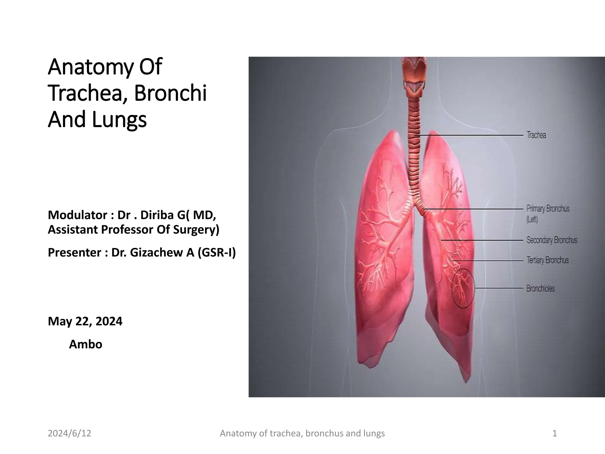
Anatomy Of
Trachea, Bronchi
And Lungs
Modulator : Dr . Diriba G( MD,
Assistant Professor Of Surgery)
Presenter : Dr. Gizachew A (GSR-I)
May 22, 2024
Ambo
2024/6/12 Anatomy of trachea, bronchus and lungs 1
Outline
• Introduction
• Surgical Embryology
• Anatomy of Trachea
• Anatomy of Bronchi
• Anatomy of Pleura and Lung
• Surgical Importance of the Anatomy and Clinical
Correlations
2024/6/12 Anatomy of trachea, bronchus and lungs 2
Introduction
• Development of lower respiratory tract begins to form
during 4th weeks (laryngo tracheal groove).
• The trachea is a midline structure and lies just anterior
the esophagus.
• The trachea divides into the left and right main stem
bronchi at junction point carina.
• The lungs are the vital organs of respiration.
2024/6/12 Anatomy of trachea, bronchus and lungs 3
Embryology
Lower respiratory tract has five milestones of
development.
• Embryologic period (3 - 6 weeks)
• Pseudo glandular period (5 -17 weeks)
• Canalicular period (17 - 25 weeks)
• Terminal Sac period (24 weeks - birth)
• Alveolar period (32 weeks- 8 years)
2024/6/12 Anatomy of trachea, bronchus and lungs 4
Trachea
• AKA the windpipe
• Is a cartilaginous tube that connects the larynx to
the bronchi of the lungs.
• Has an inner diameter of about 1.5 to 2cm.
• Length of about 10 to 11 cm.
• Wider in males than females.
2024/6/12 Anatomy of trachea, bronchus and lungs 5
2024/6/12 Anatomy of trachea, bronchus and lungs 6
Cont...
• It begins at the lower edge of the cricoid cartilage
of the larynx C6 and
• Ends at the carina, the point where the trachea
branches into left and right main bronchi T4
• Is surrounded by 16–20 rings of hyaline cartilage;
• These 'rings' are 4mm high in the adult, incomplete
and C-shaped.
• Ligaments connect the rings.
2024/6/12 Anatomy of trachea, bronchus and lungs 7
Cont...
• The trachealis muscle connects the ends of the
incomplete rings
• Adventitia, which is the outermost layer of
connective tissue that surrounds the hyaline
cartilage
• Can be displaced normally to the right by the aortic
arch.
2024/6/12 Anatomy of trachea, bronchus and lungs 8
2024/6/12 Anatomy of trachea, bronchus and lungs 9
Relations of trachea in the neck
• Anteriorly: skin, fascia, isthmus of the thyroid gland
(ring 2-4), inferior thyroid vein, jugular arch, thyroid
ima artery, the left brachiocephalic veins in
children, overlapped by the sternothyroid and
sternohyoid muscles.
• Posteriorly: right and left recurrent laryngeal
nerves and esophagus
• Laterally: lobes of the thyroid glands and the
carotid sheath and its contents.
2024/6/12 Anatomy of trachea, bronchus and lungs 10
Relations of trachea in the thorax
• Anteriorly: the sternum, the thymus, he left
bracheocephalic vein, the origins of
bracheocephalic and left common carotid arteries
and the arch of aorta.
• Posteriorly: the esophagus and the left recurrent
laryngeal nerve
• Right side: the azygos vein, the right vagus nerve
and the pleura
• Left side: the arch of the aorta, the left common
carotid and left subclavian arteries, the left vagus
and left phrenic nerves and the pleura.
2024/6/12 Anatomy of trachea, bronchus and lungs 11
2024/6/12 Anatomy of trachea, bronchus and lungs 12
Blood supply
• The proximal trachea supplied by the inferior thyroid
artery and innominate artery.
• The distal trachea, carina, and bronchi are supplied
by the bronchial arteries.
2024/6/12 Anatomy of trachea, bronchus and lungs 13
2024/6/12 Anatomy of trachea, bronchus and lungs 14
Nerve supply
Para sympathetic:
• Vagus and recurrent laryngeal nerves
Sympathetic:
• Middle cervical ganglion (vasomotor)
Lymphatic Drainage
• Pretracheal
• Para tracheal lymph node
2024/6/12 Anatomy of trachea, bronchus and lungs 15
2024/6/12 Anatomy of trachea, bronchus and lungs 16
Cont...
Development
• In the fourth week the tracheoesophageal septum is
formed.
• Divides the foregut tube into the laryngotracheal tube.
• By the start of the fifth week, the left and right main
bronchi have begin to form, initially as buds at the
terminal end of the trachea.
• The trachea is no more than 4 mm in diameter during the
first year of life, expanding to its adult diameter of
approximately 2cm by late childhood.
• The trachea is more circular and more vertical in children
2024/6/12 Anatomy of trachea, bronchus and lungs 17
Microanatomy
• The trachea is lined by psedostratified ciliated
columnar cells.
• The epithelium contains goblet cells, which are
glandular, column-shaped cells that produce
mucins, the main component of mucus.
• Mucociliary clearance.
2024/6/12 Anatomy of trachea, bronchus and lungs 18
2024/6/12 Anatomy of trachea, bronchus and lungs 19
Clinical significance of trachea
• The insertion of an endotracheal tube is a procedure that
may be required for resuscitation of the newborn at birth
and subsequently to enable artificial ventilation.
• The tube is introduced usually orally initially and guided
through the vocal cords with the help of a laryngoscope.
• The length of the trachea in the neonate can be as short as
3.1 cm in premature infants, and the T1-to-carina distance
ranges from 1.4 cm in babies weighing 500–1000 g, to 1.8
cm in those weighing 3001–3500 g.
• Once in place, the tip of the tube should be in the mid
trachea, above the carina.
2024/6/12 Anatomy of trachea, bronchus and lungs 20
Cont...
• The required length of the tube can be estimated according to
birth weight.
• Confirmation of correct positioning of the endotracheal tube is
obtained from a chest X-ray.
• The anatomical reference points used for the X-ray to assess the
position
 the clavicles,
 the bodies of the vertebrae and
 the carina (although not always visible on X-ray).
• Previously, it was advised that the tip of the endotracheal tube
should be placed just below the clavicles, at the level of the first
rib or 1–2 cm above the carina.
• It is now suggested that the body of the first thoracic vertebra
(T1) is a more stable reference point as the target for the tip of
the endotracheal tube.
2024/6/12 Anatomy of trachea, bronchus and lungs 21
Clinical Correlations Of Trachea
Congenital & others
• Tracheoesophageal fistula
• Tracheal injury
• Tracheal stenosis
• Tracheomalacia
• Infectious disease( tbc,
histoplasmosis,..)
• Burn trauma
• Post-intubation and post-
tracheostomy injuries
• Tracheal resection complication
• Tracheal agenesis/ atrsia
External lesions vs trachea
• Goiter( compressive effect)
• Vascular compression(vascur
rings)
• Mediastinal masses(e.g m.
neoplasms)
• Postpneumectomy syndrome
2024/6/12 Anatomy of trachea, bronchus and lungs 22
Anatomy of Bronchus
• Is a passage or airway in the lower respiratory tract
that conducts air into the lungs.
• The first or primary bronchi to branch from the
trachea at the carina are the right main bronchus
and the left main bronchus.
2024/6/12 Anatomy of trachea, bronchus and lungs 23
Anatomical differences of The right main
bronchus and left main bronchus
Right main Bronchus
• Shorter(2,5cm)
• Larger diameter (5cm)
• More vertical (susceptible
to aspiration)
• Passes directly to the hilum
of the lung.
• Gives 3 lobar bronchi(RSLB,
RMLB, RILB), RSLB branches
before entering hilum
• Enters hilum @ opposite to
T5.
Left main Bronchus
• Longer (5cm)
• Smaller diameter
• More horizontal relative to
trachea
• Gives 2 lobar bronchi after
entering hilum @ T6
• Passes to the left inferior to
the aortic arch, it crosses
anterior to the oesophagus,
thoracic duct and
descending aorta; the LPA is
at first anterior and then
superior to it.
2024/6/12 Anatomy of trachea, bronchus and lungs 24
Cont....
• Within the lungs, the bronchi branch in a constant
fashion to form the branches of the tracheobronchial
tree.
• The main bronchi branch into narrower secondary
bronchi or lobar bronchi.
• Each lobar bronchus divides into several tertiary or
segmental bronchi that supply the bronchopulmonary
segments.
2024/6/12 Anatomy of trachea, bronchus and lungs 25
Cont...
The bronchopulmonary segments are:
• The largest subdivisions of a lobe. Usually 18-20 in
number (10 in the right lung; 8-10 in the left lung).
• Separated from the rest of the lung by a septum of
connective tissue.
• Surgically resectable.
• Drained by intersegmental parts of the pulmonary
veins that lie in the connective tissue between and
drain adjacent segments.
2024/6/12 Anatomy of trachea, bronchus and lungs 26
2024/6/12 Anatomy of trachea, bronchus and lungs 27
Cont...
• Beyond the tertiary segmental bronchi there are 20 to
25 generations of branching conducting bronchioles
that eventually end as terminal bronchioles.
• Bronchioles lack cartilage in their walls.
• Conducting bronchioles transport air but lack glands or
alveoli.
• Each terminal bronchiole gives rise to several
generations of respiratory bronchioles, characterized
by scattered, thin-walled out pocketings (alveoli) that
extend from their lumens.
2024/6/12 Anatomy of trachea, bronchus and lungs 28
Cont...
• The pulmonary alveolus: is the basic structural unit of gas
exchange in the lung.
• Due to the presence of the alveoli, the respiratory
bronchioles are involved both in air transportation and
gas exchange.
• Each respiratory bronchiole gives rise to 2-11 alveolar
ducts, each of which gives rise to 5-6 alveolar sacs.
• Alveolar ducts are elongated airways densely lined with
alveoli
• New alveoli continue to develop until about age 8 years,
by which time there are approximately 300 million
alveoli.
2024/6/12 Anatomy of trachea, bronchus and lungs 29
2024/6/12 Anatomy of trachea, bronchus and lungs 30
Blood supply
The bronchial arteries:
• Provide 1% of total blood flow to the lungs and
• Supply the visceral pleura, intrapulmonary blood
vessel walls, and lymphatic system.
The pulmonary arteries:
• Provide the other 99% of the total blood flow to
the lung
Venous drainage
• Into the azygous and hemiazygous systems
2024/6/12 Anatomy of trachea, bronchus and lungs 31
2024/6/12 Anatomy of trachea, bronchus and lungs 32
Bronchi cilinical cosideration
• Foreign body aspiration
• Broncoscopy
2024/6/12 Anatomy of trachea, bronchus and lungs 33
Anatomy of Lungs
The lungs are separated from each other by the
mediastinum.
• Each lung has:
An apex and a base.
Two or three lobes, created by one or two fissures.
Three surfaces (costal, mediastinal, and diaphragmatic).
Three borders (anterior, inferior, and posterior).
2024/6/12 Anatomy of trachea, bronchus and lungs 34
Cont...
• The anterior borders of the lungs lie between the
2nd and 4th costal cartilages.
• The anterior border of the left lung is more deeply
indented by its cardiac notch.
• The inferior margins of the lungs reach:
MCL at the level of the 6th rib
MAL at the 8th rib, and
 SL at the 10th rib, proceeding toward the spinous
process of T10 vertebra.
2024/6/12 Anatomy of trachea, bronchus and lungs 35
Cont...
• The oblique fissure of the lungs extends from T2
vertebra posteriorly to the 6th costal cartilage
anteriorly.
• The horizontal fissure of the right lung extends
from the oblique fissure along the 4th rib and
costal cartilage anteriorly.
2024/6/12 Anatomy of trachea, bronchus and lungs 36
2024/6/12 Anatomy of trachea, bronchus and lungs 37
Cont...
• The lungs are attached to the mediastinum by the
roots of the lungs—that are :
The bronchi (and associated bronchial vessels)
Pulmonary arteries
Superior and inferior pulmonary veins
The pulmonary plexuses of nerves
Lymphatic vessels .
2024/6/12 Anatomy of trachea, bronchus and lungs 38
Blood supply of the lungs
Arterial supply
1. Bronchial arteries:
• Supplies the bronchial tree, the connective tissue stroma,
and visceral pleura
• The two left bronchial arteries usually arise directly from
the thoracic aorta.
• The single right bronchial artery may also arise directly
from the aorta
2. Pulmonary arteries:
• Arise from the pulmonary trunk.
• Each lung receives one pulmonary artery
2024/6/12 Anatomy of trachea, bronchus and lungs 39
Cont...
Veneous
Bronchial veins:
• They drain into the azygos (rt) and hemiazygos
veins (lt)
Pulmonary veins:
• Superior and inferior
• The middle lobe vein is a tributary of the right
superior pulmonary vein.
2024/6/12 Anatomy of trachea, bronchus and lungs 40
Cont...
• The arteries and bronchi are paired in the lung,
branching simultaneously and running parallel
courses.
• The pulmonary veins run independently of the
arteries and bronchi in the lung.
• Each pulmonary artery divides to secondary lobar
arteries
• Lobar arteries divide into tertiary segmental
arteries.
2024/6/12 Anatomy of trachea, bronchus and lungs 41
Cont...
• The veins from the visceral pleura and the
bronchial venous circulation drain into the
pulmonary veins
• Veins from the parietal pleura join systemic veins
in adjacent parts of the thoracic wall.
2024/6/12 Anatomy of trachea, bronchus and lungs 42
2024/6/12 Anatomy of trachea, bronchus and lungs 43
Lymphatic drainage
• The lymph from the lung is drained by two sets of
lymph vessels.
Superficial vessels
Deep vessels
• All the lymph is drained into tracheobronchial lymph
nodes (located at the hilum), which in turn drain into
bronchomediastinal lymph nodes.
• The lymphatic drainage of the lung is clinically
important because lung cancer spreads by lymphatic
path.
2024/6/12 Anatomy of trachea, bronchus and lungs 44
2024/6/12 Anatomy of trachea, bronchus and lungs 45
Nerve supply
Nerves of Lungs and Pleurae
• Are derived from the pulmonary plexuses anterior and
(mainly) posterior to the roots of the lungs.
• These nerve networks contains :
• Parasympathetic ,sympathetic and visceral afferent
fibers.
The parasympathetic fibers
• Are presynaptic fibers from the vagus nerve (CN X).
• Are bronchoconstrictor, vasodilator and
secretomotor
2024/6/12 Anatomy of trachea, bronchus and lungs 46
Cont...
The sympathetic fibers
• Derived from T2 to T5 spinal segments.
• Are bronchodilator, vasoconstrictor and secretory
The visceral afferent fibers
• Are either reflexive (conducting subconscious
sensations associated with reflexes that control
function) or nociceptive (conducting pain impulses
generated in response to painful or injurious stimuli,
such as chemical irritants, ischemia, or excessive
stretch).
2024/6/12 Anatomy of trachea, bronchus and lungs 47
2024/6/12 Anatomy of trachea, bronchus and lungs 48
Anatomy of pleura
1. The visceral pleura and 2. The parietal pleura
The visceral pleura
• Invests all surfaces of the lungs
• Is pulmonary pleura
• Produce and reabsorb fluid.
• Insensitive to pain.
• Cannot usually be dissected
• It provides the lung with a smooth slippery surface,
• Is continuous with the parietal pleura at the hilum of the
lung.
2024/6/12 Anatomy of trachea, bronchus and lungs 49
Cont...
The parietal pleura
• Lines the pulmonary cavities.
• It is thicker than the visceral pleura .
• Can be dissected or separated from the surfaces it covers.
• Limit fluid loss and reduce friction.
• Sensetive to pain
• Consists of three parts—
Costal
 Mediastinal, and
 Diaphragmatic—and the cervical pleura.
2024/6/12 Anatomy of trachea, bronchus and lungs 50
Cont...
The costal part of the parietal pleura
• It is separated from the internal surface of the thoracic
wall by endothoracic fascia.
The mediastinal part of the parietal pleura
• (mediastinal pleura) covers the lateral aspects of the
mediastinum.
• It continues superiorly into the root of the neck as
cervical pleura.
The diaphragmatic part of the parietal pleura
• covers the superior (thoracic) surface of the diaphragm
on each side of the mediastinum
2024/6/12 Anatomy of trachea, bronchus and lungs 51
Cont...
The cervical pleura
• Is reinforced by a fibrous extension of the
endothoracic fascia, the supra pleural membrane
(Sibson fascia). The membrane attaches to the
internal border of the 1st rib and the transverse
process of C7 vertebra .
2024/6/12 Anatomy of trachea, bronchus and lungs 52
Cont...
Pleural Recesses
• An area where adjacent surfaces of the partial
pleura come in to contact.
• Are two in numbers
The costo diaphragmatic recesses
The costo mediastinal recesses
2024/6/12 Anatomy of trachea, bronchus and lungs 53
2024/6/12 Anatomy of trachea, bronchus and lungs 54
Surgical Importance and Clinical
Correlations
Lung and Pleura
• Thoracostomy
• Pneumothorax
• Hemothorax
• Pneumohemothorax
• Lung resection
• Lung cancer
2024/6/12 Anatomy of trachea, bronchus and lungs 55
Reference
• Moore, K. L., & Dalley, A. F. (2018). Clinically
oriented anatomy. Wolters kluwer india Pvt Ltd.
• Illman, J. (2020). Seymour Schwartz: surgeon,
editor, cartographer, and historian.
• Lee John Skandalakis - Surgical Anatomy: The
Embryologic and Anatomic Basis of Modern
Surgery-Paschalidis (2004)
2024/6/12 Anatomy of trachea, bronchus and lungs 56
Thank you for you
Attention!
2024/6/12 Anatomy of trachea, bronchus and lungs 57

Anatomy of trachea, bronchi, & lungs.pptx

  • 1.
    Anatomy Of Trachea, Bronchi AndLungs Modulator : Dr . Diriba G( MD, Assistant Professor Of Surgery) Presenter : Dr. Gizachew A (GSR-I) May 22, 2024 Ambo 2024/6/12 Anatomy of trachea, bronchus and lungs 1
  • 2.
    Outline • Introduction • SurgicalEmbryology • Anatomy of Trachea • Anatomy of Bronchi • Anatomy of Pleura and Lung • Surgical Importance of the Anatomy and Clinical Correlations 2024/6/12 Anatomy of trachea, bronchus and lungs 2
  • 3.
    Introduction • Development oflower respiratory tract begins to form during 4th weeks (laryngo tracheal groove). • The trachea is a midline structure and lies just anterior the esophagus. • The trachea divides into the left and right main stem bronchi at junction point carina. • The lungs are the vital organs of respiration. 2024/6/12 Anatomy of trachea, bronchus and lungs 3
  • 4.
    Embryology Lower respiratory tracthas five milestones of development. • Embryologic period (3 - 6 weeks) • Pseudo glandular period (5 -17 weeks) • Canalicular period (17 - 25 weeks) • Terminal Sac period (24 weeks - birth) • Alveolar period (32 weeks- 8 years) 2024/6/12 Anatomy of trachea, bronchus and lungs 4
  • 5.
    Trachea • AKA thewindpipe • Is a cartilaginous tube that connects the larynx to the bronchi of the lungs. • Has an inner diameter of about 1.5 to 2cm. • Length of about 10 to 11 cm. • Wider in males than females. 2024/6/12 Anatomy of trachea, bronchus and lungs 5
  • 6.
    2024/6/12 Anatomy oftrachea, bronchus and lungs 6
  • 7.
    Cont... • It beginsat the lower edge of the cricoid cartilage of the larynx C6 and • Ends at the carina, the point where the trachea branches into left and right main bronchi T4 • Is surrounded by 16–20 rings of hyaline cartilage; • These 'rings' are 4mm high in the adult, incomplete and C-shaped. • Ligaments connect the rings. 2024/6/12 Anatomy of trachea, bronchus and lungs 7
  • 8.
    Cont... • The trachealismuscle connects the ends of the incomplete rings • Adventitia, which is the outermost layer of connective tissue that surrounds the hyaline cartilage • Can be displaced normally to the right by the aortic arch. 2024/6/12 Anatomy of trachea, bronchus and lungs 8
  • 9.
    2024/6/12 Anatomy oftrachea, bronchus and lungs 9
  • 10.
    Relations of tracheain the neck • Anteriorly: skin, fascia, isthmus of the thyroid gland (ring 2-4), inferior thyroid vein, jugular arch, thyroid ima artery, the left brachiocephalic veins in children, overlapped by the sternothyroid and sternohyoid muscles. • Posteriorly: right and left recurrent laryngeal nerves and esophagus • Laterally: lobes of the thyroid glands and the carotid sheath and its contents. 2024/6/12 Anatomy of trachea, bronchus and lungs 10
  • 11.
    Relations of tracheain the thorax • Anteriorly: the sternum, the thymus, he left bracheocephalic vein, the origins of bracheocephalic and left common carotid arteries and the arch of aorta. • Posteriorly: the esophagus and the left recurrent laryngeal nerve • Right side: the azygos vein, the right vagus nerve and the pleura • Left side: the arch of the aorta, the left common carotid and left subclavian arteries, the left vagus and left phrenic nerves and the pleura. 2024/6/12 Anatomy of trachea, bronchus and lungs 11
  • 12.
    2024/6/12 Anatomy oftrachea, bronchus and lungs 12
  • 13.
    Blood supply • Theproximal trachea supplied by the inferior thyroid artery and innominate artery. • The distal trachea, carina, and bronchi are supplied by the bronchial arteries. 2024/6/12 Anatomy of trachea, bronchus and lungs 13
  • 14.
    2024/6/12 Anatomy oftrachea, bronchus and lungs 14
  • 15.
    Nerve supply Para sympathetic: •Vagus and recurrent laryngeal nerves Sympathetic: • Middle cervical ganglion (vasomotor) Lymphatic Drainage • Pretracheal • Para tracheal lymph node 2024/6/12 Anatomy of trachea, bronchus and lungs 15
  • 16.
    2024/6/12 Anatomy oftrachea, bronchus and lungs 16
  • 17.
    Cont... Development • In thefourth week the tracheoesophageal septum is formed. • Divides the foregut tube into the laryngotracheal tube. • By the start of the fifth week, the left and right main bronchi have begin to form, initially as buds at the terminal end of the trachea. • The trachea is no more than 4 mm in diameter during the first year of life, expanding to its adult diameter of approximately 2cm by late childhood. • The trachea is more circular and more vertical in children 2024/6/12 Anatomy of trachea, bronchus and lungs 17
  • 18.
    Microanatomy • The tracheais lined by psedostratified ciliated columnar cells. • The epithelium contains goblet cells, which are glandular, column-shaped cells that produce mucins, the main component of mucus. • Mucociliary clearance. 2024/6/12 Anatomy of trachea, bronchus and lungs 18
  • 19.
    2024/6/12 Anatomy oftrachea, bronchus and lungs 19
  • 20.
    Clinical significance oftrachea • The insertion of an endotracheal tube is a procedure that may be required for resuscitation of the newborn at birth and subsequently to enable artificial ventilation. • The tube is introduced usually orally initially and guided through the vocal cords with the help of a laryngoscope. • The length of the trachea in the neonate can be as short as 3.1 cm in premature infants, and the T1-to-carina distance ranges from 1.4 cm in babies weighing 500–1000 g, to 1.8 cm in those weighing 3001–3500 g. • Once in place, the tip of the tube should be in the mid trachea, above the carina. 2024/6/12 Anatomy of trachea, bronchus and lungs 20
  • 21.
    Cont... • The requiredlength of the tube can be estimated according to birth weight. • Confirmation of correct positioning of the endotracheal tube is obtained from a chest X-ray. • The anatomical reference points used for the X-ray to assess the position  the clavicles,  the bodies of the vertebrae and  the carina (although not always visible on X-ray). • Previously, it was advised that the tip of the endotracheal tube should be placed just below the clavicles, at the level of the first rib or 1–2 cm above the carina. • It is now suggested that the body of the first thoracic vertebra (T1) is a more stable reference point as the target for the tip of the endotracheal tube. 2024/6/12 Anatomy of trachea, bronchus and lungs 21
  • 22.
    Clinical Correlations OfTrachea Congenital & others • Tracheoesophageal fistula • Tracheal injury • Tracheal stenosis • Tracheomalacia • Infectious disease( tbc, histoplasmosis,..) • Burn trauma • Post-intubation and post- tracheostomy injuries • Tracheal resection complication • Tracheal agenesis/ atrsia External lesions vs trachea • Goiter( compressive effect) • Vascular compression(vascur rings) • Mediastinal masses(e.g m. neoplasms) • Postpneumectomy syndrome 2024/6/12 Anatomy of trachea, bronchus and lungs 22
  • 23.
    Anatomy of Bronchus •Is a passage or airway in the lower respiratory tract that conducts air into the lungs. • The first or primary bronchi to branch from the trachea at the carina are the right main bronchus and the left main bronchus. 2024/6/12 Anatomy of trachea, bronchus and lungs 23
  • 24.
    Anatomical differences ofThe right main bronchus and left main bronchus Right main Bronchus • Shorter(2,5cm) • Larger diameter (5cm) • More vertical (susceptible to aspiration) • Passes directly to the hilum of the lung. • Gives 3 lobar bronchi(RSLB, RMLB, RILB), RSLB branches before entering hilum • Enters hilum @ opposite to T5. Left main Bronchus • Longer (5cm) • Smaller diameter • More horizontal relative to trachea • Gives 2 lobar bronchi after entering hilum @ T6 • Passes to the left inferior to the aortic arch, it crosses anterior to the oesophagus, thoracic duct and descending aorta; the LPA is at first anterior and then superior to it. 2024/6/12 Anatomy of trachea, bronchus and lungs 24
  • 25.
    Cont.... • Within thelungs, the bronchi branch in a constant fashion to form the branches of the tracheobronchial tree. • The main bronchi branch into narrower secondary bronchi or lobar bronchi. • Each lobar bronchus divides into several tertiary or segmental bronchi that supply the bronchopulmonary segments. 2024/6/12 Anatomy of trachea, bronchus and lungs 25
  • 26.
    Cont... The bronchopulmonary segmentsare: • The largest subdivisions of a lobe. Usually 18-20 in number (10 in the right lung; 8-10 in the left lung). • Separated from the rest of the lung by a septum of connective tissue. • Surgically resectable. • Drained by intersegmental parts of the pulmonary veins that lie in the connective tissue between and drain adjacent segments. 2024/6/12 Anatomy of trachea, bronchus and lungs 26
  • 27.
    2024/6/12 Anatomy oftrachea, bronchus and lungs 27
  • 28.
    Cont... • Beyond thetertiary segmental bronchi there are 20 to 25 generations of branching conducting bronchioles that eventually end as terminal bronchioles. • Bronchioles lack cartilage in their walls. • Conducting bronchioles transport air but lack glands or alveoli. • Each terminal bronchiole gives rise to several generations of respiratory bronchioles, characterized by scattered, thin-walled out pocketings (alveoli) that extend from their lumens. 2024/6/12 Anatomy of trachea, bronchus and lungs 28
  • 29.
    Cont... • The pulmonaryalveolus: is the basic structural unit of gas exchange in the lung. • Due to the presence of the alveoli, the respiratory bronchioles are involved both in air transportation and gas exchange. • Each respiratory bronchiole gives rise to 2-11 alveolar ducts, each of which gives rise to 5-6 alveolar sacs. • Alveolar ducts are elongated airways densely lined with alveoli • New alveoli continue to develop until about age 8 years, by which time there are approximately 300 million alveoli. 2024/6/12 Anatomy of trachea, bronchus and lungs 29
  • 30.
    2024/6/12 Anatomy oftrachea, bronchus and lungs 30
  • 31.
    Blood supply The bronchialarteries: • Provide 1% of total blood flow to the lungs and • Supply the visceral pleura, intrapulmonary blood vessel walls, and lymphatic system. The pulmonary arteries: • Provide the other 99% of the total blood flow to the lung Venous drainage • Into the azygous and hemiazygous systems 2024/6/12 Anatomy of trachea, bronchus and lungs 31
  • 32.
    2024/6/12 Anatomy oftrachea, bronchus and lungs 32
  • 33.
    Bronchi cilinical cosideration •Foreign body aspiration • Broncoscopy 2024/6/12 Anatomy of trachea, bronchus and lungs 33
  • 34.
    Anatomy of Lungs Thelungs are separated from each other by the mediastinum. • Each lung has: An apex and a base. Two or three lobes, created by one or two fissures. Three surfaces (costal, mediastinal, and diaphragmatic). Three borders (anterior, inferior, and posterior). 2024/6/12 Anatomy of trachea, bronchus and lungs 34
  • 35.
    Cont... • The anteriorborders of the lungs lie between the 2nd and 4th costal cartilages. • The anterior border of the left lung is more deeply indented by its cardiac notch. • The inferior margins of the lungs reach: MCL at the level of the 6th rib MAL at the 8th rib, and  SL at the 10th rib, proceeding toward the spinous process of T10 vertebra. 2024/6/12 Anatomy of trachea, bronchus and lungs 35
  • 36.
    Cont... • The obliquefissure of the lungs extends from T2 vertebra posteriorly to the 6th costal cartilage anteriorly. • The horizontal fissure of the right lung extends from the oblique fissure along the 4th rib and costal cartilage anteriorly. 2024/6/12 Anatomy of trachea, bronchus and lungs 36
  • 37.
    2024/6/12 Anatomy oftrachea, bronchus and lungs 37
  • 38.
    Cont... • The lungsare attached to the mediastinum by the roots of the lungs—that are : The bronchi (and associated bronchial vessels) Pulmonary arteries Superior and inferior pulmonary veins The pulmonary plexuses of nerves Lymphatic vessels . 2024/6/12 Anatomy of trachea, bronchus and lungs 38
  • 39.
    Blood supply ofthe lungs Arterial supply 1. Bronchial arteries: • Supplies the bronchial tree, the connective tissue stroma, and visceral pleura • The two left bronchial arteries usually arise directly from the thoracic aorta. • The single right bronchial artery may also arise directly from the aorta 2. Pulmonary arteries: • Arise from the pulmonary trunk. • Each lung receives one pulmonary artery 2024/6/12 Anatomy of trachea, bronchus and lungs 39
  • 40.
    Cont... Veneous Bronchial veins: • Theydrain into the azygos (rt) and hemiazygos veins (lt) Pulmonary veins: • Superior and inferior • The middle lobe vein is a tributary of the right superior pulmonary vein. 2024/6/12 Anatomy of trachea, bronchus and lungs 40
  • 41.
    Cont... • The arteriesand bronchi are paired in the lung, branching simultaneously and running parallel courses. • The pulmonary veins run independently of the arteries and bronchi in the lung. • Each pulmonary artery divides to secondary lobar arteries • Lobar arteries divide into tertiary segmental arteries. 2024/6/12 Anatomy of trachea, bronchus and lungs 41
  • 42.
    Cont... • The veinsfrom the visceral pleura and the bronchial venous circulation drain into the pulmonary veins • Veins from the parietal pleura join systemic veins in adjacent parts of the thoracic wall. 2024/6/12 Anatomy of trachea, bronchus and lungs 42
  • 43.
    2024/6/12 Anatomy oftrachea, bronchus and lungs 43
  • 44.
    Lymphatic drainage • Thelymph from the lung is drained by two sets of lymph vessels. Superficial vessels Deep vessels • All the lymph is drained into tracheobronchial lymph nodes (located at the hilum), which in turn drain into bronchomediastinal lymph nodes. • The lymphatic drainage of the lung is clinically important because lung cancer spreads by lymphatic path. 2024/6/12 Anatomy of trachea, bronchus and lungs 44
  • 45.
    2024/6/12 Anatomy oftrachea, bronchus and lungs 45
  • 46.
    Nerve supply Nerves ofLungs and Pleurae • Are derived from the pulmonary plexuses anterior and (mainly) posterior to the roots of the lungs. • These nerve networks contains : • Parasympathetic ,sympathetic and visceral afferent fibers. The parasympathetic fibers • Are presynaptic fibers from the vagus nerve (CN X). • Are bronchoconstrictor, vasodilator and secretomotor 2024/6/12 Anatomy of trachea, bronchus and lungs 46
  • 47.
    Cont... The sympathetic fibers •Derived from T2 to T5 spinal segments. • Are bronchodilator, vasoconstrictor and secretory The visceral afferent fibers • Are either reflexive (conducting subconscious sensations associated with reflexes that control function) or nociceptive (conducting pain impulses generated in response to painful or injurious stimuli, such as chemical irritants, ischemia, or excessive stretch). 2024/6/12 Anatomy of trachea, bronchus and lungs 47
  • 48.
    2024/6/12 Anatomy oftrachea, bronchus and lungs 48
  • 49.
    Anatomy of pleura 1.The visceral pleura and 2. The parietal pleura The visceral pleura • Invests all surfaces of the lungs • Is pulmonary pleura • Produce and reabsorb fluid. • Insensitive to pain. • Cannot usually be dissected • It provides the lung with a smooth slippery surface, • Is continuous with the parietal pleura at the hilum of the lung. 2024/6/12 Anatomy of trachea, bronchus and lungs 49
  • 50.
    Cont... The parietal pleura •Lines the pulmonary cavities. • It is thicker than the visceral pleura . • Can be dissected or separated from the surfaces it covers. • Limit fluid loss and reduce friction. • Sensetive to pain • Consists of three parts— Costal  Mediastinal, and  Diaphragmatic—and the cervical pleura. 2024/6/12 Anatomy of trachea, bronchus and lungs 50
  • 51.
    Cont... The costal partof the parietal pleura • It is separated from the internal surface of the thoracic wall by endothoracic fascia. The mediastinal part of the parietal pleura • (mediastinal pleura) covers the lateral aspects of the mediastinum. • It continues superiorly into the root of the neck as cervical pleura. The diaphragmatic part of the parietal pleura • covers the superior (thoracic) surface of the diaphragm on each side of the mediastinum 2024/6/12 Anatomy of trachea, bronchus and lungs 51
  • 52.
    Cont... The cervical pleura •Is reinforced by a fibrous extension of the endothoracic fascia, the supra pleural membrane (Sibson fascia). The membrane attaches to the internal border of the 1st rib and the transverse process of C7 vertebra . 2024/6/12 Anatomy of trachea, bronchus and lungs 52
  • 53.
    Cont... Pleural Recesses • Anarea where adjacent surfaces of the partial pleura come in to contact. • Are two in numbers The costo diaphragmatic recesses The costo mediastinal recesses 2024/6/12 Anatomy of trachea, bronchus and lungs 53
  • 54.
    2024/6/12 Anatomy oftrachea, bronchus and lungs 54
  • 55.
    Surgical Importance andClinical Correlations Lung and Pleura • Thoracostomy • Pneumothorax • Hemothorax • Pneumohemothorax • Lung resection • Lung cancer 2024/6/12 Anatomy of trachea, bronchus and lungs 55
  • 56.
    Reference • Moore, K.L., & Dalley, A. F. (2018). Clinically oriented anatomy. Wolters kluwer india Pvt Ltd. • Illman, J. (2020). Seymour Schwartz: surgeon, editor, cartographer, and historian. • Lee John Skandalakis - Surgical Anatomy: The Embryologic and Anatomic Basis of Modern Surgery-Paschalidis (2004) 2024/6/12 Anatomy of trachea, bronchus and lungs 56
  • 57.
    Thank you foryou Attention! 2024/6/12 Anatomy of trachea, bronchus and lungs 57