SlideShare a Scribd company logo
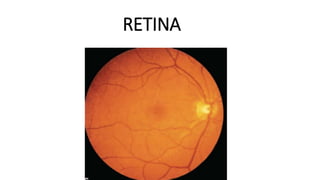
RETINA
• Inner nervous coat of eyeball
• FUNDUS
• Thin, transparent membrane
• purplish red in colour
• Formation of optical image
• Thickness- 0.56mm near optic disc to 0.1mm at ora serrata
• Thinnest at centre of fovea
EXTENSION
• Outer surface- Bruchs membrane of
choroid
• Inner surface- vitreous
• Posteriorly- optic nerve
• Anteriorly- epithelium of iris and
• ciliary body(ora serrata)
• Ant- extends more on nasal side
• Firmly attached- margins of optic disc
& ora serrata
Optic disc
Macula lutea
Peripheral retina
OPTIC DISC
• 1.5mm in diameter & 3mm nasal
to macula
• Pale pinK
• Depression in centre- physiological cup
• Central retinal vessels
• Optic nerve- lamina cribrosa
• Blind spot
• MACULA –
• oval,yellowish,at centre of posterior part
• 5mm in diameter & 3mm temporal to optic disc
• carotenoid pigment-xanthophyll(present from outer nuclear layer
onward)
• Central depression – fovea centralis
• Sides of depression – Clivus
• Floor of depression – foveola
• 1,47,000 per sq.mm
• Umbo- foveolar reflex
• Central bouquet of cones
• Parafovea-0.5mm surrounding fovea
• Perifovea-1.5mmsurrounding parafovea
• Near periphery – 1.5mm ring peripheral to temporal vascular
Arcades
• Equatorial retina- retina around the equator
• Peripheral retina – region anterior to it
• OUTER PIGMENTED LAYER
• Retinal pigment epithelium (RPE)
• INNER NEUROSENSORY LAYER
• Photoreceptors
• External limiting membrane
• Outer nuclear layer
• Outer plexiform layer
• Inner nuclear layer
• Inner plexiform layer
• Ganglion cell layer
• Nerve fibre layer
• Internal limiting membrane
• LAYERS OF RETINA
• EMRYOLOGICAL DEVELOPMENT
• Outer pigmented layer- outer layer of optic cup
• Neurosensory layer- inner layer of optic cup
• anterior one fifth- columnar
• Posterior four fifth
• outer nuclear zone & inner marginal zone
• Inner neuroblastic layer –ganglion cells,amacrine cells,Muller cells
• Outer neuroblastic layer –rods,cones,bipolar cells
• Macular area- superimposed nuclei of ganglion cell layer, lateral to optic disc
• DEVELOPMENT
• RETINAL PIGMENTED EPITHELIUM
• Single layer of hexagonal cells
• Narrow & tall Posteriorly ,flattened near ora serrata
• Apical end- microvilli – project between outer segment of rods & cone
• Embedded in glycosaminoglycans
• Cell nucei- basal part
• Melanin granules
• Basal region- zona adherens,apical region- zona occludens
• FUNCTION
• Absorption of light
• Turn over of outer segment of rods& cones
• Formation of rhodopsin& iodopsin( vitamin A)
• SUB RETINAL SPACE
• - Retinal detachment
• Ocular albinism
• -transillumination of iris
NEURAL RETINA
• PHOTO RECEPTORS
• BIPOLAR CELLS
• GANGLION CELLS
• PHOTORECEPTORS
• RODS
• 110 to 125 million
• Rhodopsin- scotopic vision
• 40to 60micrometre long
• STRUCTURE
• Outer segment – cylindrical & contains lamellar disc
• Eccentric cilium- 9 doublet microtubules with no Central pair
• Inner segment – ellipsoid& myoid
• Ellipsoid (outer segment) – mitochondria
• Myoid- (inner segment- ER,Golgi apparatus,ribosomes
• Outer rod fibre
• Rod granule( cell body with nucleus)
• Inner rod fibre
• Rod spherule
• CONES
• 6.3 to 6.8 million
• Iodopsin, scotopic vision
• 65to 75micrometre long
• Outer segment – conical,wider base &
• rounded tip
• Continuous with extra cellular space
• Cilium
• Inner segment – ellipsoid ( very plump)& myoid
• Cell body with nucleus
• Inner cone fibre
• Cone pedicle
BIPOLAR CELLS
• First order neurons (neurons in posterior root ganglion)
• Rod bipolar cells several rod cells to 1to 4 ganglion cells
• Flat or diffuse bipolar cells –many cone cells with many ganglion cells
• Midget bipolar cell- single cone cell with single midget ganglion cell
• GANGLION CELLS
• Second order neuron-nervous ganglion)
• Increases from periphery to macula(10 layers)
• Fovea- absent
• HORIZONTAL CELLS
• Associated with cones- 7
• Associateed with rods – 10 to 12
AMACRINE CELLS
No axons,large cell body,abundant cytoplasm,lobulated nuclei
Synapse with dendrites of ganglion cells& axons of bipolar cells
• Supporting cells- MULLER CELL
• Fill in most of the space of neural retina,not occupied by neurons
• On outer surface – row of zona adherents between photoreceptor cell&
radial process of Muller cells- OUTER LIMITING MEMBRANE
• On inner surface –muller cells have expanded termination covered by
basement membrane – INNER LIMITING MEMBRANE
•
• BLOOD SUPPLY
• Outer 4 layers-
chorio capillaries
• Inner 6 layers-
• Central retinal artery & vein
• Course of Central retinal
artery
• circle of zinn or Haller
• Arterial branches run in nerve fibre layer
• Foveal avascular zone- 500micronmetre in diameter
• BLOOD RETINAL BARRIER
• Tight junctions of RPE
• Nonfenestrated endothelium of capillaries
• CENTRAL RETINAL VEIN
• Accompany CRA& leaves through lamina cribrosa
• Lie on lateral side of artery in optic nerve
• Longer course in subarachnoid space
• Drains into cavernous sinus or superior ophthalmic vein
• ARRANGEMENT OF NERVE FIBRES IN RETINA
• THICKNESS OF NERVE FIBRE LAYER AT DISC
• Most lateral quadrant(thinnest)
• Upper and lower temporal quadrant
• Most medial quadrant
• Upper and lower nasal quadrant( thickest)
• CLINICAL SIGNIFANCE
• Papilledema- first in thickest quadrant
• Glaucomatous damage- arcuate fibres(more sensitive)
. Macular fibres(resistant)
CONGENITAL ANAMOLIES
• Medullated nerve fibres
• COLOBOMA of optic disc-
• Failure of closure of embryonic fissure
• Minor defect or fully developed coloboma
• HYPOPLASIA of optic disc
• Small disc surrounded by yellowish ring- double ring sign
• PERSISTENT HYALOID ARTERY
• Bregmesters papillae
• Mittendorf dot
• DRUSEN of optic disc
• Intracapillary refractile bodies
• Children- present as pseudopapilledema
• Adults- waxy pea like irregular refractilebodies
ANATOMY OF RETINA.pptx

More Related Content

Similar to ANATOMY OF RETINA.pptx

Eyeball
EyeballEyeball
Eyeball
Dr. Armaan Singh
 
anatomy of eyeball assignment presentation
anatomy of eyeball  assignment presentationanatomy of eyeball  assignment presentation
anatomy of eyeball assignment presentation
AmanuelIbrahim
 
Suraj chhetri retina
Suraj chhetri retinaSuraj chhetri retina
Suraj chhetri retina
Suraj Chhetri
 
Anatomy and Visual field defects of optic nerve and chiasma
Anatomy and Visual field defects of optic nerve and chiasmaAnatomy and Visual field defects of optic nerve and chiasma
Anatomy and Visual field defects of optic nerve and chiasma
Sadhwini Harish
 
Anatomy of the retina
Anatomy of the retinaAnatomy of the retina
Anatomy of the retina
Othman Al-Abbadi
 
ocular anatomy of eye for optometry and optalomolgy
ocular anatomy of eye for optometry and optalomolgyocular anatomy of eye for optometry and optalomolgy
ocular anatomy of eye for optometry and optalomolgy
devaramgehlot
 
ocular_anatomy.pptx
ocular_anatomy.pptxocular_anatomy.pptx
ocular_anatomy.pptx
HimanshuHarsh22ZOO00
 
anatomy of retina
 anatomy of retina anatomy of retina
anatomy of retina
Nikita Jaiswal
 
Orbit-&-it’s-content.pdf
Orbit-&-it’s-content.pdfOrbit-&-it’s-content.pdf
Orbit-&-it’s-content.pdf
Qman3
 
Retina and layers
Retina and layersRetina and layers
Retina and layers
Lhacha
 
Neuro-Ophthalmology_Dr. Bastola.pptx
Neuro-Ophthalmology_Dr. Bastola.pptxNeuro-Ophthalmology_Dr. Bastola.pptx
Neuro-Ophthalmology_Dr. Bastola.pptx
Dr. Pradeep Bastola
 
ANATOMY OF VISUAL PATHWAY - DR.RUTHRA.pptx
ANATOMY OF VISUAL PATHWAY - DR.RUTHRA.pptxANATOMY OF VISUAL PATHWAY - DR.RUTHRA.pptx
ANATOMY OF VISUAL PATHWAY - DR.RUTHRA.pptx
Ruthra Suresh
 
Retina
RetinaRetina
ANATOMY OF RETINA.pptx
ANATOMY OF RETINA.pptxANATOMY OF RETINA.pptx
ANATOMY OF RETINA.pptx
9459654457
 
ANATOMY OF EYEBALL MBBS OPTHALMOLOGY CLASS
ANATOMY OF EYEBALL MBBS OPTHALMOLOGY CLASSANATOMY OF EYEBALL MBBS OPTHALMOLOGY CLASS
ANATOMY OF EYEBALL MBBS OPTHALMOLOGY CLASS
Maxpayne485184
 
Anatomy of Retina
Anatomy of RetinaAnatomy of Retina
Anatomy of Retina
Sai Sandeep
 
Anatomy of optic nerve and its clinical significance
Anatomy of optic nerve and its clinical significanceAnatomy of optic nerve and its clinical significance
Anatomy of optic nerve and its clinical significance
Pabita Dhungel
 
HNS PART IX SPECIAL SENSES - VISION
HNS PART IX SPECIAL SENSES - VISION HNS PART IX SPECIAL SENSES - VISION
HNS PART IX SPECIAL SENSES - VISION
Dr. Aniket Shilwant
 

Similar to ANATOMY OF RETINA.pptx (20)

Eyeball
EyeballEyeball
Eyeball
 
anatomy of eyeball assignment presentation
anatomy of eyeball  assignment presentationanatomy of eyeball  assignment presentation
anatomy of eyeball assignment presentation
 
Suraj chhetri retina
Suraj chhetri retinaSuraj chhetri retina
Suraj chhetri retina
 
Anatomy and Visual field defects of optic nerve and chiasma
Anatomy and Visual field defects of optic nerve and chiasmaAnatomy and Visual field defects of optic nerve and chiasma
Anatomy and Visual field defects of optic nerve and chiasma
 
Anatomy of the retina
Anatomy of the retinaAnatomy of the retina
Anatomy of the retina
 
ocular anatomy of eye for optometry and optalomolgy
ocular anatomy of eye for optometry and optalomolgyocular anatomy of eye for optometry and optalomolgy
ocular anatomy of eye for optometry and optalomolgy
 
ocular_anatomy.pptx
ocular_anatomy.pptxocular_anatomy.pptx
ocular_anatomy.pptx
 
retina.ppt
retina.pptretina.ppt
retina.ppt
 
anatomy of retina
 anatomy of retina anatomy of retina
anatomy of retina
 
Orbit-&-it’s-content.pdf
Orbit-&-it’s-content.pdfOrbit-&-it’s-content.pdf
Orbit-&-it’s-content.pdf
 
Retina and layers
Retina and layersRetina and layers
Retina and layers
 
Neuro-Ophthalmology_Dr. Bastola.pptx
Neuro-Ophthalmology_Dr. Bastola.pptxNeuro-Ophthalmology_Dr. Bastola.pptx
Neuro-Ophthalmology_Dr. Bastola.pptx
 
ANATOMY OF VISUAL PATHWAY - DR.RUTHRA.pptx
ANATOMY OF VISUAL PATHWAY - DR.RUTHRA.pptxANATOMY OF VISUAL PATHWAY - DR.RUTHRA.pptx
ANATOMY OF VISUAL PATHWAY - DR.RUTHRA.pptx
 
Retina
RetinaRetina
Retina
 
ANATOMY OF RETINA.pptx
ANATOMY OF RETINA.pptxANATOMY OF RETINA.pptx
ANATOMY OF RETINA.pptx
 
Eye
EyeEye
Eye
 
ANATOMY OF EYEBALL MBBS OPTHALMOLOGY CLASS
ANATOMY OF EYEBALL MBBS OPTHALMOLOGY CLASSANATOMY OF EYEBALL MBBS OPTHALMOLOGY CLASS
ANATOMY OF EYEBALL MBBS OPTHALMOLOGY CLASS
 
Anatomy of Retina
Anatomy of RetinaAnatomy of Retina
Anatomy of Retina
 
Anatomy of optic nerve and its clinical significance
Anatomy of optic nerve and its clinical significanceAnatomy of optic nerve and its clinical significance
Anatomy of optic nerve and its clinical significance
 
HNS PART IX SPECIAL SENSES - VISION
HNS PART IX SPECIAL SENSES - VISION HNS PART IX SPECIAL SENSES - VISION
HNS PART IX SPECIAL SENSES - VISION
 

More from MalavikaAG

ocu hiv.ppt
ocu hiv.pptocu hiv.ppt
ocu hiv.ppt
MalavikaAG
 
LEAD.pptx
LEAD.pptxLEAD.pptx
LEAD.pptx
MalavikaAG
 
Mantoux, BCG scar-1.pptx
Mantoux, BCG scar-1.pptxMantoux, BCG scar-1.pptx
Mantoux, BCG scar-1.pptx
MalavikaAG
 
4. INSTRUMENTS & X-Rays - Prof. PBS.ppt
4. INSTRUMENTS & X-Rays - Prof. PBS.ppt4. INSTRUMENTS & X-Rays - Prof. PBS.ppt
4. INSTRUMENTS & X-Rays - Prof. PBS.ppt
MalavikaAG
 
lecture_on_anterior_uveitis_new.ppt
lecture_on_anterior_uveitis_new.pptlecture_on_anterior_uveitis_new.ppt
lecture_on_anterior_uveitis_new.ppt
MalavikaAG
 
620_Ocular_Tuberculosis.pptx
620_Ocular_Tuberculosis.pptx620_Ocular_Tuberculosis.pptx
620_Ocular_Tuberculosis.pptx
MalavikaAG
 
Ptosis.pptx
Ptosis.pptxPtosis.pptx
Ptosis.pptx
MalavikaAG
 
Mantoux, BCG scar-2.pptx
Mantoux, BCG scar-2.pptxMantoux, BCG scar-2.pptx
Mantoux, BCG scar-2.pptx
MalavikaAG
 
Intermediate uveitis.pptx
Intermediate uveitis.pptxIntermediate uveitis.pptx
Intermediate uveitis.pptx
MalavikaAG
 
oculomotor nerve.pptx
oculomotor nerve.pptxoculomotor nerve.pptx
oculomotor nerve.pptx
MalavikaAG
 
ADR.pptx
ADR.pptxADR.pptx
ADR.pptx
MalavikaAG
 
anatomy of lids final.pptx
anatomy of lids final.pptxanatomy of lids final.pptx
anatomy of lids final.pptx
MalavikaAG
 
OCULAR MANIFESTATIONS OF TB.pptx
OCULAR MANIFESTATIONS OF TB.pptxOCULAR MANIFESTATIONS OF TB.pptx
OCULAR MANIFESTATIONS OF TB.pptx
MalavikaAG
 
orbit malavika NEW.pptx
orbit malavika NEW.pptxorbit malavika NEW.pptx
orbit malavika NEW.pptx
MalavikaAG
 
STEPS AND COMPLICATIONS OF CATARACT SURGERY.pptx
STEPS AND COMPLICATIONS OF CATARACT SURGERY.pptxSTEPS AND COMPLICATIONS OF CATARACT SURGERY.pptx
STEPS AND COMPLICATIONS OF CATARACT SURGERY.pptx
MalavikaAG
 

More from MalavikaAG (15)

ocu hiv.ppt
ocu hiv.pptocu hiv.ppt
ocu hiv.ppt
 
LEAD.pptx
LEAD.pptxLEAD.pptx
LEAD.pptx
 
Mantoux, BCG scar-1.pptx
Mantoux, BCG scar-1.pptxMantoux, BCG scar-1.pptx
Mantoux, BCG scar-1.pptx
 
4. INSTRUMENTS & X-Rays - Prof. PBS.ppt
4. INSTRUMENTS & X-Rays - Prof. PBS.ppt4. INSTRUMENTS & X-Rays - Prof. PBS.ppt
4. INSTRUMENTS & X-Rays - Prof. PBS.ppt
 
lecture_on_anterior_uveitis_new.ppt
lecture_on_anterior_uveitis_new.pptlecture_on_anterior_uveitis_new.ppt
lecture_on_anterior_uveitis_new.ppt
 
620_Ocular_Tuberculosis.pptx
620_Ocular_Tuberculosis.pptx620_Ocular_Tuberculosis.pptx
620_Ocular_Tuberculosis.pptx
 
Ptosis.pptx
Ptosis.pptxPtosis.pptx
Ptosis.pptx
 
Mantoux, BCG scar-2.pptx
Mantoux, BCG scar-2.pptxMantoux, BCG scar-2.pptx
Mantoux, BCG scar-2.pptx
 
Intermediate uveitis.pptx
Intermediate uveitis.pptxIntermediate uveitis.pptx
Intermediate uveitis.pptx
 
oculomotor nerve.pptx
oculomotor nerve.pptxoculomotor nerve.pptx
oculomotor nerve.pptx
 
ADR.pptx
ADR.pptxADR.pptx
ADR.pptx
 
anatomy of lids final.pptx
anatomy of lids final.pptxanatomy of lids final.pptx
anatomy of lids final.pptx
 
OCULAR MANIFESTATIONS OF TB.pptx
OCULAR MANIFESTATIONS OF TB.pptxOCULAR MANIFESTATIONS OF TB.pptx
OCULAR MANIFESTATIONS OF TB.pptx
 
orbit malavika NEW.pptx
orbit malavika NEW.pptxorbit malavika NEW.pptx
orbit malavika NEW.pptx
 
STEPS AND COMPLICATIONS OF CATARACT SURGERY.pptx
STEPS AND COMPLICATIONS OF CATARACT SURGERY.pptxSTEPS AND COMPLICATIONS OF CATARACT SURGERY.pptx
STEPS AND COMPLICATIONS OF CATARACT SURGERY.pptx
 

Recently uploaded

Local Advanced Lung Cancer: Artificial Intelligence, Synergetics, Complex Sys...
Local Advanced Lung Cancer: Artificial Intelligence, Synergetics, Complex Sys...Local Advanced Lung Cancer: Artificial Intelligence, Synergetics, Complex Sys...
Local Advanced Lung Cancer: Artificial Intelligence, Synergetics, Complex Sys...
Oleg Kshivets
 
Pharynx and Clinical Correlations BY Dr.Rabia Inam Gandapore.pptx
Pharynx and Clinical Correlations BY Dr.Rabia Inam Gandapore.pptxPharynx and Clinical Correlations BY Dr.Rabia Inam Gandapore.pptx
Pharynx and Clinical Correlations BY Dr.Rabia Inam Gandapore.pptx
Dr. Rabia Inam Gandapore
 
Novas diretrizes da OMS para os cuidados perinatais de mais qualidade
Novas diretrizes da OMS para os cuidados perinatais de mais qualidadeNovas diretrizes da OMS para os cuidados perinatais de mais qualidade
Novas diretrizes da OMS para os cuidados perinatais de mais qualidade
Prof. Marcus Renato de Carvalho
 
Pharma Pcd Franchise in Jharkhand - Yodley Lifesciences
Pharma Pcd Franchise in Jharkhand - Yodley LifesciencesPharma Pcd Franchise in Jharkhand - Yodley Lifesciences
Pharma Pcd Franchise in Jharkhand - Yodley Lifesciences
Yodley Lifesciences
 
NVBDCP.pptx Nation vector borne disease control program
NVBDCP.pptx Nation vector borne disease control programNVBDCP.pptx Nation vector borne disease control program
NVBDCP.pptx Nation vector borne disease control program
Sapna Thakur
 
Thyroid Gland- Gross Anatomy by Dr. Rabia Inam Gandapore.pptx
Thyroid Gland- Gross Anatomy by Dr. Rabia Inam Gandapore.pptxThyroid Gland- Gross Anatomy by Dr. Rabia Inam Gandapore.pptx
Thyroid Gland- Gross Anatomy by Dr. Rabia Inam Gandapore.pptx
Dr. Rabia Inam Gandapore
 
A Classical Text Review on Basavarajeeyam
A Classical Text Review on BasavarajeeyamA Classical Text Review on Basavarajeeyam
A Classical Text Review on Basavarajeeyam
Dr. Jyothirmai Paindla
 
How STIs Influence the Development of Pelvic Inflammatory Disease.pptx
How STIs Influence the Development of Pelvic Inflammatory Disease.pptxHow STIs Influence the Development of Pelvic Inflammatory Disease.pptx
How STIs Influence the Development of Pelvic Inflammatory Disease.pptx
FFragrant
 
basicmodesofventilation2022-220313203758.pdf
basicmodesofventilation2022-220313203758.pdfbasicmodesofventilation2022-220313203758.pdf
basicmodesofventilation2022-220313203758.pdf
aljamhori teaching hospital
 
Tom Selleck Health: A Comprehensive Look at the Iconic Actor’s Wellness Journey
Tom Selleck Health: A Comprehensive Look at the Iconic Actor’s Wellness JourneyTom Selleck Health: A Comprehensive Look at the Iconic Actor’s Wellness Journey
Tom Selleck Health: A Comprehensive Look at the Iconic Actor’s Wellness Journey
greendigital
 
Ozempic: Preoperative Management of Patients on GLP-1 Receptor Agonists
Ozempic: Preoperative Management of Patients on GLP-1 Receptor Agonists  Ozempic: Preoperative Management of Patients on GLP-1 Receptor Agonists
Ozempic: Preoperative Management of Patients on GLP-1 Receptor Agonists
Saeid Safari
 
Knee anatomy and clinical tests 2024.pdf
Knee anatomy and clinical tests 2024.pdfKnee anatomy and clinical tests 2024.pdf
Knee anatomy and clinical tests 2024.pdf
vimalpl1234
 
Top-Vitamin-Supplement-Brands-in-India.pptx
Top-Vitamin-Supplement-Brands-in-India.pptxTop-Vitamin-Supplement-Brands-in-India.pptx
Top-Vitamin-Supplement-Brands-in-India.pptx
SwisschemDerma
 
Vision-1.pptx, Eye structure, basics of optics
Vision-1.pptx, Eye structure, basics of opticsVision-1.pptx, Eye structure, basics of optics
Vision-1.pptx, Eye structure, basics of optics
Sai Sailesh Kumar Goothy
 
Flu Vaccine Alert in Bangalore Karnataka
Flu Vaccine Alert in Bangalore KarnatakaFlu Vaccine Alert in Bangalore Karnataka
Flu Vaccine Alert in Bangalore Karnataka
addon Scans
 
CDSCO and Phamacovigilance {Regulatory body in India}
CDSCO and Phamacovigilance {Regulatory body in India}CDSCO and Phamacovigilance {Regulatory body in India}
CDSCO and Phamacovigilance {Regulatory body in India}
NEHA GUPTA
 
Top 10 Best Ayurvedic Kidney Stone Syrups in India
Top 10 Best Ayurvedic Kidney Stone Syrups in IndiaTop 10 Best Ayurvedic Kidney Stone Syrups in India
Top 10 Best Ayurvedic Kidney Stone Syrups in India
SwastikAyurveda
 
Colonic and anorectal physiology with surgical implications
Colonic and anorectal physiology with surgical implicationsColonic and anorectal physiology with surgical implications
Colonic and anorectal physiology with surgical implications
Dr Maria Tamanna
 
Maxilla, Mandible & Hyoid Bone & Clinical Correlations by Dr. RIG.pptx
Maxilla, Mandible & Hyoid Bone & Clinical Correlations by Dr. RIG.pptxMaxilla, Mandible & Hyoid Bone & Clinical Correlations by Dr. RIG.pptx
Maxilla, Mandible & Hyoid Bone & Clinical Correlations by Dr. RIG.pptx
Dr. Rabia Inam Gandapore
 
Best Ayurvedic medicine for Gas and Indigestion
Best Ayurvedic medicine for Gas and IndigestionBest Ayurvedic medicine for Gas and Indigestion
Best Ayurvedic medicine for Gas and Indigestion
SwastikAyurveda
 

Recently uploaded (20)

Local Advanced Lung Cancer: Artificial Intelligence, Synergetics, Complex Sys...
Local Advanced Lung Cancer: Artificial Intelligence, Synergetics, Complex Sys...Local Advanced Lung Cancer: Artificial Intelligence, Synergetics, Complex Sys...
Local Advanced Lung Cancer: Artificial Intelligence, Synergetics, Complex Sys...
 
Pharynx and Clinical Correlations BY Dr.Rabia Inam Gandapore.pptx
Pharynx and Clinical Correlations BY Dr.Rabia Inam Gandapore.pptxPharynx and Clinical Correlations BY Dr.Rabia Inam Gandapore.pptx
Pharynx and Clinical Correlations BY Dr.Rabia Inam Gandapore.pptx
 
Novas diretrizes da OMS para os cuidados perinatais de mais qualidade
Novas diretrizes da OMS para os cuidados perinatais de mais qualidadeNovas diretrizes da OMS para os cuidados perinatais de mais qualidade
Novas diretrizes da OMS para os cuidados perinatais de mais qualidade
 
Pharma Pcd Franchise in Jharkhand - Yodley Lifesciences
Pharma Pcd Franchise in Jharkhand - Yodley LifesciencesPharma Pcd Franchise in Jharkhand - Yodley Lifesciences
Pharma Pcd Franchise in Jharkhand - Yodley Lifesciences
 
NVBDCP.pptx Nation vector borne disease control program
NVBDCP.pptx Nation vector borne disease control programNVBDCP.pptx Nation vector borne disease control program
NVBDCP.pptx Nation vector borne disease control program
 
Thyroid Gland- Gross Anatomy by Dr. Rabia Inam Gandapore.pptx
Thyroid Gland- Gross Anatomy by Dr. Rabia Inam Gandapore.pptxThyroid Gland- Gross Anatomy by Dr. Rabia Inam Gandapore.pptx
Thyroid Gland- Gross Anatomy by Dr. Rabia Inam Gandapore.pptx
 
A Classical Text Review on Basavarajeeyam
A Classical Text Review on BasavarajeeyamA Classical Text Review on Basavarajeeyam
A Classical Text Review on Basavarajeeyam
 
How STIs Influence the Development of Pelvic Inflammatory Disease.pptx
How STIs Influence the Development of Pelvic Inflammatory Disease.pptxHow STIs Influence the Development of Pelvic Inflammatory Disease.pptx
How STIs Influence the Development of Pelvic Inflammatory Disease.pptx
 
basicmodesofventilation2022-220313203758.pdf
basicmodesofventilation2022-220313203758.pdfbasicmodesofventilation2022-220313203758.pdf
basicmodesofventilation2022-220313203758.pdf
 
Tom Selleck Health: A Comprehensive Look at the Iconic Actor’s Wellness Journey
Tom Selleck Health: A Comprehensive Look at the Iconic Actor’s Wellness JourneyTom Selleck Health: A Comprehensive Look at the Iconic Actor’s Wellness Journey
Tom Selleck Health: A Comprehensive Look at the Iconic Actor’s Wellness Journey
 
Ozempic: Preoperative Management of Patients on GLP-1 Receptor Agonists
Ozempic: Preoperative Management of Patients on GLP-1 Receptor Agonists  Ozempic: Preoperative Management of Patients on GLP-1 Receptor Agonists
Ozempic: Preoperative Management of Patients on GLP-1 Receptor Agonists
 
Knee anatomy and clinical tests 2024.pdf
Knee anatomy and clinical tests 2024.pdfKnee anatomy and clinical tests 2024.pdf
Knee anatomy and clinical tests 2024.pdf
 
Top-Vitamin-Supplement-Brands-in-India.pptx
Top-Vitamin-Supplement-Brands-in-India.pptxTop-Vitamin-Supplement-Brands-in-India.pptx
Top-Vitamin-Supplement-Brands-in-India.pptx
 
Vision-1.pptx, Eye structure, basics of optics
Vision-1.pptx, Eye structure, basics of opticsVision-1.pptx, Eye structure, basics of optics
Vision-1.pptx, Eye structure, basics of optics
 
Flu Vaccine Alert in Bangalore Karnataka
Flu Vaccine Alert in Bangalore KarnatakaFlu Vaccine Alert in Bangalore Karnataka
Flu Vaccine Alert in Bangalore Karnataka
 
CDSCO and Phamacovigilance {Regulatory body in India}
CDSCO and Phamacovigilance {Regulatory body in India}CDSCO and Phamacovigilance {Regulatory body in India}
CDSCO and Phamacovigilance {Regulatory body in India}
 
Top 10 Best Ayurvedic Kidney Stone Syrups in India
Top 10 Best Ayurvedic Kidney Stone Syrups in IndiaTop 10 Best Ayurvedic Kidney Stone Syrups in India
Top 10 Best Ayurvedic Kidney Stone Syrups in India
 
Colonic and anorectal physiology with surgical implications
Colonic and anorectal physiology with surgical implicationsColonic and anorectal physiology with surgical implications
Colonic and anorectal physiology with surgical implications
 
Maxilla, Mandible & Hyoid Bone & Clinical Correlations by Dr. RIG.pptx
Maxilla, Mandible & Hyoid Bone & Clinical Correlations by Dr. RIG.pptxMaxilla, Mandible & Hyoid Bone & Clinical Correlations by Dr. RIG.pptx
Maxilla, Mandible & Hyoid Bone & Clinical Correlations by Dr. RIG.pptx
 
Best Ayurvedic medicine for Gas and Indigestion
Best Ayurvedic medicine for Gas and IndigestionBest Ayurvedic medicine for Gas and Indigestion
Best Ayurvedic medicine for Gas and Indigestion
 

ANATOMY OF RETINA.pptx

  • 2. • Inner nervous coat of eyeball • FUNDUS • Thin, transparent membrane • purplish red in colour • Formation of optical image • Thickness- 0.56mm near optic disc to 0.1mm at ora serrata • Thinnest at centre of fovea
  • 3. EXTENSION • Outer surface- Bruchs membrane of choroid • Inner surface- vitreous • Posteriorly- optic nerve • Anteriorly- epithelium of iris and • ciliary body(ora serrata) • Ant- extends more on nasal side • Firmly attached- margins of optic disc & ora serrata
  • 4.
  • 5. Optic disc Macula lutea Peripheral retina OPTIC DISC • 1.5mm in diameter & 3mm nasal to macula • Pale pinK • Depression in centre- physiological cup • Central retinal vessels • Optic nerve- lamina cribrosa • Blind spot
  • 6. • MACULA – • oval,yellowish,at centre of posterior part • 5mm in diameter & 3mm temporal to optic disc • carotenoid pigment-xanthophyll(present from outer nuclear layer onward) • Central depression – fovea centralis • Sides of depression – Clivus • Floor of depression – foveola • 1,47,000 per sq.mm
  • 7. • Umbo- foveolar reflex • Central bouquet of cones • Parafovea-0.5mm surrounding fovea • Perifovea-1.5mmsurrounding parafovea
  • 8. • Near periphery – 1.5mm ring peripheral to temporal vascular Arcades • Equatorial retina- retina around the equator • Peripheral retina – region anterior to it
  • 9. • OUTER PIGMENTED LAYER • Retinal pigment epithelium (RPE) • INNER NEUROSENSORY LAYER • Photoreceptors • External limiting membrane • Outer nuclear layer • Outer plexiform layer • Inner nuclear layer • Inner plexiform layer • Ganglion cell layer • Nerve fibre layer • Internal limiting membrane
  • 11. • EMRYOLOGICAL DEVELOPMENT • Outer pigmented layer- outer layer of optic cup • Neurosensory layer- inner layer of optic cup • anterior one fifth- columnar • Posterior four fifth • outer nuclear zone & inner marginal zone • Inner neuroblastic layer –ganglion cells,amacrine cells,Muller cells • Outer neuroblastic layer –rods,cones,bipolar cells • Macular area- superimposed nuclei of ganglion cell layer, lateral to optic disc
  • 13. • RETINAL PIGMENTED EPITHELIUM • Single layer of hexagonal cells • Narrow & tall Posteriorly ,flattened near ora serrata • Apical end- microvilli – project between outer segment of rods & cone • Embedded in glycosaminoglycans • Cell nucei- basal part • Melanin granules • Basal region- zona adherens,apical region- zona occludens
  • 14. • FUNCTION • Absorption of light • Turn over of outer segment of rods& cones • Formation of rhodopsin& iodopsin( vitamin A) • SUB RETINAL SPACE • - Retinal detachment • Ocular albinism • -transillumination of iris
  • 15. NEURAL RETINA • PHOTO RECEPTORS • BIPOLAR CELLS • GANGLION CELLS
  • 16. • PHOTORECEPTORS • RODS • 110 to 125 million • Rhodopsin- scotopic vision • 40to 60micrometre long • STRUCTURE • Outer segment – cylindrical & contains lamellar disc • Eccentric cilium- 9 doublet microtubules with no Central pair • Inner segment – ellipsoid& myoid • Ellipsoid (outer segment) – mitochondria • Myoid- (inner segment- ER,Golgi apparatus,ribosomes
  • 17. • Outer rod fibre • Rod granule( cell body with nucleus) • Inner rod fibre • Rod spherule
  • 18. • CONES • 6.3 to 6.8 million • Iodopsin, scotopic vision • 65to 75micrometre long • Outer segment – conical,wider base & • rounded tip • Continuous with extra cellular space • Cilium • Inner segment – ellipsoid ( very plump)& myoid • Cell body with nucleus • Inner cone fibre • Cone pedicle
  • 19. BIPOLAR CELLS • First order neurons (neurons in posterior root ganglion) • Rod bipolar cells several rod cells to 1to 4 ganglion cells • Flat or diffuse bipolar cells –many cone cells with many ganglion cells • Midget bipolar cell- single cone cell with single midget ganglion cell
  • 20. • GANGLION CELLS • Second order neuron-nervous ganglion) • Increases from periphery to macula(10 layers) • Fovea- absent • HORIZONTAL CELLS • Associated with cones- 7 • Associateed with rods – 10 to 12 AMACRINE CELLS No axons,large cell body,abundant cytoplasm,lobulated nuclei Synapse with dendrites of ganglion cells& axons of bipolar cells
  • 21. • Supporting cells- MULLER CELL • Fill in most of the space of neural retina,not occupied by neurons • On outer surface – row of zona adherents between photoreceptor cell& radial process of Muller cells- OUTER LIMITING MEMBRANE • On inner surface –muller cells have expanded termination covered by basement membrane – INNER LIMITING MEMBRANE •
  • 22. • BLOOD SUPPLY • Outer 4 layers- chorio capillaries • Inner 6 layers- • Central retinal artery & vein • Course of Central retinal artery • circle of zinn or Haller
  • 23. • Arterial branches run in nerve fibre layer • Foveal avascular zone- 500micronmetre in diameter • BLOOD RETINAL BARRIER • Tight junctions of RPE • Nonfenestrated endothelium of capillaries • CENTRAL RETINAL VEIN • Accompany CRA& leaves through lamina cribrosa • Lie on lateral side of artery in optic nerve • Longer course in subarachnoid space • Drains into cavernous sinus or superior ophthalmic vein
  • 24. • ARRANGEMENT OF NERVE FIBRES IN RETINA
  • 25. • THICKNESS OF NERVE FIBRE LAYER AT DISC • Most lateral quadrant(thinnest) • Upper and lower temporal quadrant • Most medial quadrant • Upper and lower nasal quadrant( thickest) • CLINICAL SIGNIFANCE • Papilledema- first in thickest quadrant • Glaucomatous damage- arcuate fibres(more sensitive) . Macular fibres(resistant)
  • 27. • COLOBOMA of optic disc- • Failure of closure of embryonic fissure • Minor defect or fully developed coloboma • HYPOPLASIA of optic disc • Small disc surrounded by yellowish ring- double ring sign • PERSISTENT HYALOID ARTERY • Bregmesters papillae • Mittendorf dot
  • 28.
  • 29. • DRUSEN of optic disc • Intracapillary refractile bodies • Children- present as pseudopapilledema • Adults- waxy pea like irregular refractilebodies