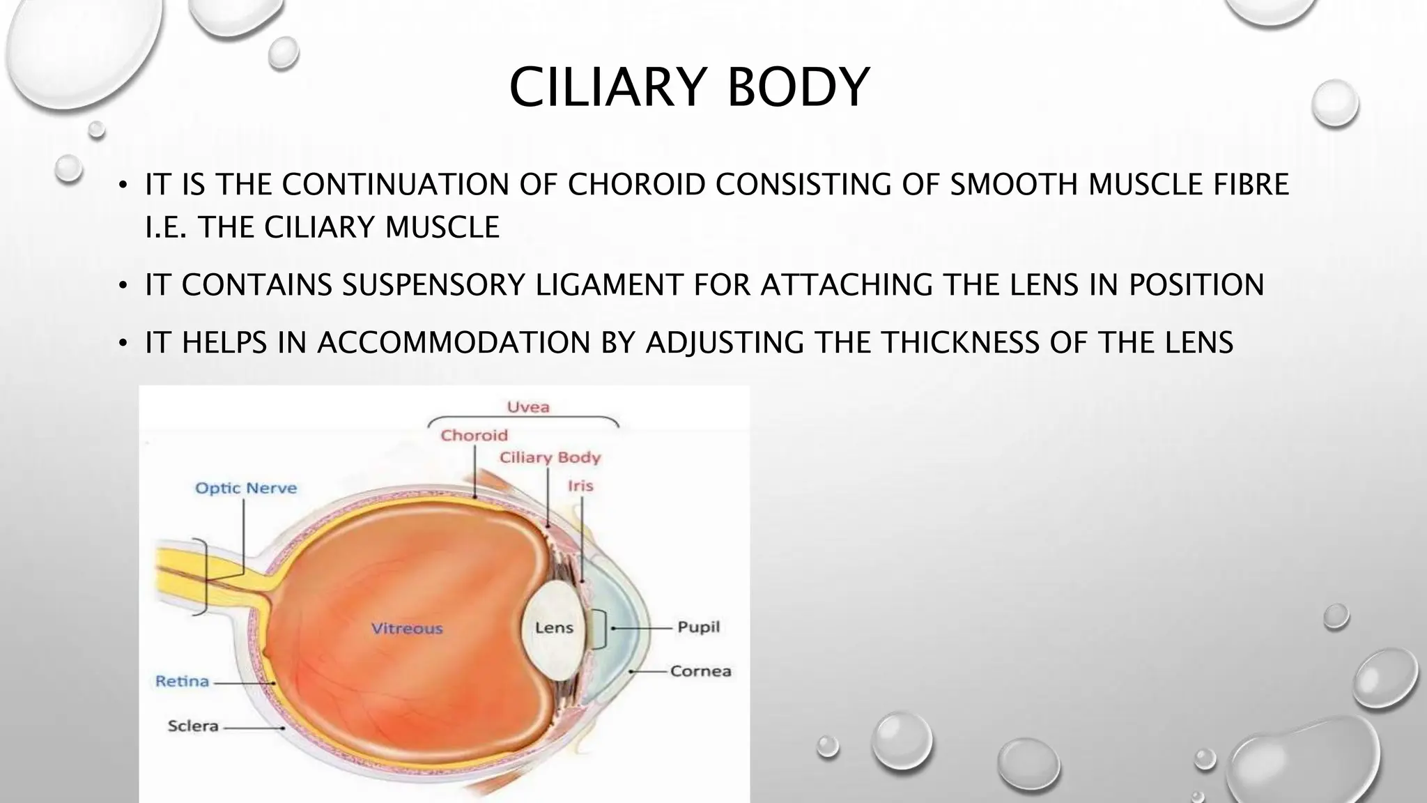
SlideShare a Scribd company logo
1 of 31
ANATOMY OF EYE
 THE EYE IS SITUATED IN THE ORBITAL CAVITY OF
THE SKULL
 IT IS WELL PROTECTED BY THE BONY WALLS OF THE
ORBIT
 ORBIT ALSO CONTAINS MUSCLES OF EYEBALL, THEIR
NERVES, BLOOD VESSELS, AND LACRIMAL GLAND
STRUCTURE OF THE EYEBALL
• WALL OF THE EYEBALL
• REFRACTIVE MEDIA
WALL OF THE EYEBALL
 OUTER LAYER(FIBROUS COAT) : SCLERA AND
CORNEA
 MIDDLE LAYER(VASCULAR COAT) : CHOROID,
CILIARY BODY AND IRIS
 INNER LAYER(NERVOUS COAT) : RETINA
SCLERA AND CORNEA
• SCLERA FORMS THE OUTERMOST LAYER OF THE EYEBALL
• THE ANTERIOR 1/6TH PART OF THE SCLERA IS TRANSPARENT AND
IS KNOWN AS CORNEA
• LIGHT RAYS PASS THROUGH THE CORNEA TO REACH THE RETINA
FUNCTIONS OF SCLERA
• IT PROVIDES PROTECTION TO THE DELICATE
STRUCTURES WITHIN THE EYE
• IT RESIST INTRAOCULAR PRESSURE
• IT MAINTAINS THE SHAPE OF THE EYEBALL
• THE SMOOTH EXTERNAL SURFACE ALLOW EASY EYE
MOVEMENT
CHOROID
• CHOROID IS A THIN PIGMENTED MEMBRANE, DARK
BROWN IN COLOR WHICH IS SITUATED IN BETWEEN
SCLERA AND RETINA
CILIARY BODY
• IT IS THE CONTINUATION OF CHOROID CONSISTING OF SMOOTH MUSCLE FIBRE
I.E. THE CILIARY MUSCLE
• IT CONTAINS SUSPENSORY LIGAMENT FOR ATTACHING THE LENS IN POSITION
• IT HELPS IN ACCOMMODATION BY ADJUSTING THE THICKNESS OF THE LENS
IRIS
• IRIS IS THE PIGMENTED MEMBRANE SURROUND THE PUPIL
• IT ARISES FROM THE MARGIN OF CILIARY BODY AND FORMS A DARK
CENTERED OPENING CALLED PUPIL
• THE SPACE BETWEEN CORNEA AND THE LENS IS THE ANTERIOR SEGMENT
• IT IS FURTHER DIVIDED INTO TWO PARTS BY THE IRIS
• THE SPACE BETWEEN THE IRIS AND THE CORNEA IS THE ANTERIOR
CHAMBER
• THE SPACE BETWEEN THE IRIS AND THE LENS IS THE POSTERIOR
CHAMBER
• THEY ARE FILLED WITH AQUEOUS HUMOR
RETINA
• IT IS A THIN DELICATE LAYER CONTINUOUS POSTERIORLY WITH OPTIC NERVE
• THE OUTER SURFACE OF THE RETINA, FORMED BY THE PIGMENT CELLS, IS ATTACHED
TO THE CHOROID
• ITS INNER SURFACE IS IN CONTACT WITH HYOID MEMBRANE OF THE VITREOUS
• THE SMALL AREA OF THE RETINA WHERE THE OPTIC NERVE LEAVES THE EYE IS THE
OPTIC DISC OR THE BLIND SPOT
• RETINA HAS THREE PARTS
1) OPTIC
2) CILIARY
3) IRIDIAL
• THE OPTIC PART CONTAINS NERVOUS TISSUE AND IT IS SENSITIVE TO LIGHT
• IT EXTENDS FROM THE OPTIC DISC TO THE CILIARY BODY
• THE THIN NON NERVOUS AND NON SENSITIVE LAYER COVERS THE CILIARY BODY AND
IRIS IS CALLED CILIARY AND IRIDIAL PART OF RETINA
• LATERAL TO THE OPTIC DISC A DEPRESSION CALLED
MACULA LUTEA
• THE CENTER OF MACULA IS AGAIN DEPRESSED TO FORM
FOVEA CENTRALIS
• IT CONTAINS CONES ONLY AND IS THE SITE OF MAXIMUM
ACUITY OF VISION
• THE RODS AND CONES ARE THE RECEPTORS OF LIGHT
AND SIGHT
• THESE CELLS CONTAIN PHOTOSENSITIVE PIGMENTS
INVOLVED IN THE CONVERSION OF LIGHT RAYS INTO
NERVE IMPULSES
• INTERFERENCE WITH DRAINAGE OF AQUEOUS HUMOR
RESULTS IN AN INCREASE IS INTRAOCULAR PRESSURE
(GLAUCOMA)
(NORMAL IOP : 10-20 MMHG)
• THIS LEADS TO ATROPHY OF RETINA ,LEADING TO BLINDNESS
THANK YOU

More Related Content

Similar to Anatomy of eye POWER POINT PRESENTATION

structureofeyeppt-190302154704 (1).pdf
structureofeyeppt-190302154704 (1).pdfstructureofeyeppt-190302154704 (1).pdf
structureofeyeppt-190302154704 (1).pdf
Shivamsharma15812
 

Similar to Anatomy of eye POWER POINT PRESENTATION (20)

Anatomy of iris
Anatomy of irisAnatomy of iris
Anatomy of iris
 
ANAT PPT.pptx
ANAT PPT.pptxANAT PPT.pptx
ANAT PPT.pptx
 
anatomy of the eyeball
anatomy of the eyeballanatomy of the eyeball
anatomy of the eyeball
 
ANATOMY OF EYEBALL MBBS OPTHALMOLOGY CLASS
ANATOMY OF EYEBALL MBBS OPTHALMOLOGY CLASSANATOMY OF EYEBALL MBBS OPTHALMOLOGY CLASS
ANATOMY OF EYEBALL MBBS OPTHALMOLOGY CLASS
 
eye ball anat.pptx
eye ball anat.pptxeye ball anat.pptx
eye ball anat.pptx
 
The eyeball anatomy
The eyeball anatomyThe eyeball anatomy
The eyeball anatomy
 
eye.pptx
eye.pptxeye.pptx
eye.pptx
 
Anatomy of Eye.pptx
Anatomy of Eye.pptxAnatomy of Eye.pptx
Anatomy of Eye.pptx
 
structure of eye
structure of eyestructure of eye
structure of eye
 
Trigeminal nerve
Trigeminal nerveTrigeminal nerve
Trigeminal nerve
 
Anatomy of eye ppt
Anatomy of eye pptAnatomy of eye ppt
Anatomy of eye ppt
 
structureofeyeppt-190302154704 (1).pdf
structureofeyeppt-190302154704 (1).pdfstructureofeyeppt-190302154704 (1).pdf
structureofeyeppt-190302154704 (1).pdf
 
Anatomy of eye
Anatomy of eyeAnatomy of eye
Anatomy of eye
 
Presentation on eye
Presentation on eyePresentation on eye
Presentation on eye
 
Ocular anatomy DR TEK
Ocular anatomy DR TEKOcular anatomy DR TEK
Ocular anatomy DR TEK
 
ocular anatomy of eye for optometry and optalomolgy
ocular anatomy of eye for optometry and optalomolgyocular anatomy of eye for optometry and optalomolgy
ocular anatomy of eye for optometry and optalomolgy
 
ocular_anatomy.pptx
ocular_anatomy.pptxocular_anatomy.pptx
ocular_anatomy.pptx
 
385bceaa-1128-4537-b6a1-feee42cce7be.pdf
385bceaa-1128-4537-b6a1-feee42cce7be.pdf385bceaa-1128-4537-b6a1-feee42cce7be.pdf
385bceaa-1128-4537-b6a1-feee42cce7be.pdf
 
The vascular coat of the eye
The vascular coat of the eyeThe vascular coat of the eye
The vascular coat of the eye
 
2 eye bony orbic and ball
2 eye bony orbic and ball2 eye bony orbic and ball
2 eye bony orbic and ball
 

More from SandeepKrishnan42

Copy of EYE DONATION, EYE BANKING, VISION 2020, 9596.pptx
Copy of EYE DONATION, EYE BANKING, VISION 2020, 9596.pptxCopy of EYE DONATION, EYE BANKING, VISION 2020, 9596.pptx
Copy of EYE DONATION, EYE BANKING, VISION 2020, 9596.pptx
SandeepKrishnan42
 
ANATOMY OF CORNEA AND MAINTENANCE OF CORNEAL TRANSPARENCY-1.pptx
ANATOMY OF CORNEA AND MAINTENANCE OF CORNEAL TRANSPARENCY-1.pptxANATOMY OF CORNEA AND MAINTENANCE OF CORNEAL TRANSPARENCY-1.pptx
ANATOMY OF CORNEA AND MAINTENANCE OF CORNEAL TRANSPARENCY-1.pptx
SandeepKrishnan42
 
SCIENTIFIC COMMITTEE Presentation for thesis
SCIENTIFIC COMMITTEE Presentation for thesisSCIENTIFIC COMMITTEE Presentation for thesis
SCIENTIFIC COMMITTEE Presentation for thesis
SandeepKrishnan42
 

More from SandeepKrishnan42 (15)

blindness_programs ppt presentation ophthal
blindness_programs ppt presentation ophthalblindness_programs ppt presentation ophthal
blindness_programs ppt presentation ophthal
 
OCT ANGIOGRAPHY.PPT PRESENTATION OPHTHALMOLOGY
OCT ANGIOGRAPHY.PPT PRESENTATION OPHTHALMOLOGYOCT ANGIOGRAPHY.PPT PRESENTATION OPHTHALMOLOGY
OCT ANGIOGRAPHY.PPT PRESENTATION OPHTHALMOLOGY
 
Extraocular muscles. presentation ophthal
Extraocular muscles. presentation ophthalExtraocular muscles. presentation ophthal
Extraocular muscles. presentation ophthal
 
primaary open angle glaucoma presentation
primaary open angle glaucoma presentationprimaary open angle glaucoma presentation
primaary open angle glaucoma presentation
 
anatomy of sclera ophthalmology presentation
anatomy of sclera ophthalmology presentationanatomy of sclera ophthalmology presentation
anatomy of sclera ophthalmology presentation
 
Copy of EYE DONATION, EYE BANKING, VISION 2020, 9596.pptx
Copy of EYE DONATION, EYE BANKING, VISION 2020, 9596.pptxCopy of EYE DONATION, EYE BANKING, VISION 2020, 9596.pptx
Copy of EYE DONATION, EYE BANKING, VISION 2020, 9596.pptx
 
ANATOMY OF CORNEA AND MAINTENANCE OF CORNEAL TRANSPARENCY-1.pptx
ANATOMY OF CORNEA AND MAINTENANCE OF CORNEAL TRANSPARENCY-1.pptxANATOMY OF CORNEA AND MAINTENANCE OF CORNEAL TRANSPARENCY-1.pptx
ANATOMY OF CORNEA AND MAINTENANCE OF CORNEAL TRANSPARENCY-1.pptx
 
ANATOMY OF VITREOUS CHAMBER-ophthalmology
ANATOMY OF VITREOUS CHAMBER-ophthalmologyANATOMY OF VITREOUS CHAMBER-ophthalmology
ANATOMY OF VITREOUS CHAMBER-ophthalmology
 
tearfilmdynamics-presentation opthalmology
tearfilmdynamics-presentation opthalmologytearfilmdynamics-presentation opthalmology
tearfilmdynamics-presentation opthalmology
 
SCIENTIFIC COMMITTEE Presentation for thesis
SCIENTIFIC COMMITTEE Presentation for thesisSCIENTIFIC COMMITTEE Presentation for thesis
SCIENTIFIC COMMITTEE Presentation for thesis
 
Medical Retina Fellowship Presentation.pptx
Medical Retina Fellowship Presentation.pptxMedical Retina Fellowship Presentation.pptx
Medical Retina Fellowship Presentation.pptx
 
angleofanteriorchamber ophthalmology ctopic
angleofanteriorchamber ophthalmology ctopicangleofanteriorchamber ophthalmology ctopic
angleofanteriorchamber ophthalmology ctopic
 
RETINA-retinal detatchment powerpoint presentation
RETINA-retinal detatchment powerpoint presentationRETINA-retinal detatchment powerpoint presentation
RETINA-retinal detatchment powerpoint presentation
 
anatomy of eyelid ophthalmology presentation
anatomy of eyelid ophthalmology presentationanatomy of eyelid ophthalmology presentation
anatomy of eyelid ophthalmology presentation
 
vocal cord paralysis presentation larynx
vocal cord paralysis presentation larynxvocal cord paralysis presentation larynx
vocal cord paralysis presentation larynx
 

Recently uploaded

Failure to thrive in neonates and infants + pediatric case.pptx
Failure to thrive in neonates and infants  + pediatric case.pptxFailure to thrive in neonates and infants  + pediatric case.pptx
Failure to thrive in neonates and infants + pediatric case.pptx
claviclebrown44
 
CAS 110-63-4 BDO Liquid 1,4-Butanediol 1 4 BDO Warehouse Supply For Excellent...
CAS 110-63-4 BDO Liquid 1,4-Butanediol 1 4 BDO Warehouse Supply For Excellent...CAS 110-63-4 BDO Liquid 1,4-Butanediol 1 4 BDO Warehouse Supply For Excellent...
CAS 110-63-4 BDO Liquid 1,4-Butanediol 1 4 BDO Warehouse Supply For Excellent...
ocean4396
 
CAD CAM DENTURES IN PROSTHODONTICS : Dental advancements
CAD CAM DENTURES IN PROSTHODONTICS : Dental advancementsCAD CAM DENTURES IN PROSTHODONTICS : Dental advancements
CAD CAM DENTURES IN PROSTHODONTICS : Dental advancements
Naveen Gokul Dr
 
Histology of Epithelium - Dr Muhammad Ali Rabbani - Medicose Academics
Histology of Epithelium - Dr Muhammad Ali Rabbani - Medicose AcademicsHistology of Epithelium - Dr Muhammad Ali Rabbani - Medicose Academics
Histology of Epithelium - Dr Muhammad Ali Rabbani - Medicose Academics
MedicoseAcademics
 

Recently uploaded (20)

Failure to thrive in neonates and infants + pediatric case.pptx
Failure to thrive in neonates and infants  + pediatric case.pptxFailure to thrive in neonates and infants  + pediatric case.pptx
Failure to thrive in neonates and infants + pediatric case.pptx
 
How to buy 5cladba precursor raw 5cl-adb-a raw material
How to buy 5cladba precursor raw 5cl-adb-a raw materialHow to buy 5cladba precursor raw 5cl-adb-a raw material
How to buy 5cladba precursor raw 5cl-adb-a raw material
 
Post-Cycle Therapy (PCT) in bodybuilding docx.pdf
Post-Cycle Therapy (PCT) in bodybuilding  docx.pdfPost-Cycle Therapy (PCT) in bodybuilding  docx.pdf
Post-Cycle Therapy (PCT) in bodybuilding docx.pdf
 
CONGENITAL HYPERTROPHIC PYLORIC STENOSIS by Dr M.KARTHIK EMMANUEL
CONGENITAL HYPERTROPHIC PYLORIC STENOSIS  by Dr M.KARTHIK EMMANUELCONGENITAL HYPERTROPHIC PYLORIC STENOSIS  by Dr M.KARTHIK EMMANUEL
CONGENITAL HYPERTROPHIC PYLORIC STENOSIS by Dr M.KARTHIK EMMANUEL
 
CAS 110-63-4 BDO Liquid 1,4-Butanediol 1 4 BDO Warehouse Supply For Excellent...
CAS 110-63-4 BDO Liquid 1,4-Butanediol 1 4 BDO Warehouse Supply For Excellent...CAS 110-63-4 BDO Liquid 1,4-Butanediol 1 4 BDO Warehouse Supply For Excellent...
CAS 110-63-4 BDO Liquid 1,4-Butanediol 1 4 BDO Warehouse Supply For Excellent...
 
SEMESTER-V CHILD HEALTH NURSING-UNIT-1-INTRODUCTION.pdf
SEMESTER-V CHILD HEALTH NURSING-UNIT-1-INTRODUCTION.pdfSEMESTER-V CHILD HEALTH NURSING-UNIT-1-INTRODUCTION.pdf
SEMESTER-V CHILD HEALTH NURSING-UNIT-1-INTRODUCTION.pdf
 
Tips to Choose the Best Psychiatrists in Indore
Tips to Choose the Best Psychiatrists in IndoreTips to Choose the Best Psychiatrists in Indore
Tips to Choose the Best Psychiatrists in Indore
 
VIP Pune 7877925207 WhatsApp: Me All Time Serviℂe Available Day and Night
VIP Pune 7877925207 WhatsApp: Me All Time Serviℂe Available Day and NightVIP Pune 7877925207 WhatsApp: Me All Time Serviℂe Available Day and Night
VIP Pune 7877925207 WhatsApp: Me All Time Serviℂe Available Day and Night
 
TEST BANK For Huether and McCance's Understanding Pathophysiology, Canadian 2...
TEST BANK For Huether and McCance's Understanding Pathophysiology, Canadian 2...TEST BANK For Huether and McCance's Understanding Pathophysiology, Canadian 2...
TEST BANK For Huether and McCance's Understanding Pathophysiology, Canadian 2...
 
Sell pmk powder cas 28578-16-7 from pmk supplier Telegram +85297504341
Sell pmk powder cas 28578-16-7 from pmk supplier Telegram +85297504341Sell pmk powder cas 28578-16-7 from pmk supplier Telegram +85297504341
Sell pmk powder cas 28578-16-7 from pmk supplier Telegram +85297504341
 
Gallbladder Double-Diverticular: A Case Report المرارة مزدوجة التج: تقرير حالة
Gallbladder Double-Diverticular: A Case Report  المرارة مزدوجة التج: تقرير حالةGallbladder Double-Diverticular: A Case Report  المرارة مزدوجة التج: تقرير حالة
Gallbladder Double-Diverticular: A Case Report المرارة مزدوجة التج: تقرير حالة
 
Get the best psychology treatment in Indore at Gokuldas Hospital
Get the best psychology treatment in Indore at Gokuldas HospitalGet the best psychology treatment in Indore at Gokuldas Hospital
Get the best psychology treatment in Indore at Gokuldas Hospital
 
CAD CAM DENTURES IN PROSTHODONTICS : Dental advancements
CAD CAM DENTURES IN PROSTHODONTICS : Dental advancementsCAD CAM DENTURES IN PROSTHODONTICS : Dental advancements
CAD CAM DENTURES IN PROSTHODONTICS : Dental advancements
 
Unveiling Alcohol Withdrawal Syndrome: exploring it's hidden depths
Unveiling Alcohol Withdrawal Syndrome: exploring it's hidden depthsUnveiling Alcohol Withdrawal Syndrome: exploring it's hidden depths
Unveiling Alcohol Withdrawal Syndrome: exploring it's hidden depths
 
Treatment Choices for Slip Disc at Gokuldas Hospital
Treatment Choices for Slip Disc at Gokuldas HospitalTreatment Choices for Slip Disc at Gokuldas Hospital
Treatment Choices for Slip Disc at Gokuldas Hospital
 
Tips and tricks to pass the cardiovascular station for PACES exam
Tips and tricks to pass the cardiovascular station for PACES examTips and tricks to pass the cardiovascular station for PACES exam
Tips and tricks to pass the cardiovascular station for PACES exam
 
Is Rheumatoid Arthritis a Metabolic Disorder.pptx
Is Rheumatoid Arthritis a Metabolic Disorder.pptxIs Rheumatoid Arthritis a Metabolic Disorder.pptx
Is Rheumatoid Arthritis a Metabolic Disorder.pptx
 
Quality control tests of suppository ...
Quality control tests  of suppository ...Quality control tests  of suppository ...
Quality control tests of suppository ...
 
Video capsule endoscopy (VCE ) in children
Video capsule endoscopy (VCE ) in childrenVideo capsule endoscopy (VCE ) in children
Video capsule endoscopy (VCE ) in children
 
Histology of Epithelium - Dr Muhammad Ali Rabbani - Medicose Academics
Histology of Epithelium - Dr Muhammad Ali Rabbani - Medicose AcademicsHistology of Epithelium - Dr Muhammad Ali Rabbani - Medicose Academics
Histology of Epithelium - Dr Muhammad Ali Rabbani - Medicose Academics
 

Anatomy of eye POWER POINT PRESENTATION

  • 2.  THE EYE IS SITUATED IN THE ORBITAL CAVITY OF THE SKULL  IT IS WELL PROTECTED BY THE BONY WALLS OF THE ORBIT  ORBIT ALSO CONTAINS MUSCLES OF EYEBALL, THEIR NERVES, BLOOD VESSELS, AND LACRIMAL GLAND
  • 3. STRUCTURE OF THE EYEBALL • WALL OF THE EYEBALL • REFRACTIVE MEDIA
  • 4. WALL OF THE EYEBALL  OUTER LAYER(FIBROUS COAT) : SCLERA AND CORNEA  MIDDLE LAYER(VASCULAR COAT) : CHOROID, CILIARY BODY AND IRIS  INNER LAYER(NERVOUS COAT) : RETINA
  • 5.
  • 6.
  • 7.
  • 8.
  • 9. SCLERA AND CORNEA • SCLERA FORMS THE OUTERMOST LAYER OF THE EYEBALL • THE ANTERIOR 1/6TH PART OF THE SCLERA IS TRANSPARENT AND IS KNOWN AS CORNEA • LIGHT RAYS PASS THROUGH THE CORNEA TO REACH THE RETINA
  • 10. FUNCTIONS OF SCLERA • IT PROVIDES PROTECTION TO THE DELICATE STRUCTURES WITHIN THE EYE • IT RESIST INTRAOCULAR PRESSURE • IT MAINTAINS THE SHAPE OF THE EYEBALL • THE SMOOTH EXTERNAL SURFACE ALLOW EASY EYE MOVEMENT
  • 11. CHOROID • CHOROID IS A THIN PIGMENTED MEMBRANE, DARK BROWN IN COLOR WHICH IS SITUATED IN BETWEEN SCLERA AND RETINA
  • 12. CILIARY BODY • IT IS THE CONTINUATION OF CHOROID CONSISTING OF SMOOTH MUSCLE FIBRE I.E. THE CILIARY MUSCLE • IT CONTAINS SUSPENSORY LIGAMENT FOR ATTACHING THE LENS IN POSITION • IT HELPS IN ACCOMMODATION BY ADJUSTING THE THICKNESS OF THE LENS
  • 13. IRIS • IRIS IS THE PIGMENTED MEMBRANE SURROUND THE PUPIL • IT ARISES FROM THE MARGIN OF CILIARY BODY AND FORMS A DARK CENTERED OPENING CALLED PUPIL • THE SPACE BETWEEN CORNEA AND THE LENS IS THE ANTERIOR SEGMENT • IT IS FURTHER DIVIDED INTO TWO PARTS BY THE IRIS • THE SPACE BETWEEN THE IRIS AND THE CORNEA IS THE ANTERIOR CHAMBER • THE SPACE BETWEEN THE IRIS AND THE LENS IS THE POSTERIOR CHAMBER • THEY ARE FILLED WITH AQUEOUS HUMOR
  • 14. RETINA • IT IS A THIN DELICATE LAYER CONTINUOUS POSTERIORLY WITH OPTIC NERVE • THE OUTER SURFACE OF THE RETINA, FORMED BY THE PIGMENT CELLS, IS ATTACHED TO THE CHOROID • ITS INNER SURFACE IS IN CONTACT WITH HYOID MEMBRANE OF THE VITREOUS • THE SMALL AREA OF THE RETINA WHERE THE OPTIC NERVE LEAVES THE EYE IS THE OPTIC DISC OR THE BLIND SPOT • RETINA HAS THREE PARTS 1) OPTIC 2) CILIARY 3) IRIDIAL • THE OPTIC PART CONTAINS NERVOUS TISSUE AND IT IS SENSITIVE TO LIGHT • IT EXTENDS FROM THE OPTIC DISC TO THE CILIARY BODY • THE THIN NON NERVOUS AND NON SENSITIVE LAYER COVERS THE CILIARY BODY AND IRIS IS CALLED CILIARY AND IRIDIAL PART OF RETINA
  • 15. • LATERAL TO THE OPTIC DISC A DEPRESSION CALLED MACULA LUTEA • THE CENTER OF MACULA IS AGAIN DEPRESSED TO FORM FOVEA CENTRALIS • IT CONTAINS CONES ONLY AND IS THE SITE OF MAXIMUM ACUITY OF VISION • THE RODS AND CONES ARE THE RECEPTORS OF LIGHT AND SIGHT • THESE CELLS CONTAIN PHOTOSENSITIVE PIGMENTS INVOLVED IN THE CONVERSION OF LIGHT RAYS INTO NERVE IMPULSES
  • 16.
  • 17.
  • 18.
  • 19. • INTERFERENCE WITH DRAINAGE OF AQUEOUS HUMOR RESULTS IN AN INCREASE IS INTRAOCULAR PRESSURE (GLAUCOMA) (NORMAL IOP : 10-20 MMHG) • THIS LEADS TO ATROPHY OF RETINA ,LEADING TO BLINDNESS
  • 20.
  • 21.
  • 22.
  • 23.
  • 24.
  • 25.
  • 26.
  • 27.
  • 28.
  • 29.
  • 30.