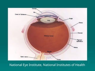
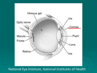
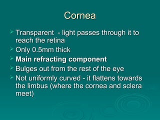
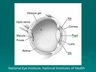
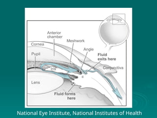
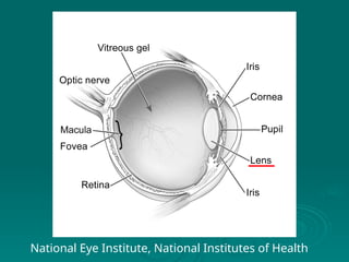
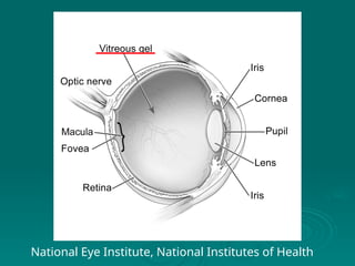
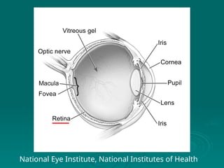
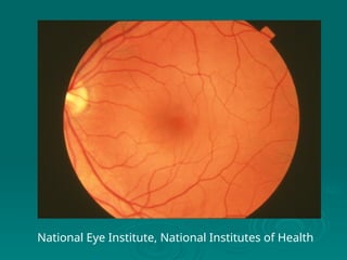
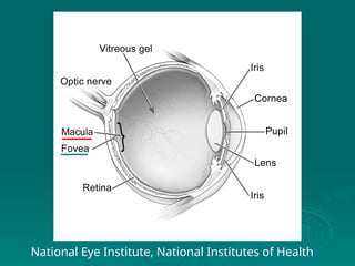
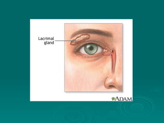
Basic Anatomy of the
Eye
Eye
National Eye Institute, National Institutes of Health
Basic Structure of the Eye
 The Eye is one part of the Visual System
The Eye is one part of the Visual System

The eye and focuses the image and detects
the light
the light

The visual pathways transfer the nerve
impulses
impulses

The visual cortex in the brain processes the
signals, and we ‘see’ the image
National Eye Institute, National Institutes of Health
Layers of the wall of the eye
 The eye has three basic coats/layers:
The eye has three basic coats/layers:

Sclera
Sclera - the white, outer-most layer
- the white, outer-most layer
• Tough layer which gives the eye its shape, and protects it
Tough layer which gives the eye its shape, and protects it

Choroid
Choroid - middle layer, contains many blood
- middle layer, contains many blood
vessels
vessels
• Nourishes the outer portions of the retina
Nourishes the outer portions of the retina

Retina
Retina - inner-most layer, contains all the
- inner-most layer, contains all the
light-sensitive cells and nerves
National Eye Institute, National Institutes of Health
Cornea
Cornea
 Transparent - light passes through it to
Transparent - light passes through it to
reach the retina
reach the retina
 Only 0.5mm thick
Only 0.5mm thick
 Main refracting component
Main refracting component
 Bulges out from the rest of the eye
Bulges out from the rest of the eye
 Not uniformly curved - it flattens towards
Not uniformly curved - it flattens towards
the limbus (where the cornea and sclera
meet)
meet)
Conjunctiva
Conjunctiva
 Two parts:
Two parts:

Thin, transparent mucous membrane which
covers the sclera over the front of the eye
from the cornea (
from the cornea (bulbar
bulbar conjunctiva)
conjunctiva)

Continuous with the skin at the lid margin
(
(palpebral
palpebral conjunctiva)
conjunctiva)

The two meet in the
The two meet in the superior
superior/
/inferior
inferior fornices
fornices
National Eye Institute, National Institutes of Health
Anterior Chamber
Anterior Chamber
 Area which lies behind the cornea, but in
Area which lies behind the cornea, but in
front of the iris and lens
 Contains a watery substance called the
Contains a watery substance called the
aqueous humour
aqueous humour

The aqueous humour is produced in the
ciliary body and circulates around the anterio
chamber, to drain through Schlemms’ Canal
Iris
Iris
 This is a circular disc, with a hole in its
This is a circular disc, with a hole in its
center - the pupil
center - the pupil

The pupil size can be made bigger or smaller
by the iris muscles (4.5-8 mm)

The pupil controls the amount of light entering
the eye
the eye
 The iris gives the eye its colour
The iris gives the eye its colour
 determined by the amount of pigment
determined by the amount of pigment
National Eye Institute, National Institutes of Health
Ciliary Body
Ciliary Body
 Positioned between the iris and the choroid
Positioned between the iris and the choroid
 It is the point of attachment for the
It is the point of attachment for the zonular
zonular
fibers
fibers, which hold the lens in place
, which hold the lens in place
 It contains many muscles - when they relax, the
It contains many muscles - when they relax, the
zonular fibers relax. This allows the lens to
change its shape, and it increase in dioptric
power - this process is called accommodation
National Eye Institute, National Institutes of Health
Posterior Chamber
Posterior Chamber
 Small area behind the iris, but in front of
Small area behind the iris, but in front of
the lens - contains aqueous humour
 Do not confuse with vitreous chamber
Do not confuse with vitreous chamber
behind the lens
behind the lens
Lens
Lens
 Lens gives approx 1/3 of the eye’s dioptric
Lens gives approx 1/3 of the eye’s dioptric
power
power
 Consists of an inner core (nucleus), surrounded
Consists of an inner core (nucleus), surrounded
by a cortex
by a cortex
 Lens is covered by an elastic capsule, to which
Lens is covered by an elastic capsule, to which
the zonular fibres are attached
 Back surface is more curved than the front
Back surface is more curved than the front
 Continues to grow throughout life, and becomes
Continues to grow throughout life, and becomes
less flexible. This reduces the accommodation
and causes presbyopia. Eventually causes a
cataract.
cataract.
National Eye Institute, National Institutes of Health
Vitreous Chamber
Vitreous Chamber
 Largest chamber
Largest chamber in the eye
in the eye
 Contains the vitreous humour (sometimes just called
Contains the vitreous humour (sometimes just called
‘vitreous’
‘vitreous’

Not watery like the aqueous, nor does it circulate

It is a clear gel-like substance, which helps to support
the other parts of the eye

Gets more watery and slightly yellow with increasing
age
age

Sometimes it shrinks in size as a person gets older –
posterior vitreous detachment

It may pull on the retina and can sometimes cause a
retinal detachment
retinal detachment
National Eye Institute, National Institutes of Health
Choroid
Choroid
 Middle coat (layer) of the eye
Middle coat (layer) of the eye
 Largely composed of blood vessels
Largely composed of blood vessels
 Function is to nourish the outer portion of
Function is to nourish the outer portion of
the underlying retina
 Very pigmented - this allows it to absorb
Very pigmented - this allows it to absorb
any stray light
any stray light
Retina
Retina
 Light sensitive layer of the eye
Light sensitive layer of the eye

The retina and visual pathways can be thought of as
an extension of the brain
 Consists of 10 layers
Consists of 10 layers, but is very thin:
, but is very thin:

0.1-0.23mm thick
0.1-0.23mm thick
 Contains two types of photoreceptors (the light
Contains two types of photoreceptors (the light
sensitive cells) called rods and cones
 Covers the innermost part of the eye - about 2/3
Covers the innermost part of the eye - about 2/3
of the surface
of the surface
National Eye Institute, National Institutes of Health
Optic Disc
Optic Disc
 The rods and cones are connected, in a complex
The rods and cones are connected, in a complex
way, with lots of other cell types in the retina
 The rods and cones connect with the ganglion cells,
The rods and cones connect with the ganglion cells,
which join together to form the optic nerve
 The optic nerve leaves the back of the eye through a
The optic nerve leaves the back of the eye through a
hole (1.5x2mm) called the optic disc
 Optic disc can be seen by ophthalmoscopy
Optic disc can be seen by ophthalmoscopy
 There are no photoreceptors in the optic disc, so this
There are no photoreceptors in the optic disc, so this
produces a blind spot in our vision
 Demonstration exercise of blind spot
Demonstration exercise of blind spot
National Eye Institute, National Institutes of Health
Macula/Fovea
Macula/Fovea
 The macula is the part of the retina that has an
The macula is the part of the retina that has an
increased number of cone cells

This area is about 5.5mm in diameter
 The fovea (foveola) is the central part of the
The fovea (foveola) is the central part of the
macula (about 0.33mm in diameter)
 contains only cones
contains only cones
 It forms a depression in the macula, and is
It forms a depression in the macula, and is
responsible for the highest acuity
 Retina is thinnest at the fovea
Retina is thinnest at the fovea
National Eye Institute, National Institutes of Health
Sclera
Sclera
 Tough, fibrous outer protective layer of the eye,
Tough, fibrous outer protective layer of the eye,
about 1mm thick
about 1mm thick
 Dense, white and continuous with the cornea
Dense, white and continuous with the cornea
 Largely responsible for giving the eye its shape
Largely responsible for giving the eye its shape
 The extra-ocular muscles are attached to the
The extra-ocular muscles are attached to the
sclera
sclera
 Sclera is thinnest where the optic nerve enters
Sclera is thinnest where the optic nerve enters
the eye - it has a sieve like appearance called
the lamina cribrosa
the lamina cribrosa
National Eye Institute, National Institutes of Health
Lids and Tears
Lids and Tears
 The lids are moveable folds of tissue that protect
The lids are moveable folds of tissue that protect
the eye
the eye
 They contain four different glands which
They contain four different glands which
contribute to the tear film

Meibomian glands - the glands of
Meibomian glands - the glands of Zeiss
Zeiss and
and Moll
Moll -
-
produce oily layer on the surface of the tear film

Accessory lacrimal glands - the glands of Krause and
Wolfring - supply most of the needed moisture to the
cornea and conjunctiva
Lacrimal Gland
Lacrimal Gland
 This is the tear secreting gland located above
This is the tear secreting gland located above
the eye
the eye
 Tears pass down over the cornea and
Tears pass down over the cornea and
conjunctiva, moistening the surface
 Tears drain into the puncta (small holes on the
Tears drain into the puncta (small holes on the
upper and lower lid margin)
 They drain through small canals (the canaliculi)
They drain through small canals (the canaliculi)
into the nasolacrimal sac, and then into the nose
1  Topic 2 Eye Structure & Function.ppth

1 Topic 2 Eye Structure & Function.ppth

  • 1.
    Basic Anatomy ofthe Basic Anatomy of the Eye Eye National Eye Institute, National Institutes of Health
  • 2.
    Basic Structure ofthe Eye Basic Structure of the Eye  The Eye is one part of the Visual System The Eye is one part of the Visual System  The eye and focuses the image and detects The eye and focuses the image and detects the light the light  The visual pathways transfer the nerve The visual pathways transfer the nerve impulses impulses  The visual cortex in the brain processes the The visual cortex in the brain processes the signals, and we ‘see’ the image signals, and we ‘see’ the image
  • 3.
    National Eye Institute,National Institutes of Health
  • 4.
    Layers of thewall of the eye Layers of the wall of the eye  The eye has three basic coats/layers: The eye has three basic coats/layers:  Sclera Sclera - the white, outer-most layer - the white, outer-most layer • Tough layer which gives the eye its shape, and protects it Tough layer which gives the eye its shape, and protects it  Choroid Choroid - middle layer, contains many blood - middle layer, contains many blood vessels vessels • Nourishes the outer portions of the retina Nourishes the outer portions of the retina  Retina Retina - inner-most layer, contains all the - inner-most layer, contains all the light-sensitive cells and nerves light-sensitive cells and nerves
  • 5.
    National Eye Institute,National Institutes of Health
  • 6.
    Cornea Cornea  Transparent -light passes through it to Transparent - light passes through it to reach the retina reach the retina  Only 0.5mm thick Only 0.5mm thick  Main refracting component Main refracting component  Bulges out from the rest of the eye Bulges out from the rest of the eye  Not uniformly curved - it flattens towards Not uniformly curved - it flattens towards the limbus (where the cornea and sclera the limbus (where the cornea and sclera meet) meet)
  • 7.
    Conjunctiva Conjunctiva  Two parts: Twoparts:  Thin, transparent mucous membrane which Thin, transparent mucous membrane which covers the sclera over the front of the eye covers the sclera over the front of the eye from the cornea ( from the cornea (bulbar bulbar conjunctiva) conjunctiva)  Continuous with the skin at the lid margin Continuous with the skin at the lid margin ( (palpebral palpebral conjunctiva) conjunctiva)  The two meet in the The two meet in the superior superior/ /inferior inferior fornices fornices
  • 8.
    National Eye Institute,National Institutes of Health
  • 9.
    Anterior Chamber Anterior Chamber Area which lies behind the cornea, but in Area which lies behind the cornea, but in front of the iris and lens front of the iris and lens  Contains a watery substance called the Contains a watery substance called the aqueous humour aqueous humour  The aqueous humour is produced in the The aqueous humour is produced in the ciliary body and circulates around the anterio ciliary body and circulates around the anterio chamber, to drain through Schlemms’ Canal chamber, to drain through Schlemms’ Canal
  • 10.
    Iris Iris  This isa circular disc, with a hole in its This is a circular disc, with a hole in its center - the pupil center - the pupil  The pupil size can be made bigger or smaller The pupil size can be made bigger or smaller by the iris muscles (4.5-8 mm) by the iris muscles (4.5-8 mm)  The pupil controls the amount of light entering The pupil controls the amount of light entering the eye the eye  The iris gives the eye its colour The iris gives the eye its colour  determined by the amount of pigment determined by the amount of pigment
  • 11.
    National Eye Institute,National Institutes of Health
  • 13.
    National Eye Institute,National Institutes of Health
  • 14.
    Ciliary Body Ciliary Body Positioned between the iris and the choroid Positioned between the iris and the choroid  It is the point of attachment for the It is the point of attachment for the zonular zonular fibers fibers, which hold the lens in place , which hold the lens in place  It contains many muscles - when they relax, the It contains many muscles - when they relax, the zonular fibers relax. This allows the lens to zonular fibers relax. This allows the lens to change its shape, and it increase in dioptric change its shape, and it increase in dioptric power - this process is called accommodation power - this process is called accommodation
  • 15.
    National Eye Institute,National Institutes of Health
  • 16.
    Posterior Chamber Posterior Chamber Small area behind the iris, but in front of Small area behind the iris, but in front of the lens - contains aqueous humour the lens - contains aqueous humour  Do not confuse with vitreous chamber Do not confuse with vitreous chamber behind the lens behind the lens
  • 17.
    Lens Lens  Lens givesapprox 1/3 of the eye’s dioptric Lens gives approx 1/3 of the eye’s dioptric power power  Consists of an inner core (nucleus), surrounded Consists of an inner core (nucleus), surrounded by a cortex by a cortex  Lens is covered by an elastic capsule, to which Lens is covered by an elastic capsule, to which the zonular fibres are attached the zonular fibres are attached  Back surface is more curved than the front Back surface is more curved than the front  Continues to grow throughout life, and becomes Continues to grow throughout life, and becomes less flexible. This reduces the accommodation less flexible. This reduces the accommodation and causes presbyopia. Eventually causes a and causes presbyopia. Eventually causes a cataract. cataract.
  • 18.
    National Eye Institute,National Institutes of Health
  • 19.
    Vitreous Chamber Vitreous Chamber Largest chamber Largest chamber in the eye in the eye  Contains the vitreous humour (sometimes just called Contains the vitreous humour (sometimes just called ‘vitreous’ ‘vitreous’  Not watery like the aqueous, nor does it circulate Not watery like the aqueous, nor does it circulate  It is a clear gel-like substance, which helps to support It is a clear gel-like substance, which helps to support the other parts of the eye the other parts of the eye  Gets more watery and slightly yellow with increasing Gets more watery and slightly yellow with increasing age age  Sometimes it shrinks in size as a person gets older – Sometimes it shrinks in size as a person gets older – posterior vitreous detachment posterior vitreous detachment  It may pull on the retina and can sometimes cause a It may pull on the retina and can sometimes cause a retinal detachment retinal detachment
  • 20.
    National Eye Institute,National Institutes of Health
  • 21.
    Choroid Choroid  Middle coat(layer) of the eye Middle coat (layer) of the eye  Largely composed of blood vessels Largely composed of blood vessels  Function is to nourish the outer portion of Function is to nourish the outer portion of the underlying retina the underlying retina  Very pigmented - this allows it to absorb Very pigmented - this allows it to absorb any stray light any stray light
  • 22.
    Retina Retina  Light sensitivelayer of the eye Light sensitive layer of the eye  The retina and visual pathways can be thought of as The retina and visual pathways can be thought of as an extension of the brain an extension of the brain  Consists of 10 layers Consists of 10 layers, but is very thin: , but is very thin:  0.1-0.23mm thick 0.1-0.23mm thick  Contains two types of photoreceptors (the light Contains two types of photoreceptors (the light sensitive cells) called rods and cones sensitive cells) called rods and cones  Covers the innermost part of the eye - about 2/3 Covers the innermost part of the eye - about 2/3 of the surface of the surface
  • 23.
    National Eye Institute,National Institutes of Health
  • 24.
    National Eye Institute,National Institutes of Health
  • 25.
    Optic Disc Optic Disc The rods and cones are connected, in a complex The rods and cones are connected, in a complex way, with lots of other cell types in the retina way, with lots of other cell types in the retina  The rods and cones connect with the ganglion cells, The rods and cones connect with the ganglion cells, which join together to form the optic nerve which join together to form the optic nerve  The optic nerve leaves the back of the eye through a The optic nerve leaves the back of the eye through a hole (1.5x2mm) called the optic disc hole (1.5x2mm) called the optic disc  Optic disc can be seen by ophthalmoscopy Optic disc can be seen by ophthalmoscopy  There are no photoreceptors in the optic disc, so this There are no photoreceptors in the optic disc, so this produces a blind spot in our vision produces a blind spot in our vision  Demonstration exercise of blind spot Demonstration exercise of blind spot
  • 27.
    National Eye Institute,National Institutes of Health
  • 28.
    Macula/Fovea Macula/Fovea  The maculais the part of the retina that has an The macula is the part of the retina that has an increased number of cone cells increased number of cone cells  This area is about 5.5mm in diameter This area is about 5.5mm in diameter  The fovea (foveola) is the central part of the The fovea (foveola) is the central part of the macula (about 0.33mm in diameter) macula (about 0.33mm in diameter)  contains only cones contains only cones  It forms a depression in the macula, and is It forms a depression in the macula, and is responsible for the highest acuity responsible for the highest acuity  Retina is thinnest at the fovea Retina is thinnest at the fovea
  • 29.
    National Eye Institute,National Institutes of Health
  • 30.
    Sclera Sclera  Tough, fibrousouter protective layer of the eye, Tough, fibrous outer protective layer of the eye, about 1mm thick about 1mm thick  Dense, white and continuous with the cornea Dense, white and continuous with the cornea  Largely responsible for giving the eye its shape Largely responsible for giving the eye its shape  The extra-ocular muscles are attached to the The extra-ocular muscles are attached to the sclera sclera  Sclera is thinnest where the optic nerve enters Sclera is thinnest where the optic nerve enters the eye - it has a sieve like appearance called the eye - it has a sieve like appearance called the lamina cribrosa the lamina cribrosa
  • 31.
    National Eye Institute,National Institutes of Health
  • 32.
    Lids and Tears Lidsand Tears  The lids are moveable folds of tissue that protect The lids are moveable folds of tissue that protect the eye the eye  They contain four different glands which They contain four different glands which contribute to the tear film contribute to the tear film  Meibomian glands - the glands of Meibomian glands - the glands of Zeiss Zeiss and and Moll Moll - - produce oily layer on the surface of the tear film produce oily layer on the surface of the tear film  Accessory lacrimal glands - the glands of Krause and Accessory lacrimal glands - the glands of Krause and Wolfring - supply most of the needed moisture to the Wolfring - supply most of the needed moisture to the cornea and conjunctiva cornea and conjunctiva
  • 34.
    Lacrimal Gland Lacrimal Gland This is the tear secreting gland located above This is the tear secreting gland located above the eye the eye  Tears pass down over the cornea and Tears pass down over the cornea and conjunctiva, moistening the surface conjunctiva, moistening the surface  Tears drain into the puncta (small holes on the Tears drain into the puncta (small holes on the upper and lower lid margin) upper and lower lid margin)  They drain through small canals (the canaliculi) They drain through small canals (the canaliculi) into the nasolacrimal sac, and then into the nose into the nasolacrimal sac, and then into the nose