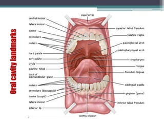
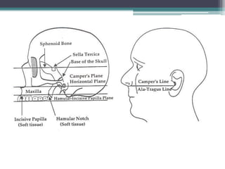
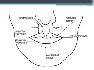
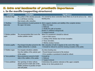
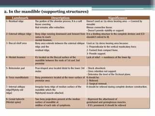
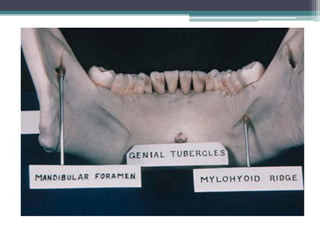
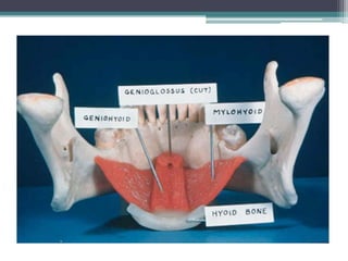
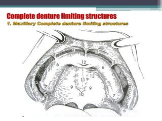
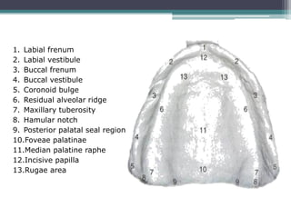
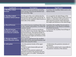
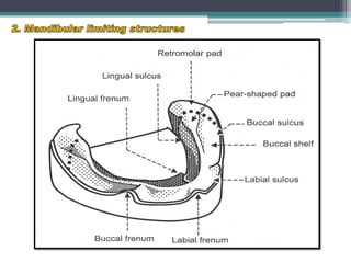
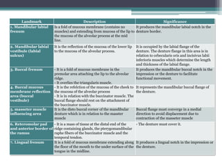
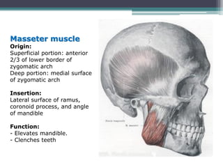
The Indian Dental Academy is the Leader in continuing dental education , training dentists in all aspects of dentistry and
offering a wide range of dental certified courses in different formats.for more details please visit
www.indiandentalacademy.com
The Indian Dental Academy is the Leader in continuing dental education , training dentists in all aspects of dentistry and
offering a wide range of dental certified courses in different formats.for more details please visit
www.indiandentalacademy.com
The anatomy of the edentulous ridge in the maxilla and mandible is very important for the design of a complete denture. Objective in fabrication of a complete denture is to provide a prosthesis that restores lost teeth and associated structures functionally, anatomically and aesthetically as much as possible with preservation of underlying structures and the knowledge landmarks help us in achieving our objective.
Anatomical landmarks for edentulous patients and facial landmarksMohammed Aziz
Anatomical landmarks for edentulous patients and facial landmarks
physiology of muscles
muscles of mastication
muscles of facial expression
physiology of saliva
Prosthodontics
dentistry
MANDIBULAR ANATOMICAL LANDMARKS
PRESENTED BY
ROSHALMARIA THOMAS
IV/II
THE ANATOMY OF EDENTULOUS RIDGES IN THE MAXILLA AND MANDIBLE IS VERY IMPORTANT FOR THE DESIGN OF THE COMPLETE DENTURE
THE TOTAL AREA OF SUPPORT FROM THE MANDIBLE IS SIGNIFICANTLY LESS THAN FROM THE MAXILLA.
THE AVERAGE AVAILABLE DENTURE BEARING AREA FOR AN EDENTULOUS MANDIBLE IS 14cm2 , WHEREAS FOR EDENTULOS MAXILLA IT IS 24cm2. THEREFORE THE MANDIBLE IS LESS CAPABLE OF RESISTING OCCLUSAL FORCES THAN THE MAXILLA.
Labial frenum
Fibrous band
Muscles incisivus and orbicularis oris
Active
Labial vestibule
Space between residual alveolar ridge and lips
Length and thickness of labial flange-influences lip support and retention
Buccal frenum
Overlies depressor anguli oris
Fibers of buccinators attached
Buccal vestibule
Extends- posteriorly from buccal frenum to retromolar pad region
Residual alveolar ridge on one side and buccinators on other
Influenced by action of masseter
Lingual frenum
Should be relieved
High lingual frenum is called tongue tie –affects stability
Alveololingual sulcus
Extends from lingual frenum to retromylohyoid curtain
Divided into 3 parts- anterior, middle and posterior
Anterior region- from lingual frenum to premylohyoid fossa
Flange is shorter anteriorly and should touch the floorof the mouth whentip of tongue touches upper incisors
Middle- extends from premylohyoid fossa to distal end of mylohyoid ridge
Shallower due to prominence of mylohyoid ridge and action of mylohyoid muscle
Posterior- retromylohyoid fossa
Typical S form of lingual sulcus
Retromolar pad
Posterior seal of mandibular denture
Pear shaped
Triangular keratinized soft pad of tissue at distal end of ridge
Bounded posteriorly by tendons of temporalis, laterally by buccinators and medially by pterygomandibular raphe and superior constrictor
Denture should extend one half to two thirds of retromolar pad
Buccal shelf area
Area between buccal frenum and anterior border of masseter
Width increases as resorption continues
Lies at right angles to occlusal forces- primary stress bearing area
Residual alveolar ridge
Edentulous mandible may become flat with concave denture bearing surface
In such cases, structures attaching on lingual side of ridge attach over the ridge
Due to resorption mandible inclines outwards and becomes progressively wider
Mylohyoid ridge
Runs along lingual surface of mandible
Anteriorly lies close to inferior border of mandible, posteriorly lies flush along the ridge
Thin mucosa- easily traumatized- hence should be relieved
Undercut present under the ridge
Mental foramen
Between first and second premolar region
Relieved- as pressure may cause paresthesia
Genial tubercles
Pair of bony tubercles
Present anteriorly on lingual side of body of mandible
Due to resorption may become increasingly prominent- denture usage difficult
A number of theories have been put forward for impressions. each having its own advantage and disadvantage.
Different spacers guide and aid in in making the desired impression with adequate pressure in the desired region of the arch in maxilla and mandible. different materials are used for spacers depending on the need.
Anatomy and clinical significance of denture bearing areasOgundiran Temidayo
A presentation on the anatomy and clinical significance of the denture bearing areas by Ogundiran Temidayo who is a dental student at OBAFEMI AWOLOWO UNIVERSITY ILE-IFE
The Pt. adaptation on his complete dentures are based on the ability of the dentures to restore the missed functions due to the loss of the teeth. Good impression is the first step in the success of the complete dentures. A trial to review all the basics necessary to have a good impression is exposed in this lecture.
Indian Dental Academy: will be one of the most relevant and exciting training center with best faculty and flexible training programs for dental professionals who wish to advance in their dental practice,Offers certified courses in Dental implants,Orthodontics,Endodontics,Cosmetic Dentistry, Prosthetic Dentistry, Periodontics and General Dentistry.
Anatomical landmarks of edentulous mandibular arch ppt (prosthodontics) easil...Shazlana Raheem
(prosthodontics) anatomic landmarks of edentulous mandible arch for dental students in brief.
very easily understandable.
before exam study purpose. there are 17 slides in it totally.
share - Lions, tigers, AI and health misinformation, oh my!.pptxTina Purnat
• Pitfalls and pivots needed to use AI effectively in public health
• Evidence-based strategies to address health misinformation effectively
• Building trust with communities online and offline
• Equipping health professionals to address questions, concerns and health misinformation
• Assessing risk and mitigating harm from adverse health narratives in communities, health workforce and health system
Antimicrobial stewardship to prevent antimicrobial resistanceGovindRankawat1
India is among the nations with the highest burden of bacterial infections.
India is one of the largest consumers of antibiotics worldwide.
India carries one of the largest burdens of drug‑resistant pathogens worldwide.
Highest burden of multidrug‑resistant tuberculosis,
Alarmingly high resistance among Gram‑negative and Gram‑positive bacteria even to newer antimicrobials such as carbapenems.
NDM‑1 ( New Delhi Metallo Beta lactamase 1, an enzyme which inactivates majority of Beta lactam antibiotics including carbapenems) was reported in 2008
DISSERTATION on NEW DRUG DISCOVERY AND DEVELOPMENT STAGES OF DRUG DISCOVERYNEHA GUPTA
The process of drug discovery and development is a complex and multi-step endeavor aimed at bringing new pharmaceutical drugs to market. It begins with identifying and validating a biological target, such as a protein, gene, or RNA, that is associated with a disease. This step involves understanding the target's role in the disease and confirming that modulating it can have therapeutic effects. The next stage, hit identification, employs high-throughput screening (HTS) and other methods to find compounds that interact with the target. Computational techniques may also be used to identify potential hits from large compound libraries.
Following hit identification, the hits are optimized to improve their efficacy, selectivity, and pharmacokinetic properties, resulting in lead compounds. These leads undergo further refinement to enhance their potency, reduce toxicity, and improve drug-like characteristics, creating drug candidates suitable for preclinical testing. In the preclinical development phase, drug candidates are tested in vitro (in cell cultures) and in vivo (in animal models) to evaluate their safety, efficacy, pharmacokinetics, and pharmacodynamics. Toxicology studies are conducted to assess potential risks.
Before clinical trials can begin, an Investigational New Drug (IND) application must be submitted to regulatory authorities. This application includes data from preclinical studies and plans for clinical trials. Clinical development involves human trials in three phases: Phase I tests the drug's safety and dosage in a small group of healthy volunteers, Phase II assesses the drug's efficacy and side effects in a larger group of patients with the target disease, and Phase III confirms the drug's efficacy and monitors adverse reactions in a large population, often compared to existing treatments.
After successful clinical trials, a New Drug Application (NDA) is submitted to regulatory authorities for approval, including all data from preclinical and clinical studies, as well as proposed labeling and manufacturing information. Regulatory authorities then review the NDA to ensure the drug is safe, effective, and of high quality, potentially requiring additional studies. Finally, after a drug is approved and marketed, it undergoes post-marketing surveillance, which includes continuous monitoring for long-term safety and effectiveness, pharmacovigilance, and reporting of any adverse effects.
Title: Sense of Taste
Presenter: Dr. Faiza, Assistant Professor of Physiology
Qualifications:
MBBS (Best Graduate, AIMC Lahore)
FCPS Physiology
ICMT, CHPE, DHPE (STMU)
MPH (GC University, Faisalabad)
MBA (Virtual University of Pakistan)
Learning Objectives:
Describe the structure and function of taste buds.
Describe the relationship between the taste threshold and taste index of common substances.
Explain the chemical basis and signal transduction of taste perception for each type of primary taste sensation.
Recognize different abnormalities of taste perception and their causes.
Key Topics:
Significance of Taste Sensation:
Differentiation between pleasant and harmful food
Influence on behavior
Selection of food based on metabolic needs
Receptors of Taste:
Taste buds on the tongue
Influence of sense of smell, texture of food, and pain stimulation (e.g., by pepper)
Primary and Secondary Taste Sensations:
Primary taste sensations: Sweet, Sour, Salty, Bitter, Umami
Chemical basis and signal transduction mechanisms for each taste
Taste Threshold and Index:
Taste threshold values for Sweet (sucrose), Salty (NaCl), Sour (HCl), and Bitter (Quinine)
Taste index relationship: Inversely proportional to taste threshold
Taste Blindness:
Inability to taste certain substances, particularly thiourea compounds
Example: Phenylthiocarbamide
Structure and Function of Taste Buds:
Composition: Epithelial cells, Sustentacular/Supporting cells, Taste cells, Basal cells
Features: Taste pores, Taste hairs/microvilli, and Taste nerve fibers
Location of Taste Buds:
Found in papillae of the tongue (Fungiform, Circumvallate, Foliate)
Also present on the palate, tonsillar pillars, epiglottis, and proximal esophagus
Mechanism of Taste Stimulation:
Interaction of taste substances with receptors on microvilli
Signal transduction pathways for Umami, Sweet, Bitter, Sour, and Salty tastes
Taste Sensitivity and Adaptation:
Decrease in sensitivity with age
Rapid adaptation of taste sensation
Role of Saliva in Taste:
Dissolution of tastants to reach receptors
Washing away the stimulus
Taste Preferences and Aversions:
Mechanisms behind taste preference and aversion
Influence of receptors and neural pathways
Impact of Sensory Nerve Damage:
Degeneration of taste buds if the sensory nerve fiber is cut
Abnormalities of Taste Detection:
Conditions: Ageusia, Hypogeusia, Dysgeusia (parageusia)
Causes: Nerve damage, neurological disorders, infections, poor oral hygiene, adverse drug effects, deficiencies, aging, tobacco use, altered neurotransmitter levels
Neurotransmitters and Taste Threshold:
Effects of serotonin (5-HT) and norepinephrine (NE) on taste sensitivity
Supertasters:
25% of the population with heightened sensitivity to taste, especially bitterness
Increased number of fungiform papillae
Basavarajeeyam is a Sreshta Sangraha grantha (Compiled book ), written by Neelkanta kotturu Basavaraja Virachita. It contains 25 Prakaranas, First 24 Chapters related to Rogas& 25th to Rasadravyas.
- Video recording of this lecture in English language: https://youtu.be/kqbnxVAZs-0
- Video recording of this lecture in Arabic language: https://youtu.be/SINlygW1Mpc
- Link to download the book free: https://nephrotube.blogspot.com/p/nephrotube-nephrology-books.html
- Link to NephroTube website: www.NephroTube.com
- Link to NephroTube social media accounts: https://nephrotube.blogspot.com/p/join-nephrotube-on-social-media.html
263778731218 Abortion Clinic /Pills In Harare ,sisternakatoto
263778731218 Abortion Clinic /Pills In Harare ,ABORTION WOMEN’S CLINIC +27730423979 IN women clinic we believe that every woman should be able to make choices in her pregnancy. Our job is to provide compassionate care, safety,affordable and confidential services. That’s why we have won the trust from all generations of women all over the world. we use non surgical method(Abortion pills) to terminate…Dr.LISA +27730423979women Clinic is committed to providing the highest quality of obstetrical and gynecological care to women of all ages. Our dedicated staff aim to treat each patient and her health concerns with compassion and respect.Our dedicated group ABORTION WOMEN’S CLINIC +27730423979 IN women clinic we believe that every woman should be able to make choices in her pregnancy. Our job is to provide compassionate care, safety,affordable and confidential services. That’s why we have won the trust from all generations of women all over the world. we use non surgical method(Abortion pills) to terminate…Dr.LISA +27730423979women Clinic is committed to providing the highest quality of obstetrical and gynecological care to women of all ages. Our dedicated staff aim to treat each patient and her health concerns with compassion and respect.Our dedicated group of receptionists, nurses, and physicians have worked together as a teamof receptionists, nurses, and physicians have worked together as a team wwww.lisywomensclinic.co.za/
These simplified slides by Dr. Sidra Arshad present an overview of the non-respiratory functions of the respiratory tract.
Learning objectives:
1. Enlist the non-respiratory functions of the respiratory tract
2. Briefly explain how these functions are carried out
3. Discuss the significance of dead space
4. Differentiate between minute ventilation and alveolar ventilation
5. Describe the cough and sneeze reflexes
Study Resources:
1. Chapter 39, Guyton and Hall Textbook of Medical Physiology, 14th edition
2. Chapter 34, Ganong’s Review of Medical Physiology, 26th edition
3. Chapter 17, Human Physiology by Lauralee Sherwood, 9th edition
4. Non-respiratory functions of the lungs https://academic.oup.com/bjaed/article/13/3/98/278874
Muktapishti is a traditional Ayurvedic preparation made from Shoditha Mukta (Purified Pearl), is believed to help regulate thyroid function and reduce symptoms of hyperthyroidism due to its cooling and balancing properties. Clinical evidence on its efficacy remains limited, necessitating further research to validate its therapeutic benefits.
3. The knowledge of oral anatomy and physiology will help the
operator and provides enough landmarks to act as positive guide
during denture construction.
This subject can be discussed under:
I. Extra-oral landmarks of prosthetic importance.
II. Intra-oral landmarks of prosthetic importance:
In the maxilla.
In the mandible.
III. Border structures that limit the periphery of the denture:
In the maxilla.
In the mandible.
IV. Anatomy and physiology of the T.M.J.
15. Landmark Description Significance
1. Maxillary labial
frenum
Is a fold of mucous membrane extending
from mucosa of the lip and attached near
the crest of the ridge.
It produces the maxillary labial notch in the
denture border.
2. Maxillary labial
vestibule (labial sulcus)
It is the part of the oral vestibule that lies
between the maxillary ridge and the lips. It
extends from the maxillary labial frenum on
both sides of the midline.
It is occupied by the labial flange of the
denture. The denture flange in this area is in
relation to orbecularis oris and incisivus
labii superioris muscles which determine
the length and thickness of the labial flange.
3. Buccal frenum It is a fold of mucous membrane that varies
in size, number and position. It overlies the
canninus muscle
It produces the maxillary buccal notch in
the impression or the denture to facilitate
functional movement.
4. Buccal mucous
membrane reflection
area (buccal vestibule)
It is the reflection of mucous membrane
from the cheek to the alveolar ridge distal to
the buccal frenum. The denture border in
this area is in relation to buccinator muscle
which limits the height and thickness of the
denture border.
It represents the maxillary buccal flange of
the denture.
5. Pterygo-maxillary
notch (hamular notch)
It is a narrow cleft between the maxillary
tuberosity and the peterygoid hamulus and
filled with connective tissue.
6. Soft palate - It is the posterior border of the upper
denture.
- It has two parts (immovable part and
movable part).
- The area of the posterior palatal seal
should be located at the junction between
these two parts. It is called “vibrating line”
or “Ah line”.
- It determines the posterior extension of
the denture.
- the posterior palatal seal is responsible for
the posterior retention of the denture.
16.
17. Landmark Description Significance
1. Mandibular labial
frenum
Is a fold of mucous membrane (contains no
muscles) and extending from mucosa of the lip to
the mucosa of the alveolar process at the mid
line.
It produces the mandibular labial notch in the
denture border.
2. Mandibular labial
vestibule (labial
sulcus)
It is the reflection of the mucosa of the lower lip
to the mucosa of the alveolar process.
It is occupied by the labial flange of the
denture. The denture flange in this area is in
relation to orbecularis oris and incisivus labii
inferioris muscles which determine the length
and thickness of the labial flange.
3. Buccal frenum - It is a fold of mucous membrane in the
premolar area attaching the lip to the alveolar
ridge.
- It overlies the triangularis muscle.
It produces the mandibular buccal notch in the
impression or the denture to facilitate
functional movement.
4. Buccal mucous
membrane reflection
area (buccal
vestibule)
- It is the refelction of the mucosa of the cheek to
the mucosa of the alveolar process
- It is in relation with the buccinator muscle. The
buccal flange should rest on the attachment of
the buccinator muscle.
It represents the mandibular buccal flange of
the denture.
5. masseter muscle
influencing area
It is the disto buccal corner of the mandibular
denture which is in relation to the masster
muscle
Buccal flange must converge in a medial
direction to avoid displacement due to
contraction of the masseter muscle
6. Reteromolar pad
and anterior border of
the ramus
- It is a mass of tissue at the distal end of the
ridge containing glands, the pterygomandibular
raphe fibers of the buccinator muscle and the
temporal tendon.
- The denture must cover it.
7. Lingual frenum It is a fold of mucous membrane extending along
the floor of the mouth to the under surface of the
tongue in the midline.
It produces a lingual notch in the impression or
the denture.
18. Muscles of Mastication
Temporalis Muscle
Origin:
Temporal fossa
Insertion:
Coronoid process and anterior
border of ramus
Function:
1. Elevates and retracts mandible
2. Clenches teeth
19. Masseter muscle
Origin:
Superficial portion: anterior
2/3 of lower border of
zygomatic arch
Deep portion: medial surface
of zygomatic arch
Insertion:
Lateral surface of ramus,
coronoid process, and angle
of mandible
Function:
- Elevates mandible.
- Clenches teeth
20. Medial pterygoid muscle
• Origin:
Medial surface of lateral pterygoid
plate
• Insertion:
Posterior and lower part of medial
surface of ramus, angle of mandible
• Function:
•Elevates, protrudes and latero-trudes
the mandible.
•Clenches teeth
Lateral pterygoid muscle
• Origin:
•Superior head: infratemporal surface
of greater wing of sphenoid bone
•Inferior head: lateral surface of
lateral pterygoid plate
• Insertion:
Anterior portion of condylar neck and
articular disc
• Function:
•Protrudes and laterotrudes the
mandible.
Pulls articular disc forward