Downloaded 279 times
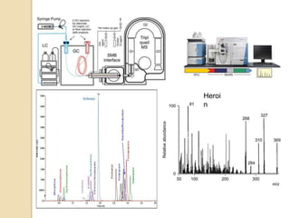
![ GC-MS
The GC-MS analysis of polar compounds, such as metabolites, from biological
matrices requires analytes extraction into a volatile organic solvent.
time-consuming sample preparation including derivatization to become stable,
volatile and amenable to the ionization technique.
Capillary electrophoresis (CE)
CE in many instances can have distinct advantages over HPLC in terms of
simplicity, rapid method development, solvent saving and minimal sample
requirement [10-30 nL injected] making this technique very interesting for rapid
and practical analyses in the biomedical field.
But have less sensitivity.
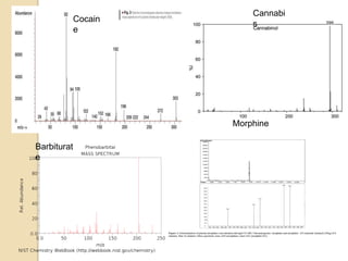
Mass
spectrometry
Separate ion according to m/z ratio.
Currently, the QQQ using single or multiple reaction monitoring is most often
used for quantitative analysis of metabolites.
SIM suffers from insufficient selectivity in comparison with MRM.
And also much lower sensitivity.
IT and TOF analyzers are also used for metabolite determination or use
combined with QQQ (Qtrap, Q-TOF).](https://image.slidesharecdn.com/analyticaltoxicology-140204090530-phpapp02/85/Analytical-toxicology-9-320.jpg)
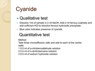
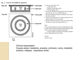
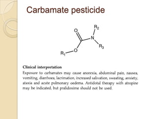
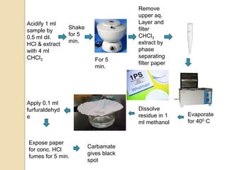


The document discusses various analytical techniques in toxicology for the analysis and identification of drug metabolites and toxic substances. Key methods include protein precipitation, liquid-liquid extraction, solid-phase extraction, and several chromatography techniques such as LC-MS/MS and GC-MS, each with their advantages and applications in biomedical analyses. It also covers qualitative and quantitative tests for specific toxins like arsenic, mercury, cyanide, and pesticides, highlighting the clinical implications of toxic exposure.


