The document discusses methods for reducing radiation doses in computed tomography (CT) scans, emphasizing both hardware and software techniques. Key hardware methods include adjusting exposure parameters like tube voltage and current, while software techniques focus on image denoising algorithms to improve image clarity. The research highlights the importance of minimizing radiation exposure, especially for children, to reduce the risk of cancer while maintaining diagnostic image quality.
![International Journal of Electrical and Computer Engineering (IJECE)
Vol. 13, No. 1, February 2023, pp. 1169~1179
ISSN: 2088-8708, DOI: 10.11591/ijece.v13i1.pp1169-1179 1169
Journal homepage: http://ijece.iaescore.com
An approach for radiation dose reduction in computerized
tomography
Shama Bekal Narayan, Savitha Halkare Mahabaleshwara
Department of Electronics and Communication Engineering, Faculty of Engineering, St Joseph Engineering College, Mangalore, India
Article Info ABSTRACT
Article history:
Received Feb 2, 2022
Revised Sep 23, 2022
Accepted Oct 8, 2022
Minimization of radiation dose plays an important role in human wellbeing.
Excess of radiation dose leads to cancer. Radiation greatly affects young
children less than 10 years of age as their life span is longer. Radiation can
be reduced by hardware and/or by software techniques. Hardware methods
deal with variation of parameters such as tube voltage, tube current,
exposure time, focal distance and filter type. Software techniques include
image processing methods. The originally acquired X-ray images may be
contaminated with noise due to the fact of instability in the case of sensors,
electrical power or X-ray source, that is responsible for the degradation of
the image attributes. An enhanced image denoising algorithm has been
proposed which decreases Gaussian noise combined with salt and pepper
noise that retains most information particulars.
Keywords:
Hardware
Image
Radiation
Software
X-ray This is an open access article under the CC BY-SA license.
Corresponding Author:
Shama Bekal Narayan
Department of Electronics and Communication Engineering, Faculty of Engineering, St Joseph
Engineering College, Affiliated to Visveshwaraya Technological University
Vamanjoor, Mangalore, Karnataka 575028
Email: shamabn@sjec.ac.in
1. INTRODUCTION
Diagnostic examination is a medical specialty procedure which involves medical procedures to
identify and treat diseases. Non-invasive methods like X-ray radiography and computed tomography (CT) are
used to diagnose or treat diseases. The X-rays pass across the human body, creating a computer display.
Direct digital radiography produces an image instantly on a computer screen. Radiologists can access the
image quickly. CT uses X-rays to recognize and note the radiation absorbed by different organs. Radiology
technicians/doctors use these images to analyze bone fractures and other illnesses in the human body [1].
These medical examinations present both benefits and threats. The benefits of these examinations
far exceed the risks. They are fast, painless and non-invasive. Examinations provide detailed information to
identify the issue, treatment plan, and assess many complications in adults and children. In addition, the
accurate images provided by CT scans may waive off the surgery [2]. Dental radiology plays a vital role in
pediatric patients. Recognizing the variety of retraction of chin is critical for distinct investigation in
research. Dentofacial skeletal features differ based on the type of malocclusion [3]. The investigation done on
the juvenile idiopathic arthritis (JIA) recommends children and youngsters are influenced with unilateral or
bilateral balanced to serious temporomandibular joint (TMJ) issues [4]. The cephalometric X-ray supports the
dentist to acquire an entire radiographed picture of the side of the face. Cephalometric regularizing quantum
values for the 8-to-12-year-old South Italian children population was referred to in study. Important
characteristic features among boys and girls in the range of the anterior cranial base and ratio obtained are
recorded [5].](https://image.slidesharecdn.com/v11227348emr23sep222feb22n-221124063330-97a3721a/85/An-approach-for-radiation-dose-reduction-in-computerized-tomography-1-320.jpg)
![International Journal of Electrical and Computer Engineering (IJECE)
Vol. 13, No. 1, February 2023, pp. 1169~1179
ISSN: 2088-8708, DOI: 10.11591/ijece.v13i1.pp1169-1179 1169
Journal homepage: http://ijece.iaescore.com
An approach for radiation dose reduction in computerized
tomography
Shama Bekal Narayan, Savitha Halkare Mahabaleshwara
Department of Electronics and Communication Engineering, Faculty of Engineering, St Joseph Engineering College, Mangalore, India
Article Info ABSTRACT
Article history:
Received Feb 2, 2022
Revised Sep 23, 2022
Accepted Oct 8, 2022
Minimization of radiation dose plays an important role in human wellbeing.
Excess of radiation dose leads to cancer. Radiation greatly affects young
children less than 10 years of age as their life span is longer. Radiation can
be reduced by hardware and/or by software techniques. Hardware methods
deal with variation of parameters such as tube voltage, tube current,
exposure time, focal distance and filter type. Software techniques include
image processing methods. The originally acquired X-ray images may be
contaminated with noise due to the fact of instability in the case of sensors,
electrical power or X-ray source, that is responsible for the degradation of
the image attributes. An enhanced image denoising algorithm has been
proposed which decreases Gaussian noise combined with salt and pepper
noise that retains most information particulars.
Keywords:
Hardware
Image
Radiation
Software
X-ray This is an open access article under the CC BY-SA license.
Corresponding Author:
Shama Bekal Narayan
Department of Electronics and Communication Engineering, Faculty of Engineering, St Joseph
Engineering College, Affiliated to Visveshwaraya Technological University
Vamanjoor, Mangalore, Karnataka 575028
Email: shamabn@sjec.ac.in
1. INTRODUCTION
Diagnostic examination is a medical specialty procedure which involves medical procedures to
identify and treat diseases. Non-invasive methods like X-ray radiography and computed tomography (CT) are
used to diagnose or treat diseases. The X-rays pass across the human body, creating a computer display.
Direct digital radiography produces an image instantly on a computer screen. Radiologists can access the
image quickly. CT uses X-rays to recognize and note the radiation absorbed by different organs. Radiology
technicians/doctors use these images to analyze bone fractures and other illnesses in the human body [1].
These medical examinations present both benefits and threats. The benefits of these examinations
far exceed the risks. They are fast, painless and non-invasive. Examinations provide detailed information to
identify the issue, treatment plan, and assess many complications in adults and children. In addition, the
accurate images provided by CT scans may waive off the surgery [2]. Dental radiology plays a vital role in
pediatric patients. Recognizing the variety of retraction of chin is critical for distinct investigation in
research. Dentofacial skeletal features differ based on the type of malocclusion [3]. The investigation done on
the juvenile idiopathic arthritis (JIA) recommends children and youngsters are influenced with unilateral or
bilateral balanced to serious temporomandibular joint (TMJ) issues [4]. The cephalometric X-ray supports the
dentist to acquire an entire radiographed picture of the side of the face. Cephalometric regularizing quantum
values for the 8-to-12-year-old South Italian children population was referred to in study. Important
characteristic features among boys and girls in the range of the anterior cranial base and ratio obtained are
recorded [5].](https://image.slidesharecdn.com/v11227348emr23sep222feb22n-221124063330-97a3721a/75/An-approach-for-radiation-dose-reduction-in-computerized-tomography-1-2048.jpg)
![ ISSN: 2088-8708
Int J Elec & Comp Eng, Vol. 13, No. 1, February 2023: 1169-1179
1170
Reducing the radiation helps in reducing the risk of cancer. Radiation can be reduced by using
hardware and software techniques. Hardware approaches include the analysis of five exposure parameters
and software approaches involve implementation of distinct algorithms. The exposure to ionizing radiation
may lead to a slight raise in an individual's lifetime risk of originating malignancy or cancer [6]. As a person
is exposed to radiation, risk of developing cancer also increases. Risk is greater in children’s as they have
longer life spans. Possibility of raising cancer boosts with periodic subjection to diagnostics imaging
methodologies. Probability is twofold in case of expecting mothers and in offspring. A baby in the womb
may also be more sensitive to radiation than an adult. Scientific research facilitates better evaluation of the
risks. It is observed that as years pass on radiation dose is reduced. Dose can be reduced by both hardware and
software techniques. Hardware techniques include five important exposure confines namely tube voltage (kV),
tube current (mA), exposure time (ms), focus to detector distance (cm) and filter type. Software approach
involves distinct algorithms. Aim is to minimize noise and import clarity to the image by applying rebuilding
techniques and required filters. If the clarity in image is good, doctors can diagnose the issue of patients. If the
image is clear then radiation dose can be reduced. Objective is: i) To reduce radiation dosage in X-ray,
especially for pediatric use and reduce the risk of cancer; ii) To develop an improved image filtering
algorithm to support the reduction of radiation dose; iii) To identify and filter the various noises present in
images; and iv) To enhance the images for scene visibility, enhance the contrast and edges in the X-ray
images. Scope includes long term health benefits to individuals, health benefits to Oncology patients who are
exposed to radiation therapy, in future, state of art algorithms can be implemented by X-ray manufactures
such as Siemens, Philips and General Electric (GE) for health care application.
2. METHOD
Radiation dose reduction [7] follows two major approaches. Hardware and software methods [8].
Figure 1 illustrates different approaches to radiation reduction. Hardware technique includes modification in
the geometry of X-ray machines. As per data sheets of top medical device manufacturing company,
Radiation induced depends on tube voltage in Kilovolts, tube current in milliamperes, exposure time in
milliseconds, prefilter, and focal spot.
Figure 1. Flowchart - techniques of radiation dose reduction](https://image.slidesharecdn.com/v11227348emr23sep222feb22n-221124063330-97a3721a/85/An-approach-for-radiation-dose-reduction-in-computerized-tomography-2-320.jpg)
![Int J Elec & Comp Eng ISSN: 2088-8708
An approach for radiation dose reduction in computerized tomography (Shama Bekal Narayan)
1171
Radiation builds on a patient body mass index. Radiograph energy is directly proportional to patient
thickness. Reputed medical device manufacturing company has inbuilt automatic exposure control
adjustments. To analyze these algorithms, one needs to have an X-ray machine with access to the gateway of
network.
A graphical user interface (GUI) based LabVIEW approach is used to implement body mass index
calculations. If the body mass index is greater than the threshold, radiation dose increases. Caldose software
program is used to calculate radiation dose induced in different parts of the body for a specific X-ray profile.
Chest X-ray profile is selected for study because chest x ray is most frequently performed examination,
according to annual frequency of examination conducted during 2002 [8].
Different software’s were tried to reduce radiation dose. Some of them are Imasim, Mevislab,
Analyze 14.0, Code_Blocks, Git bash, medical image processing, analysis, and visualization (MIPAV),
DOSCTP/DOSXYZnrc. Many radiology-based apps were studied for the purpose of radiation dose reduction.
Software Techniques include removing noise in image by the use of various filters. As radiation is reduced
image becomes blurry. Hence it is ideal to minimize the noise in the X-ray image.
Data for analysis was collected from a private hospital. Two technicians were involved from
renowned healthcare technology management services. Inclusion criteria comprise general patients requiring
X-ray diagnosis. Oncology patients were excluded from the study. Statistical methods for noise analysis
include different types of filtering along with advanced image denoising algorithms.
3. TECHNIQUES IMPLEMENTED
3.1. Hardware techniques
Radiation exposure can be controlled by using hardware techniques. External parameters can be
varied for different setups for analysis purposes. LabVIEW and Caldose software were used for
implementation. Different methods of hardware techniques tried is as follows:
3.1.1. White paper of top medical device manufacturing company
According to medical device manufacturing company’s white paper [9], Radiation dose is calculated
based on patient size, density of anatomical area irradiated and C-arm regulation of x ray tube. Radiation
dose also depends on Tube voltage (kV), Tube current (mA), Exposure time (mS), Focal spot and pre-filter
(CARE filter).
As per the analysis, radiation dose cannot be reduced in already deployed machines. Image
processing was done using C++. Objective of research mainly deals with hardware [10] control. Radiation
builds on a patient body mass index. Radiograph energy is directly proportional to patient thickness. Reputed
medical device manufacturing company has inbuilt automatic exposure control adjustments. To analyze these
algorithms, one needs to have an X-ray machine with access to the gateway of network.
3.1.2. LabVIEW
Implementation of body mass index (BMI): Radiation dose depends on patient thickness.
Calculation of BMI for pediatric patients is implemented using LabVIEW as per Figure 2. Radiation dose
increases with BMI. Radiation dose needs to be adjusted with BMI. Implementation of radiation reduction
was tried in LabVIEW. Three hardware parameters were considered namely tube voltage, tube current and
exposure time. Hounsfield units using the (1).
Figure 2. Implementation of BMI using LabVIEW](https://image.slidesharecdn.com/v11227348emr23sep222feb22n-221124063330-97a3721a/85/An-approach-for-radiation-dose-reduction-in-computerized-tomography-3-320.jpg)
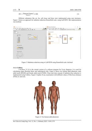
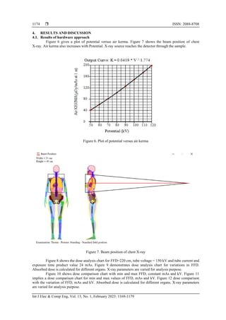
![Int J Elec & Comp Eng ISSN: 2088-8708
An approach for radiation dose reduction in computerized tomography (Shama Bekal Narayan)
1173
Figure 5. X ray examination with FFD, mAs and kV
3.2. Software techniques
As radiation is reduced the image becomes blurry [11]. Radiologists find it difficult to diagnose the
complications in the patient’s X-ray. Because of hardware restrictions and practical issues, it is impossible to
reduce dose. Low dose CT [12] with image clarity is a challenge. Aim is to identify different noise in X-ray
image, reduce noise in image and enhance image characteristics using different image processing techniques [13].
Model based Iterative reconstruction techniques [14] with high performance [15] and graphics
processing unit [16] are some of the reconstruction [17] techniques recently used for chest X-ray used in
medical imaging [18]. Filtered back projection [19] technique solves the issues of blurring. Deep learning
methods [20] can be used for image reconstruction. Convolutional neural networks play an important role in
radiation reduction [21].
The design is based profoundly on the seed particle. By dividing the image in various fragments,
each and every single pixel value of the image is contrasted with respect to a particular threshold value
determined in consideration to the gray value of the seed point [22].
a. Take a pixel in the initial input image and specify it as a seed particle. Place the particular seed pixel
value toward a blank progression.
b. Since the beginning of the progression, find consequent 8- associated neighbors of every pixel which are
not processed and for every neighbor point, see if the gray zone value of that neighbor particle is in the
range of the stated discrepancy from the seed particle’s gray zone value. This discrepancy is illustrated
by (2).
(𝑓(𝑥,𝑦)−𝑠𝑒𝑒𝑑)
𝑠𝑒𝑒𝑑
<= £ (2)
Here, f (x, y) is the gray zonal value of the current particle and £ is the origin value equal to 0.5.
c. Thirdly, the foreground section is further improved by adjusting the histogram accordingly and is
summed up to the background section.
d. Lastly, an improved image is produced by combining the grade of the initial image to the image obtained
in the C step.
e. On the basis of the seed point which is taken, the entire image is divided into foreground and background
sections.](https://image.slidesharecdn.com/v11227348emr23sep222feb22n-221124063330-97a3721a/85/An-approach-for-radiation-dose-reduction-in-computerized-tomography-5-320.jpg)
![ ISSN: 2088-8708
Int J Elec & Comp Eng, Vol. 13, No. 1, February 2023: 1169-1179
1176
Figure 11. Dose comparison chart for min and max values of FFD, mAs and kV
Figure 12 Dose Comparison with the variation of FFD, mAs and kV
4.2. Results of software approach
Software approaches were divided into 3 cases. Case 1 was analyzed when X-ray is contaminated
with Gaussian noise. Figure 13 shows the input X-ray image contaminated with Gaussian noise. Figure 14
shows the output of the filter. Figure 15 shows an enhanced output image. Case 2 was analyzed when X-ray
is contaminated with salt and pepper noise. Figure 16 shows the input X-ray image contaminated with salt
and pepper noise. Figure 17 shows the output of the filter. Figure 18 shows an enhanced output image. Case 3
was analyzed when X-ray is contaminated with both Gaussian and salt and pepper interference. Figure 19
shows the input X-ray image contaminated with both Gaussian and salt and pepper interference. Figure 20
shows the output of the filter. Figure 21 shows an enhanced output image.
The innovative noise reduction technique which involves median filtering along with threshold
filtering obtained greater signal-to-noise ratio (SNR) and peak signal-to-noise ratio (PSNR) values but,
however lower mean squared error (MSE) value [23]. This indicates that the novel interference reducing
algorithm which diminishes the interference than traditional procedures still maintain better visual details.
The human conception is no longer treated as a standard for the image quality, and therefore to estimate the
conduct of the proposed design, quality parameters were measured such as contrast to noise ratio (CNR),
SNR, PSNR, and MSE.
In hardware techniques, as focus to detector distance is increased dosage is reduced. Tube voltage,
tube current and exposure time [24] product value has to be minimum. Different algorithms can be used for
fast image processing, [25] which can be used for fluoroscopic applications [26]. Various CT reconstruction
[27] techniques may contribute to future work.
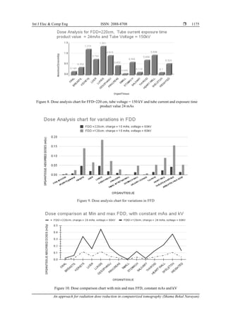
Dose analysis for FFD=120 cm, charge=10 mAs, voltage=60 kV
and FFD=220 cm, charge=24 mAs, voltage=150 kV](https://image.slidesharecdn.com/v11227348emr23sep222feb22n-221124063330-97a3721a/85/An-approach-for-radiation-dose-reduction-in-computerized-tomography-8-320.jpg)

![ ISSN: 2088-8708
Int J Elec & Comp Eng, Vol. 13, No. 1, February 2023: 1169-1179
1178
In comparison of this method with the currently prevalent methods of flexible improvement and fine
stretching and other contrast enhancement techniques, it is summarized that this innovative technique is
exhibiting much better and improved outcomes than the currently prevalent ones. Moreover, the method is
seed-dependent and therefore the determination of seed particles is very much vital in this design. A seed
selected in dark sections will yield improved outcomes compared to the seed selected in the bright sections,
as it is expected that the enhancement of the darker regions of the image is required.
Also, the innovative enhancement technique can also be combined with adaptive enhancement
Method to produce outputs with still better results and improved visual perception Hard problems include
radiation dose reduction by adjusting the X-ray machine settings. Adjusting the settings of an X-ray machine
is a challenge. This has to be done by the X-ray machine manufacturers. Non obvious mistakes include
collection of data from the hospital instead of contacting the technician of the X-ray machine.
ACKNOWLEDGEMENTS
Author thanks guide Dr. Dayakshini, Head of ECE Department, Dr. Rio D Souza, Principal, and
St Joseph Engineering College for their guidance during my research work. Author would like to express
sincere gratitude to the management of St Joseph Engineering College for constant support.
REFERENCES
[1] ITN, “Radiology imaging, imaging technology news,” Imaging Technology News, https://www.itnonline.com/channel/radiology-
imaging/Radiation (accessed Dec. 12, 2020).
[2] FDA, “Computed tomography, US food drug administration.” US Food and Drug Administration. https://www.fda.gov/radiation-
emitting-products/medical-imaging/medical-x-ray-imaging (accessed Aug. 08, 2020).
[3] L. Perillo, G. Padricelli, G. Isola, F. Femiano, P. Chiodini, and G. Matarese, “Class II malocclusion division 1: a new
classification method by cephalometric analysis,” European Journal of Paediatric Dentistry, vol. 13, no. 3, pp. 192–196, 2012.
[4] G. Isola, L. Ramaglia, G. Cordasco, A. Lucchese, L. Fiorillo, and G. Matarese, “The effect of a functional appliance in the
management of temporomandibular joint disorders in patients with juvenile idiopathic arthritis,” Minerva Stomatologica, vol. 66,
no. 1, pp. 1–8, 2017, doi: 10.23736/S0926-4970.16.03995-3.
[5] L. Perillo, G. Isola, D. Esercizio, M. Iovane, G. Triolo, and G. Matarese, “Differences in craniofacial characteristics in Southern
Italian children from Naples: a retrospective study by cephalometric analysis,” European Journal of Paediatric Dentistry, vol. 14,
no. 3, pp. 195–198, 2013.
[6] A. Berrington de González, “Projected cancer risks from computed tomographic scans performed in the United States in 2007,”
Archives of Internal Medicine, vol. 169, no. 22, pp. 2071–2077, Dec. 2009, doi: 10.1001/archinternmed.2009.440.
[7] UT Southwestern Medical Center, “Diagnostic X-ray procedures.” utswmed.org, https://utswmed.org/conditions-
treatments/diagnostic-x-ray-procedures/ (accessed Jul. 03, 2020).
[8] M. B. Freitas, “Dose measurements in chest diagnostic X rays: adult and paediatric patients,” Radiation Protection Dosimetry,
vol. 111, no. 1, pp. 73–76, Aug. 2004, doi: 10.1093/rpd/nch363.
[9] Siemens-Healthineers, “AX care clear white paper low dose.” https://www.siemens-healthineers.com/it/angio/care-clear (accessed
Nov. 02, 2020).
[10] A. Sodickson, “Strategies for reducing radiation exposure from multidetector computed tomography in the acute care setting,”
Canadian Association of Radiologists Journal, vol. 64, no. 2, pp. 119–129, May 2013, doi: 10.1016/j.carj.2013.01.002.
[11] T. ten Cate et al., “Novel X-ray image noise reduction technology reduces patient radiation dose while maintaining image quality
in coronary angiography,” Netherlands Heart Journal, vol. 23, no. 11, pp. 525–530, Nov. 2015, doi: 10.1007/s12471-015-0742-1.
[12] K. P. Murphy et al., “Feasibility of low-dose CT with model-based iterative image reconstruction in follow-up of patients with
testicular cancer,” European Journal of Radiology Open, vol. 3, pp. 38–45, 2016, doi: 10.1016/j.ejro.2016.01.002.
[13] B. Luo, Z. Sun, M. Xue, and H. Liu, “Improved noise reduction algorithms for medical X-ray images,” in 2013 3rd International
Conference on Consumer Electronics, Communications and Networks, 2013, pp. 359–362, doi: 10.1109/CECNet.2013.6703346.
[14] P. J. Pickhardt et al., “Abdominal CT with model-based iterative reconstruction (MBIR): initial results of a prospective trial
comparing ultralow-dose with standard-dose imaging,” American Journal of Roentgenology, vol. 199, no. 6, pp. 1266–1274, Dec.
2012, doi: 10.2214/AJR.12.9382.
[15] X. Wang, A. Sabne, S. Kisner, A. Raghunathan, C. Bouman, and S. Midkiff, “High performance model based image
reconstruction,” in Proceedings of the 21st ACM SIGPLAN Symposium on Principles and Practice of Parallel Programming, Feb.
2016, pp. 1–12, doi: 10.1145/2851141.2851163.
[16] A. Sabne, X. Wang, S. J. Kisner, C. A. Bouman, A. Raghunathan, and S. P. Midkiff, “Model-based iterative CT image
reconstruction on GPUs,” in Proceedings of the 22nd ACM SIGPLAN Symposium on Principles and Practice of Parallel
Programming, Jan. 2017, pp. 207–220, doi: 10.1145/3018743.3018765.
[17] M. Katsura et al., “Model-based iterative reconstruction technique for radiation dose reduction in chest CT: comparison with the
adaptive statistical iterative reconstruction technique,” European Radiology, vol. 22, no. 8, pp. 1613–1623, Aug. 2012, doi:
10.1007/s00330-012-2452-z.
[18] H. Scheffel et al., “Coronary artery plaques: Cardiac CT with model-based and adaptive-statistical iterative reconstruction
technique,” European Journal of Radiology, vol. 81, no. 3, pp. e363–e369, Mar. 2012, doi: 10.1016/j.ejrad.2011.11.051.
[19] Z. Deák et al., “Filtered back projection, adaptive statistical iterative reconstruction, and a model-based Iterative reconstruction in
abdominal CT: An experimental clinical study,” Radiology, vol. 266, no. 1, pp. 197–206, Jan. 2013, doi:
10.1148/radiol.12112707.
[20] J. Cheng et al., “Model-based deep medical Imaging: the roadmap of generalizing iterative reconstruction model using deep
learning,” arXiv:1906.08143, Jun. 2019.
[21] H. Chen et al., “Low-dose CT with a residual encoder-decoder convolutional neural network,” IEEE Transactions on Medical
Imaging, vol. 36, no. 12, pp. 2524–2535, Dec. 2017, doi: 10.1109/TMI.2017.2715284.](https://image.slidesharecdn.com/v11227348emr23sep222feb22n-221124063330-97a3721a/85/An-approach-for-radiation-dose-reduction-in-computerized-tomography-10-320.jpg)
![Int J Elec & Comp Eng ISSN: 2088-8708
An approach for radiation dose reduction in computerized tomography (Shama Bekal Narayan)
1179
[22] N. Kanwal, A. Girdhar, and S. Gupta, “Region based adaptive contrast enhancement of medical X-ray images,” in 2011 5th
International Conference on Bioinformatics and Biomedical Engineering, May 2011, pp. 1–5, doi: 10.1109/icbbe.2011.5780221.
[23] A. Neroladaki, D. Botsikas, S. Boudabbous, C. D. Becker, and X. Montet, “Computed tomography of the chest with model-based
iterative reconstruction using a radiation exposure similar to chest X-ray examination: preliminary observations,” European
Radiology, vol. 23, no. 2, pp. 360–366, Feb. 2013, doi: 10.1007/s00330-012-2627-7.
[24] D. T. Raju and K. Shanthi, “Analysis on x-ray parameters of exposure by measuring x-ray tube voltage and time of exposure,”
The International Journal of Engineering and Science (IJES), vol. 3, no. 6, pp. 69–73, 2014.
[25] H. Nien and J. A. Fessler, “Relaxed linearized algorithms for faster X-ray CT image reconstruction,” IEEE Transactions on
Medical Imaging, vol. 35, no. 4, pp. 1090–1098, Apr. 2016, doi: 10.1109/TMI.2015.2508780.
[26] A. Qadir, “Fluoroscopy dose management.” https://www.slideshare.net/airwave12/patient-radiation-dose-management (accessed
May 18, 2020).
[27] J. Zhang, Y. Hu, J. Yang, Y. Chen, J.-L. Coatrieux, and L. Luo, “Sparse-view X-ray CT reconstruction with gamma
regularization,” Neurocomputing, vol. 230, pp. 251–269, Mar. 2017, doi: 10.1016/j.neucom.2016.12.019.
BIOGRAPHIES OF AUTHORS
Shama Bekal Narayan received the B.Eng. degree in Electronics and
communication engineering from A.P.S Engineering college, Bangalore, in 2004 and the
M.Tech from N.M.A.M. Institute of Technology (NMAMIT) Nitte and pursuing Ph.D. degree
from VTU, Belagavi. Currently, she is an Assistant Professor at the Department of Electronics
and communication Engineering, St Joseph Engineering College, Mangalore, Karnataka, India.
Her research interests include Biomedical image processing, digital circuits, wireless
communication, computer communication networks, digital image processing. She can be
contacted at shamabn@sjec.ac.in, ResearchGate: https://www.researchgate.net/profile/Shama-
Bekal.
Savitha Halkare Mahabaleshwara received her B.E. degree in Electronics and
Communication Engineering from Mysore University and her M. Tech. degree in Digital
Electronics and Communication from Visvesvaraya Technological University (VTU),
Belagavi. She was awarded Doctorate from National Institute of Technology Karnataka
(NITK), Surathkal, during April 2014. She has 26 years of teaching experience. She has
published around 25 research papers in international journals, international/National
conference Proceedings. At savitha100@gmail.com.](https://image.slidesharecdn.com/v11227348emr23sep222feb22n-221124063330-97a3721a/85/An-approach-for-radiation-dose-reduction-in-computerized-tomography-11-320.jpg)