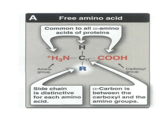
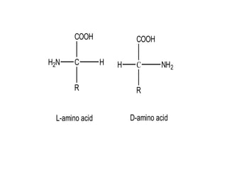
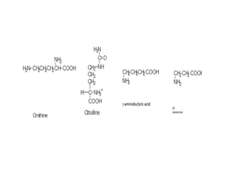
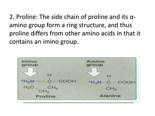
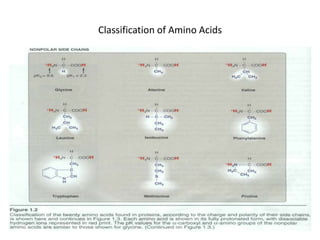
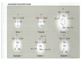
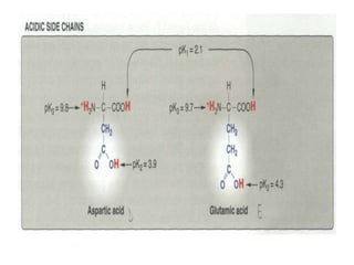
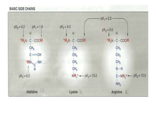
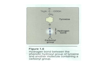
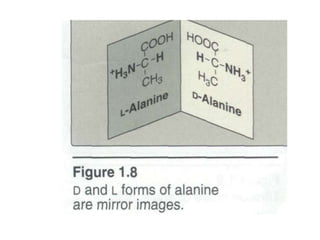
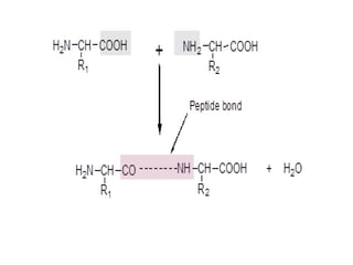
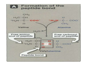
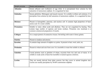
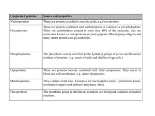
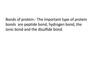
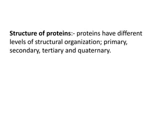
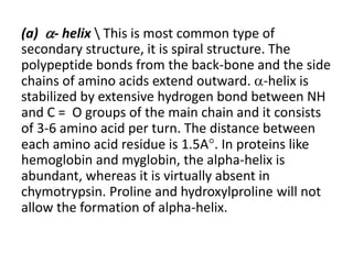
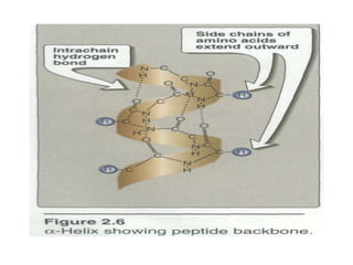
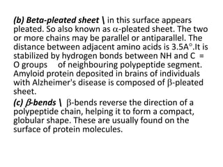
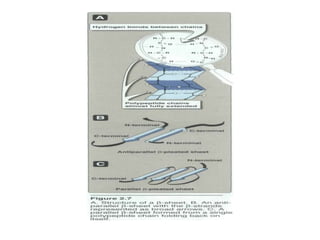
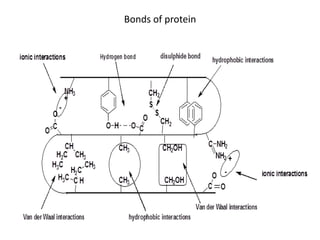
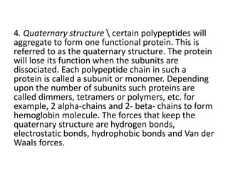
2. The aims of the lecture
-to define the amino acids.
-to know the structure of amino acids.
-to know the main functions of amino acids.
-to classify the amino acids.
3. Amino acids
- are containing amino group and carboxyl group
on the same molecule.
-Amino acids are the major units of proteins
synthesis. They are produced by hydrolysis of
proteins.
4.
5. All amino acids found in living systems,
plant and animal proteins are L--amino
acids. D-amino acids are seen in small
amount in micro-organisms and as
constituents of certain antibiotics such as
gramicidin-S, polymyxin, actinomycin-D
and valinomycin, as well as bacterial cell
wall peptidoglycans.
7. Glycine is the only amino acids, which
is optically inactive and cannot be
resolved into D-or L- form because of
symmetry on the -carbon atom. All
other amino acids are optically active
because they have asymmetric carbon
atom.
8. Function of amino acids:-
1.building block of proteins.
2.proved nitrogen for the synthesis of other nitrogen
containing compounds.
3.catabolized as fuels.
4.precursors of:-
• hormones
• purines
• pyrimidines
• porphyrins
• Vitamins.
9. Essential amino acids: -
-Those amino acids which are not synthesized in
the body and hence have to be proved in the
diet. They are leucine (Leu), isoleucine (Ile),
threonine(Thr), tryptophan (Trp),phenylalanine
(Phe), valine (Val), methionine (Met), and lysine
(Lys).
-Some amino acids, such as histidine (His) and
arginine (Arg)are synthesized in tissues but not
in adequate amounts for normal growth of small
children. These two amino acids are often
classified as ''semi essential amino acids''.
10. Deficiency of one or more essential amino acids
in the diet gives rise to depression in protein
synthesis resulting in failure in growth of child,
negative nitrogen balance in adults and fall in
plasma proteins and hemoglobin levels.
11. Non essential amino acids: - Those amino acids
which are synthesized by the body. They are
alanine (Ala), aspartic acid (Asp), asparagines
(Asn), cysteine (cys), glutamic acid (Glu),
glutamine (Gln), glycine (Gly), proline (Pro) ,
serine (Ser),and tyrosine (Tyr).
12. Classification of amino acids: - A variety of
classifications of amino acids are possible. Either
they can be classified according to the presence
of acidic, basic or neutral groups or upon their
chemical structures, i.e., presence of polar
groups, nonpolar groups, sulpher containing
group, aromatic groups, heterocyclic ring,
branched chain and so on.
13. There are number of amino acids which are
obtained in free or combined form but do not
occur in protein molecules for example
ornithine, citruline, -aminobutyric acid, -
alanine, thyroxine…etc.
15. A. Amino acids with nonpolar side
chains
-Each of these amino acids has a nonpolar side
chain that does not bind or give off protons or
participate in hydrogen or ionic bonds.
16. 2. Proline: The side chain of proline and its α-
amino group form a ring structure, and thus
proline differs from other amino acids in that it
contains an imino group.
18. B. Amino acids with uncharged polar side
chains .These amino acids have zero net charge
at neutral PH although the side chains of
cysteine and tyrosine can lose a proton at an
alkaline PH. Serine, threonine, and tyrosine each
contain a polar hydroxyl group that can
participate in hydrogen bond formation . The
side chains of asparagine and Glutamine
each contain a carbonyl group and an amide
group, both of which can also participate in
hydrogen bonds.
19.
20. - Disulfide bond: The side chain of cysteine
contains a sulfhydryl group (-SH), which is an
important component of the active site of many
enzymes. in proteins, the -SH groups of two
cysteines can become oxidized to form a dimer,
cystine, which contains a covalent cross-link
called a disulfide bond (-S-S-).
21. Side chains as sites of attachment for other
compounds: Serine, threonine, and, rarely,
tyrosine contain a polar hydroxyl group that can
serve as a site of attachment for structures such
as a phosphate group.
Note: The side chain of serine is an important
component of the active site of many enzymes.
22. in addition, the amide group of asparagine, as
well as the hydroxyl group of serine or
threonine, can serve as a site of attachment for
oligosaccharide chains in glycoproteins.
23. C. Amino acids with acidic side chains
The amino acids aspartic and glutamic acid are
proton donors. At neutral ph the side chains of
these amino acids are fully ionized, containing a
negatively charged carboxylate group (-C00").
They are, therefore, called aspartate or
glutamate. to emphasize that these amino acids
are negatively charged at physiologic PH.
24.
25. D. Amino acids with basic side chains
The side chains of the basic amino acids
accept protons. At physiologic PH the side
chains of lysine and arginine are fully ionized
and positively charged.
In contrast, histidine is weakly basic and the free
amino acid is largely uncharged at physiologic
PH.
26.
27. Optical properties of amino acids:
-The α-carbon of each amino acid is attached to
four different chemical groups and is, therefore, a
chiral or optically active carbon atom.
-Glycine is the exception because its α-carbon has
two hydrogen substituents and, therefore, is
optically inactive.
-Note: Amino acids that have an asymmetric center
at the α-carbon can exist in two forms,
designated D and L, that are mirror images of
each other.
-The two forms in each pair are termed stereo
isomers, optical isomers,
28. All amino acids found in proteins are of the L-
configuration. However, D-amino acids are
found in some antibiotics and in bacterial cell
walls.
29.
30.
31.
32. Proteins
• Proteins:- are defined as compounds of high
molecular weight made up of - amino acids
linked to one another by peptide linkage.
• Proteins are linear polymers consisting of L--
amino acids. The amino acids are joined
together by peptide bond.
33. Peptide bond:- is formed by the union of carboxyl
group of one amino acids with amino group of
other amino acids with an elimination of water
molecule. Two amino acids are combined to form a
dipeptide; three amino acids form tripeptide; four
will make an oligopeptide; and combination of 10 to
50 amino acids is called as a polypeptide. By
convention, big polypeptide chains containing more
than 50 amino acids are called proteins.
34.
35.
36. • Role of proteins:- proteins have many diverse functions
1. As catalysts, i.e., as enzymes.
2. As structural proteins (e.g. collagen, elastin, keratin)
3. As mode of transport (e.g., albumin, globulin, hemoglobin,
myoglobin).
4. As regulatory proteins or hormones (e.g., insulin, growth
hormones).
5. As protective agents, (i.e., antibodies, clotting factors).
6. Genetic proteins, e.g. histones
•
38. Simple proteins Source and properties
Albumins Serum albumin and ovalbumin of egg white. It is precipitated from solution by full
saturation of ammonium sulphate. It is coagulated by heat.
Globulins Serum globulins, fibrinogens and muscle myosin. It is soluble in dilute salt solutions. It is
precipitate from solution by half saturation of ammonium sulphate. It is coagulated by heat.
Histones Present in hemoglobin, pancreas, and nucleus cell. It contains high proportion of basic
amino acid. It is water soluble.
Protamines Soluble in water, dilute acids and alkalines. They are not coagulated by heating. They
contain large number of arginine and lysine residues. Protamine zinc insulinate is a
common commercial preparation of insulin.
Collagen It is a major protein of connective tissues. On boiling with water it forms gelatin.
Elastins Present in tendons and arteries.
Keratins It contains large amount of sulpher as cystine. It present in hair, wool, nails, etc.
Prolamins Present in wheat and zein from corn. It is insoluble in water but soluble in ethanol.
Glutelins Cereal proteins such as glutelins of wheat, oxyzenin from rice and zein of maize. It is
soluble in weak acids or bases but insoluble in neutral aqueous solutions.
Lectins They are mostly derived from plant sources; but they occur in animal kingdom also.
Lectins are usually precipitate by 30-60% ammonium sulphate.
39. Conjucated proteins Source and properties
Nucleoproteins These are proteins attached to nucleic acids, e.g.virus proteins.
Glycoproteins
These are proteins combined with carbohydrates or a derivative of carbohydrate.
When the carbohydrate content is more than 10% of the molecule; they are
sometimes known as mucoproteins or proteoglycans. Blood group antigens and
many serum proteins are glycoproteins.
Phosphoproteins The phosphoric acid is esterified to the hydroxyl groups of serine and threonine
residues of proteins. (e.g. casein of milk and vitellin of egg yolk )
Lipoproteins These are proteins loosely combined with lipid components. They occur in
blood and cell membranes. e.g. serum lipoproteins.
Metalloproteins They contain metal ions. Examples are haemoglobin (iron), cytochrome (iron),
tyrosinase (copper) and carbonic anhydrase (zinc).
Flavoproteins The prosthetic group is riboflavin; examples are biological oxidation reduction
reactions.
40. Bonds of protein:- The important type of protein
bonds are peptide bond, hydrogen bond, the
ionic bond and the disulfide bond.
41. Structure of proteins:- proteins have different
levels of structural organization; primary,
secondary, tertiary and quaternary.
42. Primary structureThe sequence of amino acids in a
protein is called the primary structure of the
protein. Understanding the primary structure of
proteins is important because many genetic
diseases result in proteins with abnormal amino
acid sequences, which cause improper folding and
loss or impairment of normal function. If the
primary structures of the normal and the mutated
proteins are known, this information may be used
to diagnose or study the disease.
43. A protein with a specific primary structure, when put in
solution, will automatically form its natural three
dimensional shapes. So the higher levels of
organization are dependent on the primary structure.
Even a single amino acid change (mutation) in the
linear sequence may have profound biological effects
on the function. For example, in HbA (normal
hemoglobin) the 6th amino acid in beta chain is
glutamic acid; it is changed to valine in HbS (sickle cell
anemia).
44. A second example of small differences in protein structure is
shown with two protein type hormones called oxytocin and
vasopressin. These two substances are very similar in structure,
differing only in the identified of two amino acid. They are
similar in structure but differ in function. Oxytocin causes
contraction of uterus and milk ejection from mammary glands.
Vasopressin causes water to be reabsorbed from urine as it passes
through distils tubules of kidney and affects blood pressure, it is
administrated to treat diabetes insipidus (excess urine
production).
45. 2.Secondary structure: - The polypeptide back-
bone does not assume a random three dimensional
structure, but instead generally forms regular
arrangements of amino acids that are located near
to each other in linear sequence. These
arrangements are called as secondary structure of
proteins. Secondary and tertiary levels of protein
structure are preserved by non covalent forces
bonds like hydrogen bonds, electrostatic bonds,
hydrophobic interactions and van der Waals forces.
Some of the proteins or portions of the protein
exhibit regularly repeating types of secondary
structure,; e.g. alpha helix, beta pleated sheet, and
beta bends.
46. (a) - helix This is most common type of
secondary structure, it is spiral structure. The
polypeptide bonds from the back-bone and the side
chains of amino acids extend outward. -helix is
stabilized by extensive hydrogen bond between NH
and C = O groups of the main chain and it consists
of 3-6 amino acid per turn. The distance between
each amino acid residue is 1.5A. In proteins like
hemoglobin and myglobin, the alpha-helix is
abundant, whereas it is virtually absent in
chymotrypsin. Proline and hydroxylproline will not
allow the formation of alpha-helix.
47.
48. (b) Beta-pleated sheet in this surface appears
pleated. So also known as -pleated sheet. The two
or more chains may be parallel or antiparallel. The
distance between adjacent amino acids is 3.5A.It is
stabilized by hydrogen bonds between NH and C =
O groups of neighbouring polypeptide segment.
Amyloid protein deposited in brains of individuals
with Alzheimer's disease is composed of -pleated
sheet.
(c) -bends -bends reverse the direction of a
polypeptide chain, helping it to form a compact,
globular shape. These are usually found on the
surface of protein molecules.
49.
50. Tertiary structure refers to the coiling of
several helical portion of single helix into a three
dimensional structure of the whole protein. It
defines the steric relationship of amino acids
which are far apart from each other in the linear
sequence but are opposed each other by non-
covalent interactions. The tertiary structure of
proteins is stabilized by :
a. Hydrogen bonding b. Disulphide bonding.
C.Ionic bond or salt bridges. D.Hydrophobic
interactions. e. Van der Waal's forces
52. • Denaturation of proteins:- heating, X-ray,
ultraviolet rays, high pressure, vigorous
shaking, pH change, organic solvents, and
such other physicochemical agents
produce non-specific alterations in
secondary, tertiary and quaternary
structures of protein molecules. This is
called denaturation. Primary structure is
not altered during the process of
denaturation. It generally decreases the
solubility, increases precipitability and
often causes loss of biological activity.
53. Denatured proteins are sometimes re-natured
when the physical agent is removed.
Ribonuclease is a good example for such
reversible denaturation. Protein kept in solution
for long periods may denature, and lose their
biological or enzymatic properties. Such loss of
activity can be minimized when the solution is
kept at low temperature.
54. 4. Quaternary structure certain polypeptides will
aggregate to form one functional protein. This is
referred to as the quaternary structure. The protein
will lose its function when the subunits are
dissociated. Each polypeptide chain in such a
protein is called a subunit or monomer. Depending
upon the number of subunits such proteins are
called dimmers, tetramers or polymers, etc. for
example, 2 alpha-chains and 2- beta- chains to form
hemoglobin molecule. The forces that keep the
quaternary structure are hydrogen bonds,
electrostatic bonds, hydrophobic bonds and Van der
Waals forces.