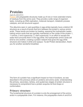
![the hormone insulin are depicted in the diagram below. (The insulin molecule
pictured here is actually cow insulin, but it shares a lot of similarities with
human insulin in terms of structure.) Each chain has a unique collection of
amino acids that are put together in a certain order. For instance, the A
chain’s sequence differs from the B chain’s because it begins with glycine at
the N-terminus and finishes with asparagine at the C-terminus. [Why are
those S-S bonds there?]
The DNA of the gene that codes for a protein (or for a portion of a protein in
the case of multi-subunit proteins) determines the protein’s sequence. The
amino acid sequence of the protein may change if the DNA sequence of the
gene changes. The overall structure and function of a protein can be impacted
by changing even a single amino acid in its sequence.
For instance, sickle cell anaemia, an inherited condition that affects red blood
cells, is linked to a single amino acid alteration. Haemoglobin, the protein that
delivers oxygen in the blood, is made up of polypeptide chains, and one of
these chains has a small sequence variation in sickle cell anaemia. One of the
two types of protein chains that make up haemoglobin ordinarily has glutamic
acid as the sixth amino acid; nevertheless, valine is used in its place. In the
picture below, this substitution is depicted for a section of the chain.](data:image/gif;base64,R0lGODlhAQABAIAAAAAAAP///yH5BAEAAAAALAAAAAABAAEAAAIBRAA7)
Recommended
More Related Content
Similar to Proteins.pdf
Similar to Proteins.pdf (20)
More from zainulabideen762825
More from zainulabideen762825 (20)
Recently uploaded
Recently uploaded (20)
Proteins.pdf
- 1. Proteins April 29, 2023admin Proteins are polypeptide structures made up of one or more extended chains of residues from the amino acid. They provide a wide range of organism tasks, including as DNA replication, molecule transport, metabolic process catalysis, and cell structural support. The albumins seen in vast quantities in egg whites typically have a distinct 3D structure as a result of bonds that form between the protein’s various amino acids. These bonds are broken by heating, exposing the hydrophobic (water- hating) amino acids that are typically maintained on the inside of the protein 1, 1 comma, 2 end superscript, 2, start superscript. In an effort to escape the water that surrounds them in the egg white, the hydrophobic amino acids will bind to one another, creating a protein network that gives the egg white structure and makes it white and opaque. Ta-da! Protein denaturation, thank you for another wonderful breakfast. The form of a protein has a significant impact on how it functions, as was described in the previous article on proteins and amino acids. Understanding the primary, secondary, tertiary, and quaternary stages of protein structure is necessary to comprehend how a protein acquires its final shape or conformation. Primary structure The fundamental structure of a protein is only the arrangement of the amino acids in a polypeptide chain. For instance, the polypeptide chains A and B of
- 2. the hormone insulin are depicted in the diagram below. (The insulin molecule pictured here is actually cow insulin, but it shares a lot of similarities with human insulin in terms of structure.) Each chain has a unique collection of amino acids that are put together in a certain order. For instance, the A chain’s sequence differs from the B chain’s because it begins with glycine at the N-terminus and finishes with asparagine at the C-terminus. [Why are those S-S bonds there?] The DNA of the gene that codes for a protein (or for a portion of a protein in the case of multi-subunit proteins) determines the protein’s sequence. The amino acid sequence of the protein may change if the DNA sequence of the gene changes. The overall structure and function of a protein can be impacted by changing even a single amino acid in its sequence. For instance, sickle cell anaemia, an inherited condition that affects red blood cells, is linked to a single amino acid alteration. Haemoglobin, the protein that delivers oxygen in the blood, is made up of polypeptide chains, and one of these chains has a small sequence variation in sickle cell anaemia. One of the two types of protein chains that make up haemoglobin ordinarily has glutamic acid as the sixth amino acid; nevertheless, valine is used in its place. In the picture below, this substitution is depicted for a section of the chain.
- 3. A person who only produces sickle cell haemoglobin will experience sickle cell anaemia symptoms. These happen as a result of the haemoglobin molecules assembling into long fibres as a result of the amino acid change from glutamic acid to valine. Red blood cells with disc forms are deformed into crescent shapes by the fibres. In the blood, regular, disc-shaped cells can be seen mingling with instances of “sickled” cells. As the sickled cells attempt to move through the blood vessels, they become impaled. People with sickle cell anaemia may experience serious health issues due to the stuck cells, including abdominal pain, headaches, dizziness, and shortness of breath. Secondary structure Secondary structure, the next level of protein structure, describes the local folded shapes that develop within a polypeptide as a result of interactions between the atoms in the backbone. (Since the R group atoms are not a component of secondary structure, the backbone simply refers to the polypeptide chain without them.) Helixes and pleated sheets are the two most typical forms of secondary structures. Hydrogen bonds, which develop between the amino H and carbonyl O of two different amino acids, keep both structures in place.
- 4. The amino H (N-H) of an amino acid that is four amino acids down the chain is hydrogen bound to the carbonyl (C=O) of one amino acid in a helix. (For instance, the carbonyl of amino acid 1 would join forces with amino acid 5’s N- H to form a hydrogen bond.) Each turn of the helical structure, which resembles a curled ribbon and is formed by this pattern of bonding, has 3.6 amino acids. The amino acids’ R groups protrude from the helix and are free to interact there. When two or more polypeptide chain segments are lined up next to one another, they produce a pleated sheet that is held together by hydrogen bonds. The backbone’s carbonyl and amino groups establish hydrogen bonds, while the R groups extend above and below the sheet’s plane cubed. A pleated sheet’s strands can be parallel, heading in the same direction (i.e., having matching N- and C-termini), or antiparallel, pointing in the opposite direction (i.e., having one strand’s N-terminus next to the other’s C-terminus).
- 5. There are some amino acids that are more or less likely to be present in pleated sheets or -helices. For instance, the amino acid proline is frequently referred to as a “helix breaker” because of its unique R group, which forms a ring with the amino group and bends the chain, making it unsuitable for the creation of helices. Though some proteins only contain one type of secondary structure (or don’t form either type), many proteins contain both helices and pleated sheets. Tertiary structure A polypeptide’s overall three-dimensional structure is referred to as its tertiary structure. The interactions between the R groups of the amino acids that make up the protein are principally responsible for the tertiary structure. The entire spectrum of non-covalent bonds, including hydrogen bonds, ionic bonds, dipole-dipole interactions, and London dispersion forces, all contribute to tertiary structure. For instance, R groups with opposite charges can form an ionic bond, while those with like charges repel one another. Similar to other dipole-dipole interactions, polar R groups can create hydrogen bonds. Hydrophobic interactions, in which amino acids with nonpolar, hydrophobic R groups bind together on the inside of the protein, leaving hydrophilic amino acids on the exterior to interact, are also significant for tertiary structure. The disulfide link is a unique sort of covalent bond that can contribute to tertiary structure. Disulfide bonds, covalent connections between the side chains of cysteines that contain sulphur, are substantially more powerful than the other kinds of bonds that make up tertiary structure. They serve as molecular “safety pins,” firmly connecting various polypeptide components to one another.
- 6. Quaternary structure Many proteins consist of just one polypeptide chain and only have the three levels of structure we just covered. Nevertheless, some proteins are composed of numerous polypeptide chains, also referred to as subunits. These component parts combine to form the protein’s quaternary structure. Haemoglobin is one instance of a protein having quaternary structure that we’ve already come across. As was previously discussed, haemoglobin is made up of four subunits, two of each kind, and carries oxygen in the blood. Another illustration is DNA polymerase, an enzyme with ten subunits that creates new DNA strands.
- 7. Generally speaking, the same interactions that give rise to tertiary structure—mostly weak interactions like hydrogen bonds and London dispersion forces—also
- 8. keep the subunits together to produce quaternary structure. Denaturation and Protein Folding Each protein has a distinct structure of its own. These connections may be interfered with if a protein’s surroundings undergoes changes in temperature or pH or if it is exposed to chemicals. If this occurs, the protein may lose its three-dimensional structure and revert to being an unstructured string of amino acids. A protein is considered to be denatured when its higher-order structure but not its fundamental sequence are lost. Normally, denatured proteins are not useful. Some proteins can have their denaturation reversed. The polypeptide’s primary structure is still intact (the amino acids haven’t broken apart), so if it’s put back in its usual environment, it might be able to refold into its functional form. Classification of Proteins Proteins can be divided into two categories based on their molecular structure: 1. Fibrous Proteins: The fiber-like structure is created when the parallel polypeptide chains are joined by hydrogen and disulfide bonds. These proteins typically do not dissolve in water. These proteins are insoluble in water. Example: Myosin, which is found in muscles, and keratin, which is found in hair, wool, and silk. 2. Globular Proteins: The fiber-like structure is created when the parallel polypeptide chains are joined by hydrogen and disulfide bonds. These proteins typically do not dissolve in water. These proteins are insoluble in water. Example: Myosin, which is found in muscles, and keratin, which is found in hair, wool, and silk. The amino acid composition of proteins All proteins share the trait of being composed of lengthy chains of -amino (alpha amino) acids. The diagram below depicts the general structure of -amino acids. Because the molecule’s -carbon atom carries both a carboxyl group (COOH) and an amino group (NH2), the -amino acids are so named.
- 9. When the pH of an acidic solution is lower than 4, the COO groups interact with hydrogen ions (H+) to create the uncharged form (COOH). Ammonium groups (NH+3) in alkaline solutions lose a hydrogen ion and become amino groups (NH2) at pH levels higher than 9. Amino acids have both a positive and a negative charge and do not migrate in an electrical field when the pH is between 4 and 8. These objects are referred to as dipolar ions or zwitterions (hybrid ions). Structures of common amino acids The design of their side (R) chains distinguishes the amino acids found in proteins from one another. Glycine is the most basic amino acid, and the hydrogen atom in R makes it. R stands for carbon chains that are either straight or branched in a variety of amino acids. Alanine, where R is the methyl group (CH3), is one of these amino acids. The alkyl side-chain series is finished by valine, leucine, and isoleucine, all of which have longer R groups. These amino acids’ alkyl side chains (R groups) are nonpolar, which means they have some affinity for one another but no affinity for water. Serine and cysteine, two amino acids with three carbon atoms apiece, are produced from alanine. Instead of the methyl group found in alanine, serine has an alcohol group (CH2OH), and cysteine has a mercapto group (CH2SH). Serine may be produced by animals, but neither cysteine nor cystine can. Proteins primarily contain cystine, which is cystine’s oxidised form of cysteine (oxidation in this context refers to the removal of hydrogen atoms). Cystine is made up of two cysteine molecules that are joined together by a disulfide bond (SS), which is created when a hydrogen atom is taken out of the mercapto group of each cysteine. Disulfide bonds are crucial for the creation of loops in otherwise straight protein molecules because they enable the connection of two distinct protein molecule components.
- 10. Proteins contain the four amino acids aspartic acid, asparagine, threonine, and methionine, each of which has four carbon atoms. Animals are capable of producing the abundant aspartic acid and asparagine. Since threonine and methionine cannot be synthesised, they must be obtained from the diet as essential amino acids. Methionine is a tiny component of the majority of proteins. Physicochemical properties of the amino acids The corresponding qualities of the amino acids in a protein define its physical characteristics. All amino acids, with the exception of glycine, have an asymmetric carbon atom, which implies that it is connected to four separate chemical components (atoms or groups of atoms). Each amino acid, with the exception of glycine, can therefore exist in two distinct spatial or geometric configurations (i.e., isomers), which are mirror images akin to the right and left hands. The optical rotational feature is present in these isomers. When light waves are polarised so that they only vibrate in one plane or direction, this process is known as optical rotation. Solutions of polarization-rotating compounds are referred to as optically active, and the strength of the rotation is referred to as the optical rotation of the solution. The light is typically constructed with a plus sign, or d, for dextrorotatory rotation (to the right), or a minus sign, or l, for
- 11. levorotatory rotation (to the left). Levorotatory amino acids are different from dextrorotatory amino acids. The amino acids that are present in proteins are L-amino acids, with the exception of a few tiny proteins (peptides) that are found in bacteria. D-alanine and a few other D-amino acids have been discovered in bacteria as gramicidin and bacitracin constituents. These peptides are employed as antibiotics in medicine and are harmful to other microorganisms. Additionally, D-alanine has been discovered in a few peptides from bacterial membranes. Amino acid sequence in protein molecules Since every protein molecule is made up of a long chain of amino acid residues connected to one another by peptide bonds, the hydrolytic cleavage of every peptide bond is necessary before the amino acid residues can be quantified. The most common method for achieving hydrolysis is to boil the protein in strong hydrochloric acid. The discovery that amino acids may be separated from one another by chromatography on filter paper and rendered visible by spraying the paper with ninhydrin is the foundation for the quantitative determination of the amino acids. The protein hydrolysate is passed through a column of adsorbents, which adsorb the amino acids with various affinities and release them after washing the column with buffer solutions, to separate the amino acids from one another. The protein is degraded sequentially, with one amino acid being split off in each stage, in order to learn the sequence of the amino acid residues in the protein. The N-terminal amino acid’s free -amino group (NH2) is coupled with phenyl isothiocyanate to do this; subsequent mild hydrolysis has no impact on the peptide bonds. Repeating the process, known as the Edman degradation, shows the order of the amino acids in the peptide chain. By using this method, it is impossible to establish the sequence of more than 30 to 50 amino acids due to tiny losses that are unavoidable at each stage. Due to this, trypsin, an enzyme that only cleaves peptide bonds made by the carboxyl groups of lysine and arginine, is typically used to hydrolyze proteins first. Each of the few peptides created as a result of trypsin action is subsequently subjected to the Edman degradation. By hydrolyzing a different area of the protein with a different enzyme, such as chymotrypsin, which mostly dissolves peptide bonds made of the amino acids tyrosine, phenylalanine, and tryptophan, more information can be learned. When using two or more different proteolytic (protein-degrading) enzymes, combined results were first used. Main Source of Protein:
- 12. Plant-based foods (fruits, vegetables, grains, nuts, and seeds) frequently lack one or more essential amino acids, but animal-based foods (meat, chicken, fish, eggs, and dairy products) are frequently good sources of complete protein. Protein from food comes from plant and animal sources such as: meat and fish eggs dairy products seeds and nuts legumes like beans and lentils But you can get all the protein you need from plant-based sources. These include: Nuts Seeds Legumes, like beans, peas, or lentils Grains, like wheat, rice, or corn Although protein is a necessary macronutrient, not all protein-rich foods are created equal, and you may not require as much as you might think. Learn the fundamentals of protein and how to incorporate wholesome protein meals into your diet. Eating high-protein foods has many fitness benefits, including: Speeding recovery after exercise and/or injury. Reducing muscle loss. Building lean muscle. Helping maintain a healthy weight. Curbing hunger. How Much Protein Do I Need? Adults should consume at least 0.8 grammes of protein per kilogramme of body weight each day, or little over 7 grammes for every 20 pounds of body weight, according to the National Academy of Medicine. For a 140-pound person, that means about 50 grams of protein each day.
- 13. For a 200-pound person, that means about 70 grams of protein each day. The National Academy of Medicine also establishes a broad range for the daily allowance of appropriate protein—anywhere between 10% and 35% of calories. Beyond that, there isn’t a lot of reliable information regarding the optimum protein intake or the number of calories that should come from protein in the diet. The percentage of calories from total protein intake was not linked to overall mortality or to particular causes of death in a Harvard study including more than 130,000 men and women who were followed for up to 32 years. However, the protein’s source was crucial. How Do High-Protein Diets Affect You? The Atkins Diet and the Ketogenic Diet, for example, call for high protein and fat intake while restricting carbohydrates. However, studies suggest that they primarily appear to function well only in the short-term. One explanation could be because many find it difficult to maintain this kind of eating strategy for an extended length of time. Pay attention to the diets you try. Concentrating just on protein and fat can prevent you from obtaining all the nutrients you require, which can have negative side effects. Fatigue, wooziness, headaches, poor breath, and constipation may result from that. Reference https://www.ncbi.nlm.nih.gov/books/NBK564343/#:~:text=Proteins%20are%20pol ypeptide%20structures%20consisting,providing%20structural%20support%20to %20cells. https://en.wikipedia.org/wiki/Protein_structure https://www.britannica.com/science/protein