Learning outcomes
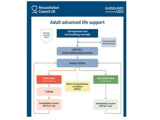
• TheALS algorithm
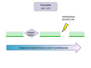
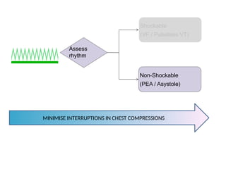
• Importance of high quality chest compressions
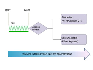
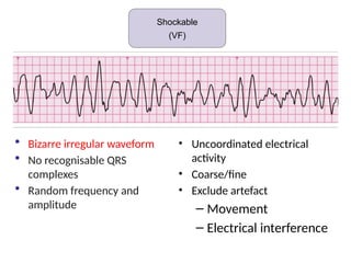
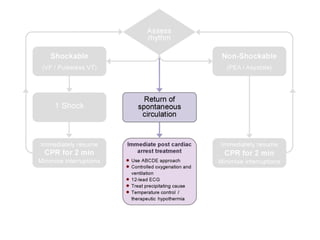
• Treatment of shockable and non-shockable rhythms
• Administration of drugs during cardiac arrest
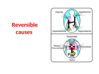
• Potentially reversible causes of cardiac arrest
• Role of resuscitation team
4.
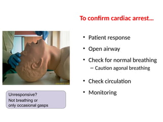
• Patient response
•Open airway
• Check for normal breathing
– Caution agonal breathing
• Check circulation
• Monitoring
To confirm cardiac arrest…
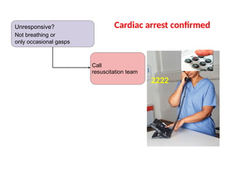
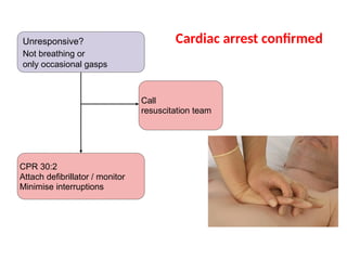
Unresponsive?
Not breathing or
only occasional gasps
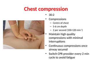
Chest compression
• 30:2
•Compressions
– Centre of chest
– 5-6 cm depth
– 2 per second (100-120 min-1
)
• Maintain high quality
compressions with minimal
interruptions
• Continuous compressions once
airway secured
• Switch CPR provider every 2 min
cycle to avoid fatigue
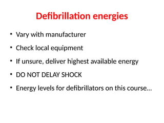
• Vary withmanufacturer
• Check local equipment
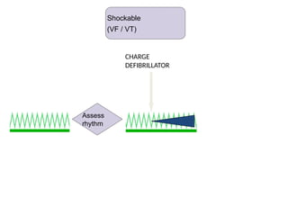
• If unsure, deliver highest available energy
• DO NOT DELAY SHOCK
• Energy levels for defibrillators on this course…
Defibrillation energies
18.
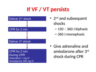
• 2nd
and subsequent
shocks
–150 – 360 J biphasic
– 360 J monophasic
• Give adrenaline and
amiodarone after 3rd
shock during CPR
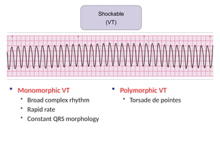
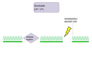
If VF / VT persists
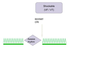
CPR for 2 min
CPR for 2 min
During CPR
Adrenaline 1 mg IV
Amiodarone 300 mg IV
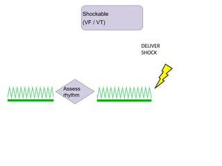
Deliver 2nd
shock
Deliver 3rd
shock
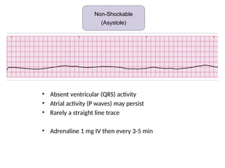
• Absent ventricular(QRS) activity
• Atrial activity (P waves) may persist
• Rarely a straight line trace
• Adrenaline 1 mg IV then every 3-5 min
Non-shockable (Asystole)
Non-Shockable
(Asystole)
21.
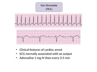
• Clinical featuresof cardiac arrest
• ECG normally associated with an output
• Adrenaline 1 mg IV then every 3-5 min
Non-shockable (Asystole)
Non-Shockable
(PEA)
23.
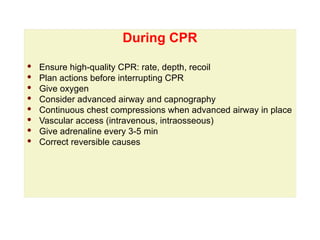
During CPR
During CPR
·Ensure high-quality CPR: rate, depth, recoil
· Plan actions before interrupting CPR
· Give oxygen
· Consider advanced airway and capnography
· Continuous chest compressions when advanced airway in place
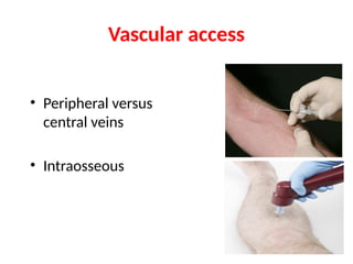
· Vascular access (intravenous, intraosseous)
· Give adrenaline every 3-5 min
· Correct reversible causes
24.
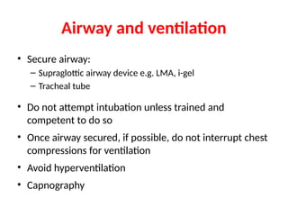
Airway and ventilation
•Secure airway:
– Supraglottic airway device e.g. LMA, i-gel
– Tracheal tube
• Do not attempt intubation unless trained and
competent to do so
• Once airway secured, if possible, do not interrupt chest
compressions for ventilation
• Avoid hyperventilation
• Capnography
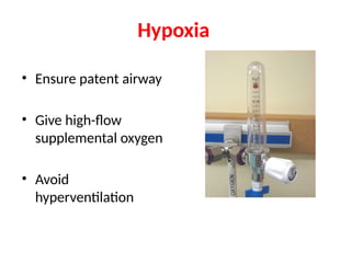
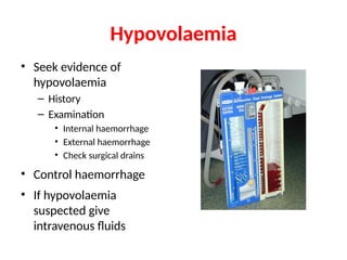
Hypovolaemia
• Seek evidenceof
hypovolaemia
– History
– Examination
• Internal haemorrhage
• External haemorrhage
• Check surgical drains
• Control haemorrhage
• If hypovolaemia
suspected give
intravenous fluids
29.
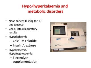
Hypo/hyperkalaemia and
metabolic disorders
•Near patient testing for K+
and glucose
• Check latest laboratory
results
• Hyperkalaemia
– Calcium chloride
– Insulin/dextrose
• Hypokalaemia/
Hypomagnesaemia
– Electrolyte
supplementation
30.
Hypothermia
• Rare ifpatient is an
in-patient
• Use low reading
thermometer
• Treat with active
rewarming techniques
• Consider
cardiopulmonary
bypass
31.
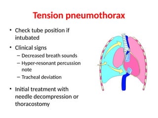
Tension pneumothorax
• Checktube position if
intubated
• Clinical signs
– Decreased breath sounds
– Hyper-resonant percussion
note
– Tracheal deviation
• Initial treatment with
needle decompression or
thoracostomy
32.
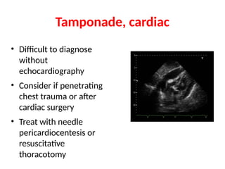
Tamponade, cardiac
• Difficultto diagnose
without
echocardiography
• Consider if penetrating
chest trauma or after
cardiac surgery
• Treat with needle
pericardiocentesis or
resuscitative
thoracotomy
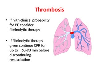
Thrombosis
• If highclinical probability
for PE consider
fibrinolytic therapy
• If fibrinolytic therapy
given continue CPR for
up to 60-90 min before
discontinuing
resuscitation
35.
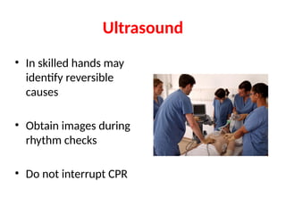
Ultrasound
• In skilledhands may
identify reversible
causes
• Obtain images during
rhythm checks
• Do not interrupt CPR
Resuscitation team
• Rolesplanned in advance
• Identify team leader
• Importance of non-technical skills
– Task management
– Team working
– Situational awareness
– Decision making
• Structured
communication
• The ALSalgorithm
• Importance of high quality chest compressions
• Treatment of shockable and non-shockable
rhythms
• Administration of drugs during cardiac arrest
• Potentially reversible causes of cardiac arrest
• Role of resuscitation team
Summary


![ONFH[AVN HIP] -TRIPLE REGIME -A NOVAL SURGICAL CONCEPT .pptx](https://cdn.slidesharecdn.com/ss_thumbnails/onfhavnhip2026koaconcalicutdrgokuldevdrmashraf-260210064517-213ec005-thumbnail.jpg?width=640&height=640&fit=bounds)


![PERI-PROSTHETIC FRACTURE NAIL-PLATE CONSTRUCT [NPC].pptx](https://cdn.slidesharecdn.com/ss_thumbnails/drarunkumardrmohamedashrafperiprostheticfrasturenail-plateconstructnpc-260209164459-7e9d15a1-thumbnail.jpg?width=640&height=640&fit=bounds)

