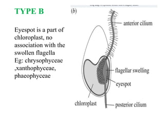
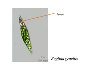
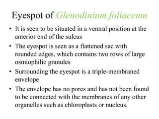
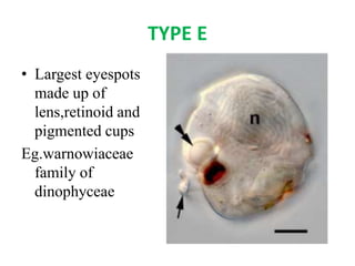
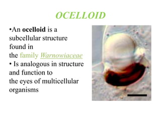
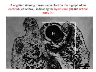
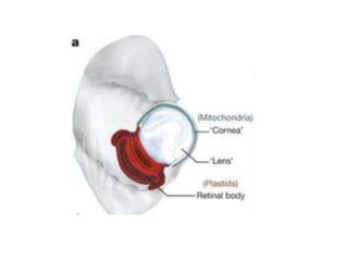
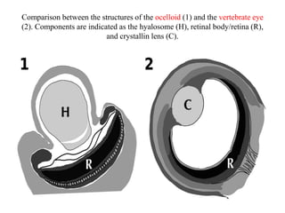
ALGAL EYESPOT
JASMINE MARIYA A J
DEPT. OF BOTANY ,CMS COLLEGE KOTTAYAM
EYESPOT
• Pigmented area in certain motile algae
involved directly or indirectly in
photoreception( Bold & Wynne )
• Is also known as stigma
• Cluster of lipid globules in red red colour of
carotenoid
• Often seen just beneath the chromatophore
membrane
Function :
• Receiving light stimuli
• Eyespot helps chlamydomonas to swim toward
the light
There are 5 different types of eyespots
based on EM studies by Dodge (1969)
TYPE A
Is a part of
chloroplast but has
no association with
flagella
Eg. Chlorophyceae
and cryptophyceae
The eyespot apparatus of Chlamydomonas reinhardtii in
the light microscope (A1, white arrow-head), in the
transmission electron microscope (A2) and as a schematic
drawing (A3). Structural intact eyespot apparatuses can be
isolated by sucrose density gradient centrifugation (B).
Eyespot apparatus of
Chlamydomonas reinhardtii
• It is composed of two highly ordered layers of
carotenoid rich lipid globules inside the
chloroplast
• Thee globules exhibit a remarkably a constant
diameter
• They are seen subtended between thylakoid
membranes
• The outer most globule layer is attached to
specialized areas of the two chloroplast envelop
membranes and the adjacent plasma membrane
EM image of eyespot of chlamydomonas
TYPE B
Eyespot is a part of
chloroplast, no
association with the
swollen flagella
Eg: chrysophyceae
,xanthophyceae,
phaeophyceae
TYPE C
• Eyespots are
independent
clusters of
osmophilic
granules
Eg.
euglenophyceae
Euglina gracilis
Eyespot
E. variabilis
TYPE D
• Osmophilic
granules of
eyespots have
membraneous
structures
Eg.Dinophyceae
Eyespot of Glenodinium foliaceum
• It is seen to be situated in a ventral position at the
anterior end of the sulcus
• The eyespot is seen as a flattened sac with
rounded edges, which contains two rows of large
osmiophilic granules
• Surrounding the eyespot is a triple-membraned
envelope
• The envelope has no pores and has not been found
to be connected with the membranes of any other
organelles such as chloroplasts or nucleus.
TYPE E
• Largest eyespots
made up of
lens,retinoid and
pigmented cups
Eg.warnowiaceae
family of
dinophyceae
OCELLOID
•An ocelloid is a
subcellular structure
found in
the family Warnowiaceae
• Is analogous in structure
and function to
the eyes of multicellular
organisms
Structure of an ocelloid
• Ocelloids contain subcomponents analogous to
eye structures including the lens, cornea, iris,
and retina
• It can be divided into two substructures, the
translucent, roundish hyalosome and the
heavily pigmented melanosome, also known as
the retinal body or pigment cup
A negative staining transmission electron micrograph of an
ocelloid (white box), indicating the hyalosome (H) and retinal
body (R)
• The hyalosome serves as the
refractive lens of the ocelloid; it is
surrounded by a layer
of mitochondria serving as the cornea and
has constrictive rings analogous to the iris
• The retinal body has internal structure
reminiscent of thylakoid membranes
in chloroplasts and contains proteins
related to bacteriorhodopsin, a light-
sensitive protein found in some archaea
Comparison between the structures of the ocelloid (1) and the vertebrate eye
(2). Components are indicated as the hyalosome (H), retinal body/retina (R),
and crystallin lens (C).

Algal eyespot

  • 1.
    ALGAL EYESPOT JASMINE MARIYAA J DEPT. OF BOTANY ,CMS COLLEGE KOTTAYAM
  • 2.
    EYESPOT • Pigmented areain certain motile algae involved directly or indirectly in photoreception( Bold & Wynne ) • Is also known as stigma • Cluster of lipid globules in red red colour of carotenoid • Often seen just beneath the chromatophore membrane
  • 3.
    Function : • Receivinglight stimuli • Eyespot helps chlamydomonas to swim toward the light There are 5 different types of eyespots based on EM studies by Dodge (1969)
  • 4.
    TYPE A Is apart of chloroplast but has no association with flagella Eg. Chlorophyceae and cryptophyceae
  • 5.
    The eyespot apparatusof Chlamydomonas reinhardtii in the light microscope (A1, white arrow-head), in the transmission electron microscope (A2) and as a schematic drawing (A3). Structural intact eyespot apparatuses can be isolated by sucrose density gradient centrifugation (B).
  • 6.
    Eyespot apparatus of Chlamydomonasreinhardtii • It is composed of two highly ordered layers of carotenoid rich lipid globules inside the chloroplast • Thee globules exhibit a remarkably a constant diameter • They are seen subtended between thylakoid membranes • The outer most globule layer is attached to specialized areas of the two chloroplast envelop membranes and the adjacent plasma membrane
  • 7.
    EM image ofeyespot of chlamydomonas
  • 8.
    TYPE B Eyespot isa part of chloroplast, no association with the swollen flagella Eg: chrysophyceae ,xanthophyceae, phaeophyceae
  • 9.
    TYPE C • Eyespotsare independent clusters of osmophilic granules Eg. euglenophyceae
  • 10.
  • 11.
  • 12.
    TYPE D • Osmophilic granulesof eyespots have membraneous structures Eg.Dinophyceae
  • 13.
    Eyespot of Glenodiniumfoliaceum • It is seen to be situated in a ventral position at the anterior end of the sulcus • The eyespot is seen as a flattened sac with rounded edges, which contains two rows of large osmiophilic granules • Surrounding the eyespot is a triple-membraned envelope • The envelope has no pores and has not been found to be connected with the membranes of any other organelles such as chloroplasts or nucleus.
  • 15.
    TYPE E • Largesteyespots made up of lens,retinoid and pigmented cups Eg.warnowiaceae family of dinophyceae
  • 16.
    OCELLOID •An ocelloid isa subcellular structure found in the family Warnowiaceae • Is analogous in structure and function to the eyes of multicellular organisms
  • 17.
    Structure of anocelloid • Ocelloids contain subcomponents analogous to eye structures including the lens, cornea, iris, and retina • It can be divided into two substructures, the translucent, roundish hyalosome and the heavily pigmented melanosome, also known as the retinal body or pigment cup
  • 18.
    A negative stainingtransmission electron micrograph of an ocelloid (white box), indicating the hyalosome (H) and retinal body (R)
  • 19.
    • The hyalosomeserves as the refractive lens of the ocelloid; it is surrounded by a layer of mitochondria serving as the cornea and has constrictive rings analogous to the iris • The retinal body has internal structure reminiscent of thylakoid membranes in chloroplasts and contains proteins related to bacteriorhodopsin, a light- sensitive protein found in some archaea
  • 21.
    Comparison between thestructures of the ocelloid (1) and the vertebrate eye (2). Components are indicated as the hyalosome (H), retinal body/retina (R), and crystallin lens (C).