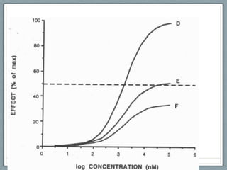
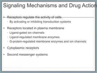
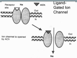
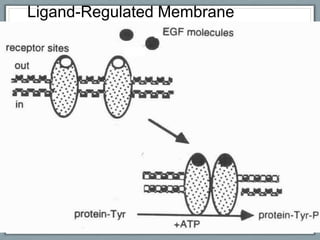
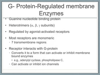
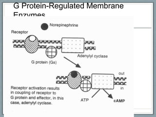
Pharmacodynamics
        University of Miami
   Advanced Practice Preparation
Biotransformation

• Preferable to metabolism – drugs do not normally
  provide new materials or energy.

• Physiochemical reactions that are not a component of
  usual metabolism.

• Prodrug – biotransformed from inactive to active state.
  • Precursor to active compound
  • Widely used to overcome problems with absorption,
    solubility, duration of action, non-compliance, site specific
    drug delivery.
Prodrugs

• Azarabine – prodrug of Azauridine
  • Intesitinal micr0-organisms block the formation of
    toxic components – azauracil.
  • Therapeutic effects beneficial.

• Levodopa, prodrug of dopamine
  • Rapidly absorbed after PO administration –
    distributed to CNS.
  • Converted to dopamine in the basal ganglia.
  • Dopamine is poorly absorbed when given PO –
    biotransformation is necessary.
Mechanisms of
             Biotransformation
• Phase I Reactions
  •   Cytochrome P 450 microsomal enzymes.
  •   Found in the endoplasmic reticulum of the liver cells.
  •   Human genome encodes 57 enzymes.
  •   14 metabolize steroids
  •   4 oxidize fat soluble vitamins
  •   9 metabolize fatty acids and eicosanoids
Biotransformation
                        Classification

• Phase I biosynthetic reaction
 • Introduce or expose a functional group
 • Results in loss of pharmacologic activity
  • Exception is prodrugs
 • Oxidations, reductions, hydroxylations, occur
Biotransformation
                  Classification

• Phase II biosynthetic reaction
 • Conjugation reactions
  • Covalent linkage with functional group on
    parent compound.
  • Highly polar conjugates
  • Generally inactive
   • Exception M-6-G
   • Alkylations, acetylations, methylations occur
     here.
Biotransformation

• Lipophilicity of drugs facilitate passage through biologic
  membranes

• This property hinders elimination and biotransformation

• Renal excretion of unchanged drug plays modest role in
  elimination
  • Lipophilic drugs mostly reabsorbed

• Biotransformation generates polar molecules for excretion
Enzyme Induction
• Repeated administration of same or similar drugs can
  “induce” cytochrome P450
• Accelerates metabolism
• Reduction in pharmacologic activity
• CYP2B1
  • Induced by phenobarbital

• CYP1A1
  • Polycyclic aromatics

• CYP3A
  • Macrolide antibiotics
  • Anticonvulsants
Enzyme Induction
• CYP2E1
  • Induced by chronic EtOH
  • Isoniazid

• Environmental pollutants have been shown to induce P450
  enzymes
  • Benzopyrene
  • Charcoal-broiled meat
  • Certain environmental chemicals
   • Polychlorinated Biphenyls (PCBs)
Enzyme Inhibition
• Inhibition of biotransformation results in elevated
  levels of the parent compound
  • Prolonged pharmacologic effects


• Inhibition of CYP2D6 by quinidine


• Cimetidine and ketoconazole inhibit oxidative drug
  metabolism
  • Bind with heme iron
Pharmacogenetics

• Genetic polymorphisms
  • Autosomal recessive traits
  • Differences in abilities to metabolize certain
    drugs
  • Extensive vs. poor (slow) metabolizers
    • Poor metabolizers are at increased risk of
      adverse effects
Pharmacogenetics
• Genetic polymorphisms
 • Affects oxidative drug metabolism
 • Autosomal recessive traits
 • Differences in abilities to metabolize
   certain drugs
 • Extensive vs. poor (slow) metabolizers
  • Poor metabolizers are at increased risk of
    adverse effects
Pharmacogenetics
• Genetic polymorphisms
 • Poor metabolizers of debrisoquin
  • 8% - 10% of Caucasions
  • 0% - 2% of Asians
 • Faulty expression of cytochrome
   P450 isozyme (P4502D6)
 • Dextromethorphan is used to monitor
   pathway
Pharmacogenetics
• Genetic polymorphisms
 • N-acetylation
  • Slow acetylation of certain arylamines
   • Procainamide
   • Hydralazine
   • Isoniazid
  • 50% of US are slow acetylators
  • 10% of Japanese and Chinese are slow
    acetylators
  • Important with drugs that have low therapeutic
    index
Genetic Polymorphisms

• Debrisoquin oxidation   • N-Acetylation
  • Alprenolol              • p-Aminobenzoic acid
  • Amitriptyline           • Aminogluthemide
  • Desipramine             • *Caffiene
  • *Dextromethorphan       • Clonazepam
  • Encainide               • Dapsone
  • Guanoxan                • Hydralazine
  • Metoprolol              • Isoniazid
                            • Procainamide
                            • Phenelzine
Elimination Kinetics
                    First-order Kinetics
• Elimination is proportional to the concentration

• A constant fraction of drug is eliminated per unit time
  • e.g., sulfisoxazole (10% /hour)

• Elimination rate varies with the first power of the
  concentration

• Most drugs follow this pattern


                                      dC(t) = -kEC(T)
                                      dt
Elimination Kinetics
                    Zero-order Kinetics

• A constant amount of drug is eliminated per unit time
  • e.g., phenytoin & ethanol

• Rate is independent of concentration

• Result of overwhelmed enzyme system

• Saturation kinetics


                                C(t) = e-kEnt 1/2
                                C0
Renal Elimination of Drug

• Glomerular filtration – GRF 125 ml/min


  • Drugs enter through capillary plexus

  • Free drug flows into Bowman’s space

  • 20% of renal plasma flow – RPF 600 ml/min

  • Lipid solubility and pH have no influence here
Renal Elimination of Drugs

• Proximal Tubular Secretion
  • Drug not transferred to glomerular filtrate enters
    through afferent arterioles into capillary plexus –
    surrounds the nephric lumen.
  • Secretion occurs by 2 active transport systems
    •   Anionic – deprotonated forms of weak acids
    •   Cationic – protonated forms of weak bases
    •   Low specificity
    •   Competitive between multiple drugs
Renal Elimination of Drugs

• Distal tubular reabsorption
  • Distal convoluted tubule receives drug
  • Concentration exceeds capacity of perivascular
    space
  • If uncharged, drug may diffuse out of nephric lumen
  • pH manipulation plays a part here – Ion Trapping
Biliary Elimination

• Most drugs eliminated by kidney.

• Some removed via enterohepatic recirculation.
  •   Lipid soluble drugs present in bile
  •   Reabsorbed
  •   Returned to liver
  •   Re-secreted into bil
  •   Increase in plasma concentration of drug
  •   Delay in elimination
Half-Life t1/2
• Time necessary for drug concentration in plasma to decrease
  by one half
  • Often related to duration of action

• If peak plasma concentration and half-life are known, then
  plasma concentration at any time can be estimated

  t1/2 = 0.693 x Vd
                  CL
Accumulation


Elimination
Half-Life t1/2
 Time after Peak     Plasma Concentration
Concentration (hr)         (mg/L)
        0                    100
        2                     50
        4                     25
        6                    12.5
        8                    6.25
       10                    3.125
Steady State

• Dosing dependent
• Accumulation vs Elimination
• Repeated dosing
• 4 to 5 half-lives are necessary for plasma drug levels
  to reach a steady state
• Steady state can be calculated by multiplying drug
  half-life by 5.
  • HL x 5 = SS
  • Approx. 90% SS value
Clearance
• Measure of the removal of drug from plasma
  • Expressed as volume/time
• A drug’s clearance and volume of distribution
  determine drug half-life
• Drugs can be cleared from plasma by several
  mechanisms
  • Hepatic transformation, renal, biliary etc.
Fig. Model for Organ Clearance of a Drug

                                     Organ of
    Cin                             Elimination                                  Cout
drug drug
                  Q              (Liver or Kidney)                      Q
drug drug                                                                        drug
drug drug


      Cin     Cout
E
            Cin

Extraction ratio (no units) is                      Drug is eliminated in the
a fraction between 0 and 1.           drug drug     bile and/or the urine
                                      drug drug
                                      drug drug


          CLorgan     Q   E
                                                  Q = blood flow (volume . time-1)
                                                  C = concentration (amount . volume-1)
Drug Modeling
• Use of modeling
 • Compartmental models
  • Used to describe drugs’ behavior in the
    body
  • Do not represent single tissue or fluid
   • Groups of similar tissues
Compartmental Models
• One-compartment model
  • Simplest
  • All body tissues and fluids
  • Instantaneous distribution is assumed
• Two-compartment model
  • Some drugs do not distribute instantaneously to all
    body parts
  • Distribute rapidly to vessel-rich groups
  • More slowly to other tissues
    • Peripheral compartment
    • Drugs move back and forth between these
      compartments
One Compartment Model
• Simplest model
• Comprises all body tissues and fluids
• Assumes instantaneous distribution of
  the dose of the drug throughout the
  body
Drug Dose   Compartment


            Elimination
Two Compartment Model

• Central Compartment            • Peripheral Compartment
  • Highly Perfused Tissues        • Less perfused tissues
  • Rapid Distribution of Drug     • Slower distribution of drug
Fig. One and Two Compartment Models


          Dose                                      Dose
       X0    kin or k0                           X0    kin or k0              Peripheral
                                                                              Compartment
     One                                 Central                    k12       (V2)
     Compartment                         Compartment
     (Vd)                                (Vc or V1)
                                                                    k21
               kout or ke                 kout or ke     or k1O


X0 = dose of drug at time zero; units are amount
kin or k0 = infusion rate constant at time zero; units are amount . time-1
k12 = rate constant for transfer of drug from the 1st (central) to the 2nd (peripheral) compartment
k21 = rate constant for transfer of drug from the 2nd (peripheral) to the 1st (central) compartment
kout or ke = first-order elimination rate constant; units are time-1
k1O = first-order elimination rate constant from the 1st (central) compartment; units are time-1
Compartmental Models – Clinical Correlate
• Digoxin
 • Two-compartment pharmacokinetics
 • Plasma concentrations rise initially
  • Decline rapidly as drug redistributes to
    muscle
 • Plasma concentration is the central
   compartment
 • Muscle is the peripheral compartment
Pharmacodynamics

• Study of biological and
  physiological effects of drugs and
  their mechanisms of actions.
Pharmacodynamics
       Properties of Drug Receptors
• Interactions with macromolecular components
  • Based on work by Ehrlich and Langley (1900s)
  • Receptor was term used to denote part of organism that
    reacted with drug
  • All drugs do not act at receptors
   •   Osmotic diuretics - mannitol, urea
   •   Non-specific membrane interactions
   •   Antacids
   •   Chelating agents
Properties of Drug Receptors
• Drugs interact with receptors
  • Receptors mediate physiologic regulators of cell function
      • Hormones, neurotransmitters, autocoids etc.

• Drugs may mimic the actions or block the physiologic
  messengers
  •   Agonists
  •   Antagonists
  •   Partial agonists
  •   Inverse agonists
Properties of Drug Receptors
• Agonists
 • Drugs that produce a response by activating
   a receptor
• Antagonists
 • Drugs that reduce or prevent the effects of
   agonists
• Partial agonists
 • Drugs that may act as either agonist or
   antagonist
Properties of Drug Receptors
• Drug receptors are often proteins
 • Soluble proteins
 • Protein in intracellular organelle (bacterial
   ribosomal protein)
 • Membrane bound protein (Na+-K+ ATPase)
Properties of Drug Receptors
• To activate a receptor a ligand
  (drug) must bind to that receptor
 • Covalent bonds
 • Ionic bonds
 • Hydrogen bonds
Properties of Drug Receptors
• Structure-Activity relationships (drug
  shape)
 • Intrinsic activity and affinity determined by
   chemical structure
   • The fit of drug with the receptor
 • Stereoisomerisms
   • One is a better fit with the receptor
   • Some drugs are racemic mixtures or individual
     isomers
Antagonists
• Agonists and antagonists both
  occupy receptors
 • Only agonists activate receptors
   • k3 is high for agonists
• Antagonists have little or no intrinisic
  activity
   • k3 is equal to zero or a very low value
• Antagonists may act competitively or
  non-competitively
Antagonists
• Competitive antagonists bind
  reversibly to the same receptor site
  as the agonists
 • Prevents agonist from binding
 • Potency of antagonist is determined by
   the rate of dissociation
  • Usually slower than the dissociation of the
    agonist
Competitive Antagonism
Antagonists
• Non-competitive antagonists can act
  in two ways.
 • Irreversibly bind (covalently) with the
   receptor site that the agonist binds
  • Cannot be competed by high concentrations
    of agonist
 • An irreversible OR reversible non-
   competitive antagonist may bind to a
   different site on the receptor
  • Can regulate affinity of agonist for that site
  • This is an allosteric interaction
Partial Agonists
• Some drugs have intermediate activity between
  full agonists and antagonists
  • Full agonists have high efficacy

• Partial agonists only activate a fraction of the
  receptors that they bind to

• Effects will depend upon other agonists or
  antagonists that are in equilibrium with the
  receptor
Partial Agonists
• Partial agonist may produce an effect of lessor
  magnitude than that of a full agonist

• Partial agonist may produce a maximum response in
  the presence of spare receptors

• Partial agonists can prevent full agonists from inducing
  a maximum effect
  • By competing with full agonist for receptors
Signaling Mechanisms and Drug Action

• Receptors regulate the activity of cells
  • By activating or inhibiting transduction systems

• Receptors located in plasma membrane
  • Ligand-gated ion channels
  • Ligand-regulated membrane enzymes
  • G-protein-regulated membrane enzymes and ion channels

• Cytoplasmic receptors

• Second messenger systems
Ligand-
Gated Ion
Channel
Ligand-Regulated Membrane
Enzymes
G- Protein-Regulated membrane
            Enzymes
• Guanine nucleotide binding protein
• Heterotrimers ( , , subunits)
• Regulated by agonist-activated receptors
• Most receptors are monomeric
  • 7 transmembrane regions

• Receptor interacts with G-protein
  • Converts it to a form that can activate or inhibit membrane
    bound enzymes
    • e.g., adenylyl cyclase, phospholipase C,
  • Can activate or inhibit ion channels
G Protein-Regulated Membrane
Enzymes
Second Messengers
• Extracellular ligands can act by increasing concentrations
  of second messengers
  • e.g., cyclic adenosine-3’5’ -monophosphate (cAMP), calcium
    ions, or phosphoinositides
Points to Consider

• Drug action occurs at the cellular level.
• Drug effects influence total body functioning.
• Receptors are specialized proteins, cell
  membranes, or enzymes – The stronger the
  affinity for a receptor, the longer the drug action.
• The intensity of response elicited by a drug is a
  function of the dose administered.
  • Dosage increases, most persons response to drug is
    increased
Points to Consider

• Drugs are agonists when they interact with a
  receptor to produce an effect of their own.

• Drugs are antagonists when they interact with a
  receptor to produce no response of their own, but
  impair the receptors ability to combine with
  effector molecule.

• Irreversible antagonists remain tightly bound to
  receptors. The binding cannot be overcome by
  reducing dosage.

Advanced practice preparation pharmacodynamics[1]

  • 1.
    Pharmacodynamics University of Miami Advanced Practice Preparation
  • 2.
    Biotransformation • Preferable tometabolism – drugs do not normally provide new materials or energy. • Physiochemical reactions that are not a component of usual metabolism. • Prodrug – biotransformed from inactive to active state. • Precursor to active compound • Widely used to overcome problems with absorption, solubility, duration of action, non-compliance, site specific drug delivery.
  • 3.
    Prodrugs • Azarabine –prodrug of Azauridine • Intesitinal micr0-organisms block the formation of toxic components – azauracil. • Therapeutic effects beneficial. • Levodopa, prodrug of dopamine • Rapidly absorbed after PO administration – distributed to CNS. • Converted to dopamine in the basal ganglia. • Dopamine is poorly absorbed when given PO – biotransformation is necessary.
  • 4.
    Mechanisms of Biotransformation • Phase I Reactions • Cytochrome P 450 microsomal enzymes. • Found in the endoplasmic reticulum of the liver cells. • Human genome encodes 57 enzymes. • 14 metabolize steroids • 4 oxidize fat soluble vitamins • 9 metabolize fatty acids and eicosanoids
  • 5.
    Biotransformation Classification • Phase I biosynthetic reaction • Introduce or expose a functional group • Results in loss of pharmacologic activity • Exception is prodrugs • Oxidations, reductions, hydroxylations, occur
  • 6.
    Biotransformation Classification • Phase II biosynthetic reaction • Conjugation reactions • Covalent linkage with functional group on parent compound. • Highly polar conjugates • Generally inactive • Exception M-6-G • Alkylations, acetylations, methylations occur here.
  • 7.
    Biotransformation • Lipophilicity ofdrugs facilitate passage through biologic membranes • This property hinders elimination and biotransformation • Renal excretion of unchanged drug plays modest role in elimination • Lipophilic drugs mostly reabsorbed • Biotransformation generates polar molecules for excretion
  • 8.
    Enzyme Induction • Repeatedadministration of same or similar drugs can “induce” cytochrome P450 • Accelerates metabolism • Reduction in pharmacologic activity • CYP2B1 • Induced by phenobarbital • CYP1A1 • Polycyclic aromatics • CYP3A • Macrolide antibiotics • Anticonvulsants
  • 9.
    Enzyme Induction • CYP2E1 • Induced by chronic EtOH • Isoniazid • Environmental pollutants have been shown to induce P450 enzymes • Benzopyrene • Charcoal-broiled meat • Certain environmental chemicals • Polychlorinated Biphenyls (PCBs)
  • 10.
    Enzyme Inhibition • Inhibitionof biotransformation results in elevated levels of the parent compound • Prolonged pharmacologic effects • Inhibition of CYP2D6 by quinidine • Cimetidine and ketoconazole inhibit oxidative drug metabolism • Bind with heme iron
  • 11.
    Pharmacogenetics • Genetic polymorphisms • Autosomal recessive traits • Differences in abilities to metabolize certain drugs • Extensive vs. poor (slow) metabolizers • Poor metabolizers are at increased risk of adverse effects
  • 12.
    Pharmacogenetics • Genetic polymorphisms • Affects oxidative drug metabolism • Autosomal recessive traits • Differences in abilities to metabolize certain drugs • Extensive vs. poor (slow) metabolizers • Poor metabolizers are at increased risk of adverse effects
  • 13.
    Pharmacogenetics • Genetic polymorphisms • Poor metabolizers of debrisoquin • 8% - 10% of Caucasions • 0% - 2% of Asians • Faulty expression of cytochrome P450 isozyme (P4502D6) • Dextromethorphan is used to monitor pathway
  • 14.
    Pharmacogenetics • Genetic polymorphisms • N-acetylation • Slow acetylation of certain arylamines • Procainamide • Hydralazine • Isoniazid • 50% of US are slow acetylators • 10% of Japanese and Chinese are slow acetylators • Important with drugs that have low therapeutic index
  • 16.
    Genetic Polymorphisms • Debrisoquinoxidation • N-Acetylation • Alprenolol • p-Aminobenzoic acid • Amitriptyline • Aminogluthemide • Desipramine • *Caffiene • *Dextromethorphan • Clonazepam • Encainide • Dapsone • Guanoxan • Hydralazine • Metoprolol • Isoniazid • Procainamide • Phenelzine
  • 17.
    Elimination Kinetics First-order Kinetics • Elimination is proportional to the concentration • A constant fraction of drug is eliminated per unit time • e.g., sulfisoxazole (10% /hour) • Elimination rate varies with the first power of the concentration • Most drugs follow this pattern dC(t) = -kEC(T) dt
  • 18.
    Elimination Kinetics Zero-order Kinetics • A constant amount of drug is eliminated per unit time • e.g., phenytoin & ethanol • Rate is independent of concentration • Result of overwhelmed enzyme system • Saturation kinetics C(t) = e-kEnt 1/2 C0
  • 19.
    Renal Elimination ofDrug • Glomerular filtration – GRF 125 ml/min • Drugs enter through capillary plexus • Free drug flows into Bowman’s space • 20% of renal plasma flow – RPF 600 ml/min • Lipid solubility and pH have no influence here
  • 20.
    Renal Elimination ofDrugs • Proximal Tubular Secretion • Drug not transferred to glomerular filtrate enters through afferent arterioles into capillary plexus – surrounds the nephric lumen. • Secretion occurs by 2 active transport systems • Anionic – deprotonated forms of weak acids • Cationic – protonated forms of weak bases • Low specificity • Competitive between multiple drugs
  • 21.
    Renal Elimination ofDrugs • Distal tubular reabsorption • Distal convoluted tubule receives drug • Concentration exceeds capacity of perivascular space • If uncharged, drug may diffuse out of nephric lumen • pH manipulation plays a part here – Ion Trapping
  • 22.
    Biliary Elimination • Mostdrugs eliminated by kidney. • Some removed via enterohepatic recirculation. • Lipid soluble drugs present in bile • Reabsorbed • Returned to liver • Re-secreted into bil • Increase in plasma concentration of drug • Delay in elimination
  • 23.
    Half-Life t1/2 • Timenecessary for drug concentration in plasma to decrease by one half • Often related to duration of action • If peak plasma concentration and half-life are known, then plasma concentration at any time can be estimated t1/2 = 0.693 x Vd CL
  • 24.
  • 25.
    Half-Life t1/2 Timeafter Peak Plasma Concentration Concentration (hr) (mg/L) 0 100 2 50 4 25 6 12.5 8 6.25 10 3.125
  • 26.
    Steady State • Dosingdependent • Accumulation vs Elimination • Repeated dosing • 4 to 5 half-lives are necessary for plasma drug levels to reach a steady state • Steady state can be calculated by multiplying drug half-life by 5. • HL x 5 = SS • Approx. 90% SS value
  • 27.
    Clearance • Measure ofthe removal of drug from plasma • Expressed as volume/time • A drug’s clearance and volume of distribution determine drug half-life • Drugs can be cleared from plasma by several mechanisms • Hepatic transformation, renal, biliary etc.
  • 29.
    Fig. Model forOrgan Clearance of a Drug Organ of Cin Elimination Cout drug drug Q (Liver or Kidney) Q drug drug drug drug drug drug drug drug Cin Cout E Cin Extraction ratio (no units) is Drug is eliminated in the a fraction between 0 and 1. drug drug bile and/or the urine drug drug drug drug CLorgan Q E Q = blood flow (volume . time-1) C = concentration (amount . volume-1)
  • 30.
    Drug Modeling • Useof modeling • Compartmental models • Used to describe drugs’ behavior in the body • Do not represent single tissue or fluid • Groups of similar tissues
  • 31.
    Compartmental Models • One-compartmentmodel • Simplest • All body tissues and fluids • Instantaneous distribution is assumed • Two-compartment model • Some drugs do not distribute instantaneously to all body parts • Distribute rapidly to vessel-rich groups • More slowly to other tissues • Peripheral compartment • Drugs move back and forth between these compartments
  • 32.
    One Compartment Model •Simplest model • Comprises all body tissues and fluids • Assumes instantaneous distribution of the dose of the drug throughout the body
  • 33.
    Drug Dose Compartment Elimination
  • 34.
    Two Compartment Model •Central Compartment • Peripheral Compartment • Highly Perfused Tissues • Less perfused tissues • Rapid Distribution of Drug • Slower distribution of drug
  • 35.
    Fig. One andTwo Compartment Models Dose Dose X0 kin or k0 X0 kin or k0 Peripheral Compartment One Central k12 (V2) Compartment Compartment (Vd) (Vc or V1) k21 kout or ke kout or ke or k1O X0 = dose of drug at time zero; units are amount kin or k0 = infusion rate constant at time zero; units are amount . time-1 k12 = rate constant for transfer of drug from the 1st (central) to the 2nd (peripheral) compartment k21 = rate constant for transfer of drug from the 2nd (peripheral) to the 1st (central) compartment kout or ke = first-order elimination rate constant; units are time-1 k1O = first-order elimination rate constant from the 1st (central) compartment; units are time-1
  • 36.
    Compartmental Models –Clinical Correlate • Digoxin • Two-compartment pharmacokinetics • Plasma concentrations rise initially • Decline rapidly as drug redistributes to muscle • Plasma concentration is the central compartment • Muscle is the peripheral compartment
  • 40.
    Pharmacodynamics • Study ofbiological and physiological effects of drugs and their mechanisms of actions.
  • 41.
    Pharmacodynamics Properties of Drug Receptors • Interactions with macromolecular components • Based on work by Ehrlich and Langley (1900s) • Receptor was term used to denote part of organism that reacted with drug • All drugs do not act at receptors • Osmotic diuretics - mannitol, urea • Non-specific membrane interactions • Antacids • Chelating agents
  • 42.
    Properties of DrugReceptors • Drugs interact with receptors • Receptors mediate physiologic regulators of cell function • Hormones, neurotransmitters, autocoids etc. • Drugs may mimic the actions or block the physiologic messengers • Agonists • Antagonists • Partial agonists • Inverse agonists
  • 43.
    Properties of DrugReceptors • Agonists • Drugs that produce a response by activating a receptor • Antagonists • Drugs that reduce or prevent the effects of agonists • Partial agonists • Drugs that may act as either agonist or antagonist
  • 44.
    Properties of DrugReceptors • Drug receptors are often proteins • Soluble proteins • Protein in intracellular organelle (bacterial ribosomal protein) • Membrane bound protein (Na+-K+ ATPase)
  • 45.
    Properties of DrugReceptors • To activate a receptor a ligand (drug) must bind to that receptor • Covalent bonds • Ionic bonds • Hydrogen bonds
  • 46.
    Properties of DrugReceptors • Structure-Activity relationships (drug shape) • Intrinsic activity and affinity determined by chemical structure • The fit of drug with the receptor • Stereoisomerisms • One is a better fit with the receptor • Some drugs are racemic mixtures or individual isomers
  • 47.
    Antagonists • Agonists andantagonists both occupy receptors • Only agonists activate receptors • k3 is high for agonists • Antagonists have little or no intrinisic activity • k3 is equal to zero or a very low value • Antagonists may act competitively or non-competitively
  • 48.
    Antagonists • Competitive antagonistsbind reversibly to the same receptor site as the agonists • Prevents agonist from binding • Potency of antagonist is determined by the rate of dissociation • Usually slower than the dissociation of the agonist
  • 49.
  • 50.
    Antagonists • Non-competitive antagonistscan act in two ways. • Irreversibly bind (covalently) with the receptor site that the agonist binds • Cannot be competed by high concentrations of agonist • An irreversible OR reversible non- competitive antagonist may bind to a different site on the receptor • Can regulate affinity of agonist for that site • This is an allosteric interaction
  • 51.
    Partial Agonists • Somedrugs have intermediate activity between full agonists and antagonists • Full agonists have high efficacy • Partial agonists only activate a fraction of the receptors that they bind to • Effects will depend upon other agonists or antagonists that are in equilibrium with the receptor
  • 52.
    Partial Agonists • Partialagonist may produce an effect of lessor magnitude than that of a full agonist • Partial agonist may produce a maximum response in the presence of spare receptors • Partial agonists can prevent full agonists from inducing a maximum effect • By competing with full agonist for receptors
  • 54.
    Signaling Mechanisms andDrug Action • Receptors regulate the activity of cells • By activating or inhibiting transduction systems • Receptors located in plasma membrane • Ligand-gated ion channels • Ligand-regulated membrane enzymes • G-protein-regulated membrane enzymes and ion channels • Cytoplasmic receptors • Second messenger systems
  • 55.
  • 56.
  • 57.
    G- Protein-Regulated membrane Enzymes • Guanine nucleotide binding protein • Heterotrimers ( , , subunits) • Regulated by agonist-activated receptors • Most receptors are monomeric • 7 transmembrane regions • Receptor interacts with G-protein • Converts it to a form that can activate or inhibit membrane bound enzymes • e.g., adenylyl cyclase, phospholipase C, • Can activate or inhibit ion channels
  • 58.
  • 59.
    Second Messengers • Extracellularligands can act by increasing concentrations of second messengers • e.g., cyclic adenosine-3’5’ -monophosphate (cAMP), calcium ions, or phosphoinositides
  • 60.
    Points to Consider •Drug action occurs at the cellular level. • Drug effects influence total body functioning. • Receptors are specialized proteins, cell membranes, or enzymes – The stronger the affinity for a receptor, the longer the drug action. • The intensity of response elicited by a drug is a function of the dose administered. • Dosage increases, most persons response to drug is increased
  • 61.
    Points to Consider •Drugs are agonists when they interact with a receptor to produce an effect of their own. • Drugs are antagonists when they interact with a receptor to produce no response of their own, but impair the receptors ability to combine with effector molecule. • Irreversible antagonists remain tightly bound to receptors. The binding cannot be overcome by reducing dosage.