
Recommended
More Related Content
What's hot
What's hot (20)
Similar to RUPTURE OF THE SPLEEN.pptx
Similar to RUPTURE OF THE SPLEEN.pptx (20)
More from DR.P.S SUDHAKAR
More from DR.P.S SUDHAKAR (20)
Recently uploaded
Recently uploaded (20)
RUPTURE OF THE SPLEEN.pptx
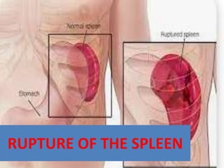
- 1. RUPTURE OF THE SPLEEN
- 2. AETIOLOGY • The spleen is the most common intra-abdominal organ injured in blunt trauma. • Only occasionally there may be spontaneous rupture. In majority of cases rupture of the spleen occurs from • 1.penetrating trauma, • 2.nonpenetrating trauma • 3.operative trauma. • 4. Spontaneous Rupture
- 3. 1.Penetrating Trauma • Gun-shot wounds, missiles and stabings may cause splenic rupture. • The penetration may occur through the anterior abdominal wall, through the flank or trans-thoracically piercing the pleural space, the lung and the diaphragm. • Surrounding organs may be injured, of which the stomach, the left kidney, the pancreas and the root of the mesentery are important.
- 4. 2. Non-penetrating Trauma. • Automobile accidents, bicycle injuries, blows and during various contact sports injury to the spleen may occur. • In blunt trauma other organs besides spleen may be injured, of which the liver, the kidneys, the chest (rib fractures), the lungs, the small intestine, the colon and the stomach are important.
- 5. 3. Operative Trauma. • Spleen is injured in about 2% of operations involving viscera of the left upper quadrant • Injury may also occur from retractors placed against this organ to get exposure to the depth in various operations.
- 6. 4. Spontaneous Rupture • Spontaneous ruptures may only occur when the spleen is pathologic. • Such rupture may occur from minor trauma. • Spleen ruptures more easily when it is enlarged in infectious mononucleosis or malaria. • In infectious mononucleosis, this complication occurs most frequently in the 2nd to 4th weeks of the disease. • In other pathologic conditions also splenic rupture has been reported e.g. sarcoidosis, acute and chronic leukaemia, congestive splenomegaly, haemolytic anaemia and polycythemia vera.
- 7. PATHOLOGY. • Splenic injuries vary from simple transverse tear of the parenchyma to transverse crack of the hilus. • There may be subcapsular haematomas only in minor cases or there may be complete disruption of the organ and its vessels in the fulminating injuries. • Majority of the injuries result in transverse rupture of the parenchyma, the direction of rupture is determined by the internal architecture of the organ which is arranged in transverse fashion in spleen.
- 8. Mainly 3 types of rupture are seen in spleen 1. ACUTE RUPTURE 2. DELAYED RUPTURE 3. OCCULT SPLENIC RUPTURE
- 9. 1. ACUTE RUPTURE • Which occurs mostly due to blunt trauma and is featured by immediate intraperitonealbleeding. • In this variety two types are seen — • In one type the patient succumbs rapidly giving no chance to initiate proper treatment. In the • 2nd type there is initial shock, from where the patient recovers by treatment revealing signs of ruptured spleen. • Fortunately the 2nd type is much more common.
- 10. 2. DELAYED RUPTURE • In this type after an interval of a few days to weeks after injury, sudden intraperitoneal bleeding starts. • This delayed rupture is reported in 10 to 15% of the cases of blunt trauma. • In about half of these cases bleeding occurs within 7 days and in 75% of cases bleeding starts within 2 weeks of the accident. Such delayed type of rupture is probably due to • (a) blood clot, temporarily sealing the rent, becomes lysed by the enzymes of the lacerated tail of the pancreas; • (b) slowly enlarging subcapsular haematoma which eventually ruptures or • (c) the greater omentum, which shuts off the injured site initially, gradually moves off.
- 11. 3. OCCULT SPLENIC RUPTURE • The term is applied when traumatic pseudocyst of the spleen is diagnosed though injury to the organ previously has not been diagnosed. • This type is seen in less than 1% of patients sustaining injury to the spleen. • It is caused by organisation of intrasplenic or parasplenic haematoma.
- 12. • Another condition related to splenic injury is known as splenosis. • It is due to autotransplantation of fragments of ruptured spleen on to the peritoneal surface. • This condition is usually asymptomatic, but patients may present with intestinal obstruction later on due to adhesions.
- 13. CLINICAL FEATURES • The clinical course of an isolated splenic injury is variable depending on severity and rapidity of intra-abdominal haemorrhage. • Laceration through the body of the spleen can extend into the splenic pedicle causing extensive and continued haemorrhage with haemoperitoneum and acute shock. • An adhesion between the spleen and its ligaments or diaphragm may seal the capsular avulsion with cessation of haemorrhage after an initial blood loss of not more than 500 ml.
- 14. • If injury is limited to the capsule or pulp and does not involve the major splenic vasculature, the patient may remain haemodynamically stable. • However subcapsular haematomas have potentiality to rupture later producing ‘delayed rupture’ of the spleen.
- 15. • If a splenic injury is suspected, admission to the hospital for monitoring is mandatory. • A careful history should be obtained regarding mechanism of the injury. • Injury to the left upper abdomen, more so with associatedb fractured ribs, may cause injury to spleen.
- 16. • The signs and symptoms of injury to the spleen depend on severity and rapidity of intra-abdom inal haemorrhage, as also on presence of other organ injuries.
- 17. • (i) Some degree of shock due to hypovolaemia, characterised by tachycardia, low blood pressure, restlessness, increasing pallor and sighing respiration may be seen in majority of cases. • (ii) Local bruising and tenderness in the left upper quadrant of the abdomen is often seen. • (iii) Patient usually complains of generalised upper abdominal pain, which in 1/3 rd of the cases is localised to the upper left quadrant.
- 18. • (iv) Pain may be referred to the tip of the left shoulder, which is known as Kehr’s sign. • Kehr’s sign can be elicited by bimanual compression of the left upper quadrant after the patient has been in Trendelenburg’s position for about 10 minutes prior to the manoeuvre. • There may be hyperaesthesia on the left shoulder. • Kehr’s sign is due to irritation of the undersurface of the diaphragm with blood and the pain is referred to the shoulder through the affected fibres of the phrenic nerve (C4 and C5).
- 20. • (v) On rare occasions a palpable tender mass can be felt in the left upper quadrant with persistent dullness. • This is known as Ballance’s sign. • This sign is due to extracapsular or subcapsular haematoma which is guarded by omentum or by early coagulation of splenic blood.
- 21. • (vi) Tenderness and rigidity of the left upper quadrant is a frequent and reliable physical sign. • (vii) Shifting dullness may be detected on the right side due to intraperitoneal haemorrhage. • (viii) Diagnostic peritoneal lavage is a useful and inexpensive manoeuvre which may reveal intraperitoneal haemorrhage.
- 22. Special Investigations • 1. Haematocrit value may be reduced if there is major bleeding, but initial readings may be normal. • Increase in W.B.C. count (moderate leukocytosis) is noticed in many cases. • 2. Routine STRAIGHT X-RAY of the abdomen often gives confirmatory evidence regarding diagnosis.
- 23. • This investigation is extremely helpful in places where sophisticated investigations are not possible. The probable findings in X-ray in case of splenic rupture are:—
- 24. • (a) Obliteration of the splenic outline, • (b) An enlarged splenic shadow, • (c) Obliteration of the psoas shadow, • (d) Indentation of the left side of the gastric shadow, • (e) Widening of the space between the splenic flexure and the properitoneal pad of fat. • (f) Elevation of the left side of the diaphragm, • (g) Free fluid between gas filled intestinal coils, • (h) Fracture of one or more lower ribs on the left side. A normal well outlined spleen indicates intact spleen on straight X-ray.
- 25. • 3. SPLENIC ANGIOGRAPHY • 4. ULTRASONOGRAPHY • 5. ISOTOPE SCANS • 6. CT SCAN
- 26. • TREATMENT • Immediate laparotomy and splenectomy is the life saving procedure and is still the standard treatment.
- 27. THANK YOU