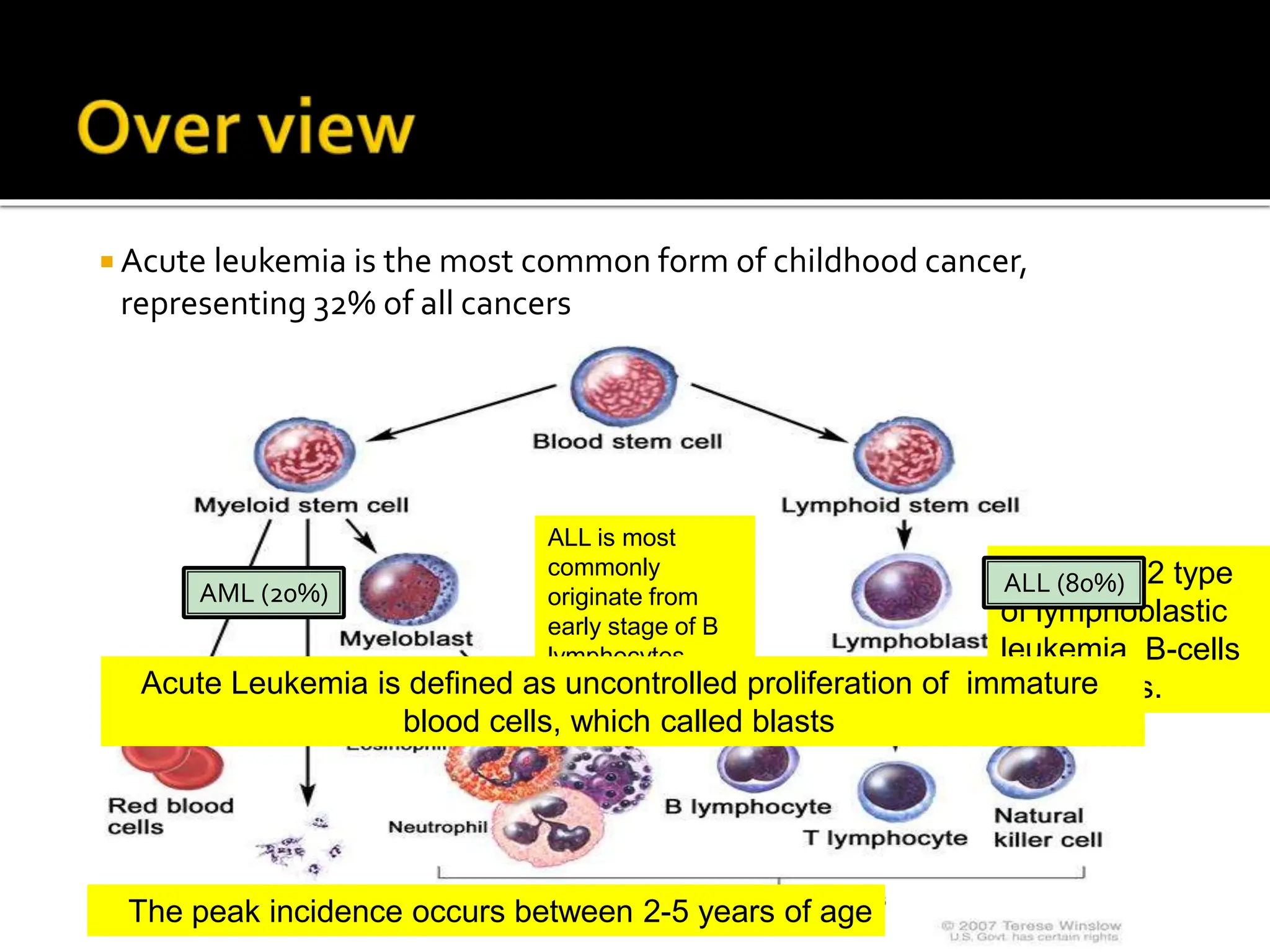
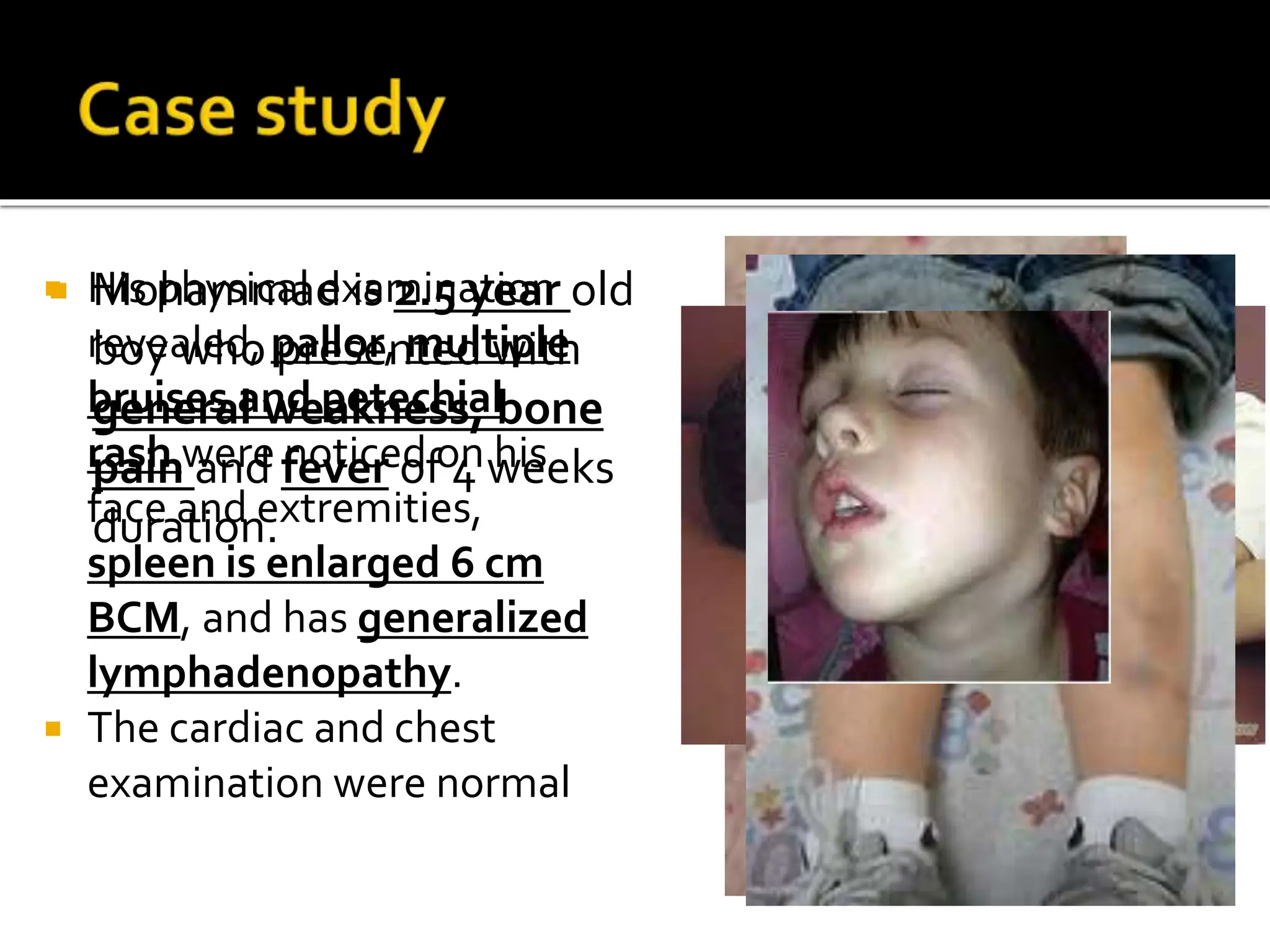
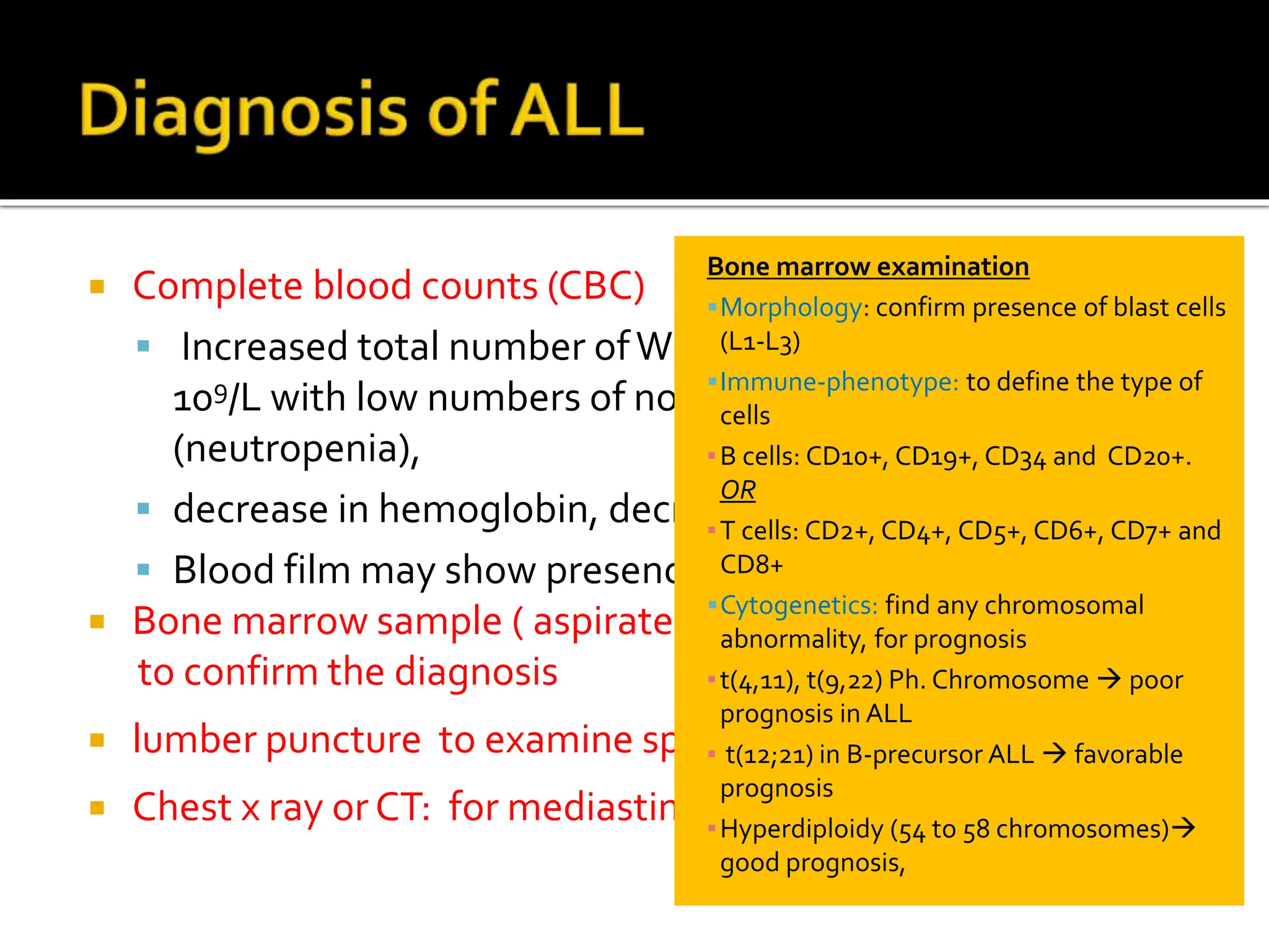
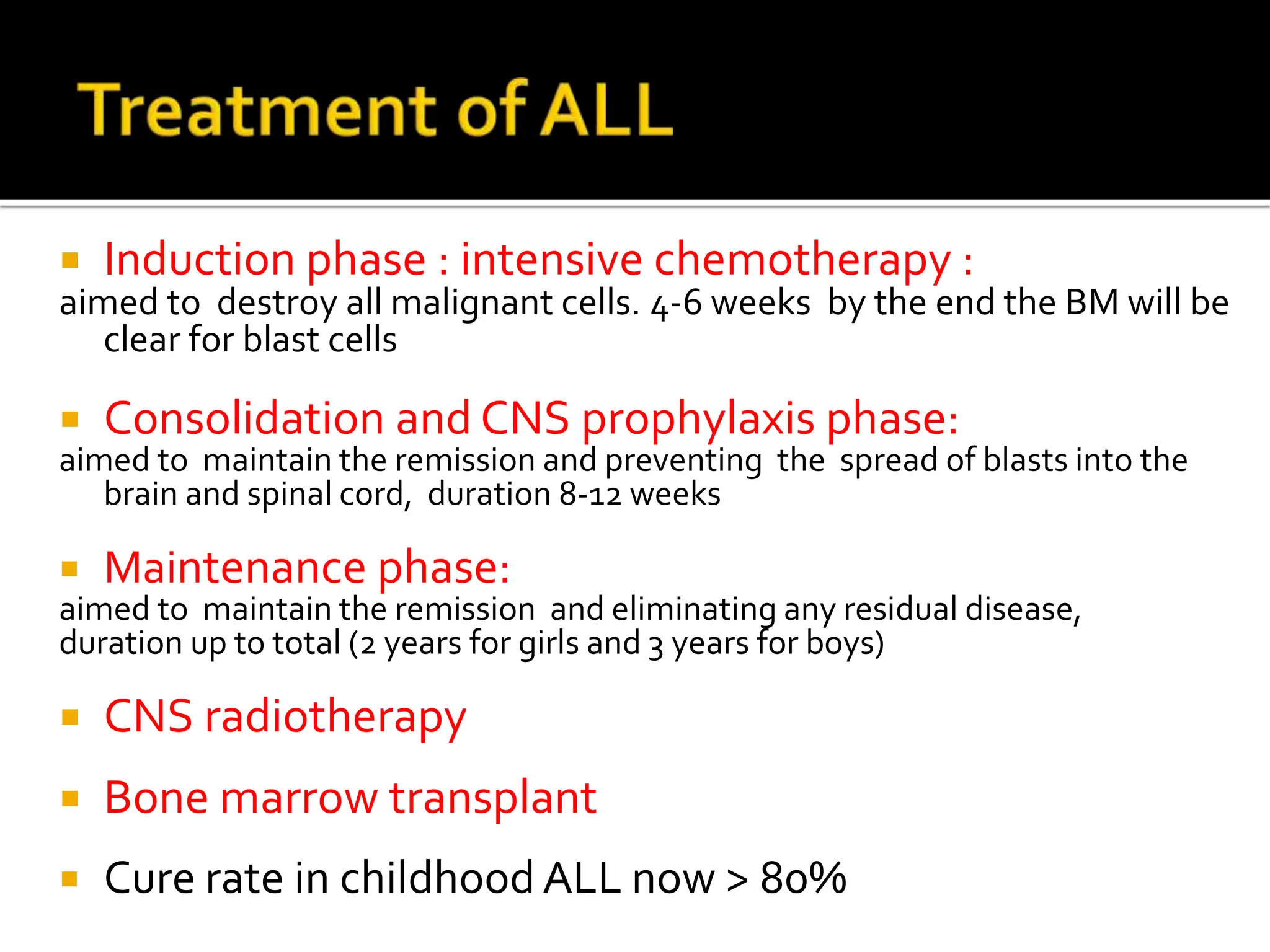
2. Over view of childhood leukemia
Discussion of a case of acute lymphoblastic
leukemia
The common signs and symptoms of ALL
The methods of diagnosis of ALL
Outline the treatment of ALL
3. Acute leukemia is the most common form of childhood cancer,
representing 32% of all cancers
ALL is most
commonly
originate from
early stage of B
lymphocytes
(pre-B)
There are 2 type
of lymphoblastic
leukemia, B-cells
and T-cells.
ALL (80%)
AML (20%)
The peak incidence occurs between 2-5 years of age
Acute Leukemia is defined as uncontrolled proliferation of immature
blood cells, which called blasts
4. His physical examination
revealed, pallor, multiple
bruises and petechial
rash were noticed on his
face and extremities,
spleen is enlarged 6 cm
BCM, and has generalized
lymphadenopathy.
The cardiac and chest
examination were normal
Mohammad is 2.5 year old
boy who presented with
general weakness, bone
pain and fever of 4 weeks
duration.
5. As Lymphoblasts reproduce out of control, crowd out
normal cells in the bone marrow
Decrease in red cells anemia , pallor, lethargy, general
weakness,
Decrease platelets bleeding tendency, bruises
petechial rash
Decrease neutrophils recurrent infection , fever
Increased pressure inside Bone marrow by malignant cells
bone pain
It can go into peripheral bloodstream and invade any
organ/tissue enlarged lymph nodes, enlarged
liver and spleen, mediastinal mass
Usually symptoms become clear after 2-4 weeks
6. Mohammad Laboratory investigation showed
CBC: WBCs 50 X 109 /L , with neutrophils of 1%,
platelets count of 16 X 109 /L , and a Hb of 7 gm/dl.
And a suspicious cells is seen in peripheral smear
(lymphoblast)
Bone marrow aspirate done
Confirm B-lineage ALL
Chest X ray no mediastinal mass
lumber puncture for spinal fluids showed
presence of few lymphoblast
7. Complete blood counts (CBC)
Increased total number ofWBCs (blasts) to more than 10 ×
109/L with low numbers of normal white blood cells
(neutropenia),
decrease in hemoglobin, decrease in platelets count
Blood film may show presence of abnormal cells (blasts)
Bone marrow sample ( aspirate and trephine) is needed
to confirm the diagnosis
no
lumber puncture to examine spinal fluid for malignant cells
Chest x ray or CT: for mediastinal mass
Bone marrow examination
Morphology: confirm presence of blast cells
(L1-L3)
Immune-phenotype: to define the type of
cells
▪B cells: CD10+, CD19+, CD34 and CD20+.
OR
▪T cells: CD2+, CD4+, CD5+, CD6+, CD7+ and
CD8+
Cytogenetics: find any chromosomal
abnormality, for prognosis
▪t(4,11), t(9,22) Ph. Chromosome poor
prognosis in ALL
▪ t(12;21) in B-precursor ALL favorable
prognosis
▪Hyperdiploidy (54 to 58 chromosomes)
good prognosis,
8. We put central venous line to Mohammad which allow us to
give chemotherapy , and we started intensive chemotherapy :
9. Induction phase : intensive chemotherapy :
aimed to destroy all malignant cells. 4-6 weeks by the end the BM will be
clear for blast cells
Consolidation and CNS prophylaxis phase:
aimed to maintain the remission and preventing the spread of blasts into the
brain and spinal cord, duration 8-12 weeks
Maintenance phase:
aimed to maintain the remission and eliminating any residual disease,
duration up to total (2 years for girls and 3 years for boys)
CNS radiotherapy
Bone marrow transplant
Cure rate in childhood ALL now > 80%