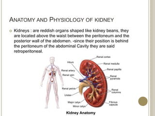
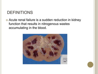
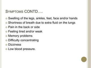
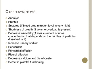
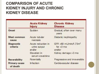
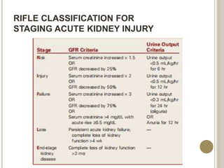
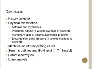
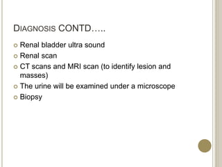
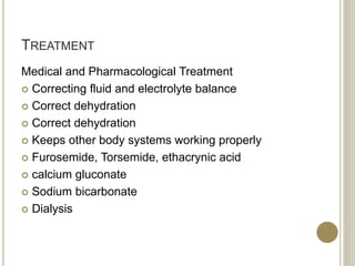
ACUTE KIDNEY FAILURE
BY
Mr. VINOD KUMAR
M.Sc NURSING 1ST YEAR
SRIHER (DU)
ANATOMY AND PHYSIOLOGY OF KIDNEY
 Kidneys : are reddish organs shaped like kidney beans, they
are located above the waist between the peritoneum and the
posterior wall of the abdomen. -since their position is behind
the peritoneum of the abdominal Cavity they are said
retroperitoneal.
KIDNEYS
 The kidneys located between the levels of the last
thoracic and third lumbar vertebrae protected by the
eleventh and twelfth pairs of ribs, the right kidney
slightly lower than the left because the liver takes a
large area on the right side superior to the kidney.
 An average adult kidney is 10-12 cm long, 5-7.5 cm
wide and 2.5 thick. It's concave medial border faces
the vertebral column.
LAYERS OF TISSUE
There are three layers of tissue surround each kidney
:
 1- renal capsule
 2- adipose capsule (perirenal fat)
 3- renal fascia
1-Renal capsule: is the deeper layer and it's smooth,
transparent, fibrous membrane that is continuous with
the coat of the ureter . It serves as a barrier against
trauma and helps to maintain the shape of the kidney.
LAYERS OF TISSUE
 2- adipose capsule: is a mass of fatty tissue
surrounding the renal capsule. It also protects the
kidney from trauma and holds it firmly in place
within the abdominal cavity.
 3- renal fascia: is the superficial layer, and it's a
thin layer of dense, irregular connective tissue that
anchors the kidney to surrounding structure and to
the abdominal wall . On the anterior surface of the
kidneys, the renal fascia is deep to the peritoneum.
ADRENAL GLANDS
 A type of endocrine gland that are triangle- shaped
and located on top of the kidneys. The outer part of
the adrenal glands is known as the cortex and
releases hormones including testosterone and
cortical. The inner area of the adrenal glands is
known as the medulla and produces the hormones
norepinephrine and epinephrine. Whe11 the
adrenal glands produce too much or too little of a
hormone, illness can result.
URINE FORMATION
 Urine formed by the nephrons ultimately drains into
large ducts called papillary ducts. they lead to
cuplike structure called minor and maior calyces.
Each kidney has 8-18 minor calyces and 2-3 major
calyces.
RENAL ARTERY
 Renal Artery: This is the artery responsible for
bringing blood to the kidney from the left side of the
heart. 200 quarts of blood passes through the
kidneys each day, coming through the renal
arteries. That means that this blood contains
glucose and oxygen. The incoming artery going in
to each kidney divides into four or five branches,
and then form arterioles. the abdominal aorta which
branches off into the renal arteries in the right and
left kidney.
RENAL VEIN
 Renal Vein: In this Vein the remainder, filtered blood
is returned to the right side of the heart after all of
the urea and impurities have been removed. the
inferior Vena Cava. It then branches off into the
renal Veins to the left and right kidney.
DEFINITIONS
 Acute renal failure is a sudden reduction in kidney
function that results in nitrogenous wastes
accumulating in the blood.
COMMON CAUSES OF ACUTE KIDNEY
INJURY
Prerenal Intrarenal Postrenal
COMMON CAUSES OF ACUTE KIDNEY
INJURY
Hypovolemia
 Dehydration
 Hemorrhage
 GI losses (diarrhea,
vomiting)
 Excessive diuresis
 Hypoalbuminemia
 Burns
Decreased Cardiac Output
 Cardiac dysrhythmias
 Cardiogenic shock
 Heart failure
 Myocardial infarction
Decreased Peripheral
Vascular Resistance
 Anaphylaxis
 Neurologic injury
 Septic shock
Decreased Renovascular
Blood Flow
 Bilateral renal vein
thrombosis
 Embolism
 Hepatorenal syndrome
 Renal artery thrombosis
Prerenal
COMMON CAUSES OF ACUTE KIDNEY
INJURY
Nephrotoxic Injury
 Drugs: aminoglycosides
(gentamicin, amikacin),
 amphotericin B
 Contrast media
 Hemolytic blood transfusion
reaction
 Severe crush injury
 Chemical exposure: ethylene
glycol, lead, arsenic,
 carbon tetrachloride
Other Causes
 Prolonged prerenal ischemia
 Acute glomerulonephritis
 Thrombotic disorders
 Toxemia of pregnancy
 Malignant hypertension
 Systemic lupus erythematosus
Interstitial Nephritis
 Allergies: antibiotics
(sulfonamides, rifampin),
 nonsteroidal antiinflammatory
drugs, ACE inhibitors
 Infections: bacterial (acute
pyelonephritis), viral (CMV),
fungal (candidiasis)
Intrarenal
COMMON CAUSES OF ACUTE KIDNEY
INJURY
 Benign prostatic
hyperplasia
 Bladder cancer
 Calculi formation
 Neuromuscular
disorders
 Prostate cancer
 Spinal cord disease
 Strictures
 Trauma (back, pelvis,
perineum)
Postrenal
RISK FACTORS
 Advanced age
 Blockages in the blood vessels in your arms or legs
 Diabetes
 High blood pressure
 Heart failure
 Kidney diseases
 Liver disease
PATHOPHYSIOLOGY
 Acute renal failure (ARF) is a sudden and almost
complete loss of kidney function (decreased GFR) over
a period of hours to days. Although ARF is often
thought of as a problem seen only in hospitalized
patients, it may occur in the outpatient setting as well.
ARF manifests with oliguria, anuria, or normal urine
volume. Oliguria (less than 400 mL/day of urine) is the
most common clinical situation seen in ARF; anuria
(less than 50 mL/day of urine) and normal urine output
are not as common. Regardless of the volume of urine
excreted, the patient with ARF experiences rising
serum creatinine and BUN levels and retention of other
metabolic waste products (azotemia) normally excreted
by the kidneys.
COMPARING TYPES OF ACUTE RENAL
FAILURE
SYMPTOMS
 Vomiting and/or diarrhea, which may lead to dehydration
 Nausea
 Weight loss
 Nocturnal urination
 Pale urine
 Less frequent urination, or in smaller amounts than usual, with
dark coloured urine
 Haematuria
 Pressure, or difficulty urinating
 Itching
 Bone damage
 Non-union in broken bones
 Muscle cramps (caused by low levels of calcium which can cause
hypocalcaemia)
 Abnormal heart rhythms
 Muscle paralysis.
SYMPTOMS CONTD….
 Swelling of the legs, ankles, feet, face and/or hands
 Shortness of breath due to extra fluid on the lungs
 Pain in the back or side
 Feeling tired and/or weak
 Memory problems
 Difficulty concentrating
 Dizziness
 Low blood pressure.
OTHER SYMPTOMS
 Anorexia
 Pruritus
 Seizures (if blood urea nitrogen level is very high)
 Shortness of breath (if volume overload is present)
 Decrease osmolality(A measurement of urine
concentration that depends on the number of particles
dissolved in it)
 Increase urinary sodium
 Pericarditis
 Pericardial effusion
 Pleural effusion
 Decrease calcium and bicarbonate
 Defect in platelet functioning
COMPARISON OF ACUTE
KIDNEY INJURY AND CHRONIC
KIDNEY DISEASE
PHASES OF ARF
 Initiating phase
 Oliguric phase
 Diuretic phase
 Recovery phase
RIFLE CLASSIFICATION FOR
STAGING ACUTE KIDNEY INJURY
DIAGNOSIS
 History collection
 Physical examination
 Asterixis and myoclonus
 Peripheral edema (if volume overload is present)
 Pulmonary rales (if volume overload is present)
 Elevated right atrial pressure (if volume overload is
present)
 Identification of precipitating cause
 Serum creatinine and BUN level .(n 7-18mg/dl)
 Serum electrolytes
 Urine analysis.
DIAGNOSIS CONTD…..
 Renal bladder ultra sound
 Renal scan
 CT scans and MRI scan (to identify lesion and
masses)
 The urine will be examined under a microscope
 Biopsy
TREATMENT
Medical and Pharmacological Treatment
 Correcting fluid and electrolyte balance
 Correct dehydration
 Keeps other body systems working properly
 Furosemide, Torsemide, ethacrynic acid
 calcium gluconate
 Sodium bicarbonate
 Dialysis
NUTRITIONAL THERAPY
 Provide protein diet.
 Calori requirements are met with high carbohydrate
meals (carbohydrates have a protein-sparing effect
 Foods and fluid containing potassium or
phosphorous (banana, coffee) are restricted.
 Patient may require parenteral nutrition.
PREVENTION
 A careful history(nephrotoxic antibiotic agent
aminoglycosides, gentamicin, tobramicine, etc.)
 Blood tests and urinalysis
 Drink enough fluids
 Difficulties urinating or blood in the urine should
prompt a visit
 Treat hypotension promptly.
 Prevent and treat infections promptly.
 Pay special attention to wound, burns and other
precursors of sepsis.
COMPLICATIONS
 ARF can affect the entire body
 Infection
 Hyperkalemia, Hyperphosphatemia, hyponatremia
 Water overload
 Pericarditis
 Pulmonary oedema.
REFERENCE
1. Lewis Medical Surgical Nursing 3rd edition 2nd
volume
2. Brunner and Suddarths Text Book of Medical
Surgical Nursing 13th edition
Acute kidney failure

Acute kidney failure

  • 1.
    ACUTE KIDNEY FAILURE BY Mr.VINOD KUMAR M.Sc NURSING 1ST YEAR SRIHER (DU)
  • 2.
    ANATOMY AND PHYSIOLOGYOF KIDNEY  Kidneys : are reddish organs shaped like kidney beans, they are located above the waist between the peritoneum and the posterior wall of the abdomen. -since their position is behind the peritoneum of the abdominal Cavity they are said retroperitoneal.
  • 3.
    KIDNEYS  The kidneyslocated between the levels of the last thoracic and third lumbar vertebrae protected by the eleventh and twelfth pairs of ribs, the right kidney slightly lower than the left because the liver takes a large area on the right side superior to the kidney.  An average adult kidney is 10-12 cm long, 5-7.5 cm wide and 2.5 thick. It's concave medial border faces the vertebral column.
  • 4.
    LAYERS OF TISSUE Thereare three layers of tissue surround each kidney :  1- renal capsule  2- adipose capsule (perirenal fat)  3- renal fascia 1-Renal capsule: is the deeper layer and it's smooth, transparent, fibrous membrane that is continuous with the coat of the ureter . It serves as a barrier against trauma and helps to maintain the shape of the kidney.
  • 5.
    LAYERS OF TISSUE 2- adipose capsule: is a mass of fatty tissue surrounding the renal capsule. It also protects the kidney from trauma and holds it firmly in place within the abdominal cavity.  3- renal fascia: is the superficial layer, and it's a thin layer of dense, irregular connective tissue that anchors the kidney to surrounding structure and to the abdominal wall . On the anterior surface of the kidneys, the renal fascia is deep to the peritoneum.
  • 6.
    ADRENAL GLANDS  Atype of endocrine gland that are triangle- shaped and located on top of the kidneys. The outer part of the adrenal glands is known as the cortex and releases hormones including testosterone and cortical. The inner area of the adrenal glands is known as the medulla and produces the hormones norepinephrine and epinephrine. Whe11 the adrenal glands produce too much or too little of a hormone, illness can result.
  • 7.
    URINE FORMATION  Urineformed by the nephrons ultimately drains into large ducts called papillary ducts. they lead to cuplike structure called minor and maior calyces. Each kidney has 8-18 minor calyces and 2-3 major calyces.
  • 8.
    RENAL ARTERY  RenalArtery: This is the artery responsible for bringing blood to the kidney from the left side of the heart. 200 quarts of blood passes through the kidneys each day, coming through the renal arteries. That means that this blood contains glucose and oxygen. The incoming artery going in to each kidney divides into four or five branches, and then form arterioles. the abdominal aorta which branches off into the renal arteries in the right and left kidney.
  • 9.
    RENAL VEIN  RenalVein: In this Vein the remainder, filtered blood is returned to the right side of the heart after all of the urea and impurities have been removed. the inferior Vena Cava. It then branches off into the renal Veins to the left and right kidney.
  • 10.
    DEFINITIONS  Acute renalfailure is a sudden reduction in kidney function that results in nitrogenous wastes accumulating in the blood.
  • 11.
    COMMON CAUSES OFACUTE KIDNEY INJURY Prerenal Intrarenal Postrenal
  • 12.
    COMMON CAUSES OFACUTE KIDNEY INJURY Hypovolemia  Dehydration  Hemorrhage  GI losses (diarrhea, vomiting)  Excessive diuresis  Hypoalbuminemia  Burns Decreased Cardiac Output  Cardiac dysrhythmias  Cardiogenic shock  Heart failure  Myocardial infarction Decreased Peripheral Vascular Resistance  Anaphylaxis  Neurologic injury  Septic shock Decreased Renovascular Blood Flow  Bilateral renal vein thrombosis  Embolism  Hepatorenal syndrome  Renal artery thrombosis Prerenal
  • 13.
    COMMON CAUSES OFACUTE KIDNEY INJURY Nephrotoxic Injury  Drugs: aminoglycosides (gentamicin, amikacin),  amphotericin B  Contrast media  Hemolytic blood transfusion reaction  Severe crush injury  Chemical exposure: ethylene glycol, lead, arsenic,  carbon tetrachloride Other Causes  Prolonged prerenal ischemia  Acute glomerulonephritis  Thrombotic disorders  Toxemia of pregnancy  Malignant hypertension  Systemic lupus erythematosus Interstitial Nephritis  Allergies: antibiotics (sulfonamides, rifampin),  nonsteroidal antiinflammatory drugs, ACE inhibitors  Infections: bacterial (acute pyelonephritis), viral (CMV), fungal (candidiasis) Intrarenal
  • 14.
    COMMON CAUSES OFACUTE KIDNEY INJURY  Benign prostatic hyperplasia  Bladder cancer  Calculi formation  Neuromuscular disorders  Prostate cancer  Spinal cord disease  Strictures  Trauma (back, pelvis, perineum) Postrenal
  • 15.
    RISK FACTORS  Advancedage  Blockages in the blood vessels in your arms or legs  Diabetes  High blood pressure  Heart failure  Kidney diseases  Liver disease
  • 16.
    PATHOPHYSIOLOGY  Acute renalfailure (ARF) is a sudden and almost complete loss of kidney function (decreased GFR) over a period of hours to days. Although ARF is often thought of as a problem seen only in hospitalized patients, it may occur in the outpatient setting as well. ARF manifests with oliguria, anuria, or normal urine volume. Oliguria (less than 400 mL/day of urine) is the most common clinical situation seen in ARF; anuria (less than 50 mL/day of urine) and normal urine output are not as common. Regardless of the volume of urine excreted, the patient with ARF experiences rising serum creatinine and BUN levels and retention of other metabolic waste products (azotemia) normally excreted by the kidneys.
  • 17.
    COMPARING TYPES OFACUTE RENAL FAILURE
  • 18.
    SYMPTOMS  Vomiting and/ordiarrhea, which may lead to dehydration  Nausea  Weight loss  Nocturnal urination  Pale urine  Less frequent urination, or in smaller amounts than usual, with dark coloured urine  Haematuria  Pressure, or difficulty urinating  Itching  Bone damage  Non-union in broken bones  Muscle cramps (caused by low levels of calcium which can cause hypocalcaemia)  Abnormal heart rhythms  Muscle paralysis.
  • 19.
    SYMPTOMS CONTD….  Swellingof the legs, ankles, feet, face and/or hands  Shortness of breath due to extra fluid on the lungs  Pain in the back or side  Feeling tired and/or weak  Memory problems  Difficulty concentrating  Dizziness  Low blood pressure.
  • 20.
    OTHER SYMPTOMS  Anorexia Pruritus  Seizures (if blood urea nitrogen level is very high)  Shortness of breath (if volume overload is present)  Decrease osmolality(A measurement of urine concentration that depends on the number of particles dissolved in it)  Increase urinary sodium  Pericarditis  Pericardial effusion  Pleural effusion  Decrease calcium and bicarbonate  Defect in platelet functioning
  • 21.
    COMPARISON OF ACUTE KIDNEYINJURY AND CHRONIC KIDNEY DISEASE
  • 22.
    PHASES OF ARF Initiating phase  Oliguric phase  Diuretic phase  Recovery phase
  • 23.
  • 24.
    DIAGNOSIS  History collection Physical examination  Asterixis and myoclonus  Peripheral edema (if volume overload is present)  Pulmonary rales (if volume overload is present)  Elevated right atrial pressure (if volume overload is present)  Identification of precipitating cause  Serum creatinine and BUN level .(n 7-18mg/dl)  Serum electrolytes  Urine analysis.
  • 25.
    DIAGNOSIS CONTD…..  Renalbladder ultra sound  Renal scan  CT scans and MRI scan (to identify lesion and masses)  The urine will be examined under a microscope  Biopsy
  • 26.
    TREATMENT Medical and PharmacologicalTreatment  Correcting fluid and electrolyte balance  Correct dehydration  Correct dehydration  Keeps other body systems working properly  Furosemide, Torsemide, ethacrynic acid  calcium gluconate  Sodium bicarbonate  Dialysis
  • 27.
    NUTRITIONAL THERAPY  Provideprotein diet.  Calori requirements are met with high carbohydrate meals (carbohydrates have a protein-sparing effect  Foods and fluid containing potassium or phosphorous (banana, coffee) are restricted.  Patient may require parenteral nutrition.
  • 28.
    PREVENTION  A carefulhistory(nephrotoxic antibiotic agent aminoglycosides, gentamicin, tobramicine, etc.)  Blood tests and urinalysis  Drink enough fluids  Difficulties urinating or blood in the urine should prompt a visit  Treat hypotension promptly.  Prevent and treat infections promptly.  Pay special attention to wound, burns and other precursors of sepsis.
  • 29.
    COMPLICATIONS  ARF canaffect the entire body  Infection  Hyperkalemia, Hyperphosphatemia, hyponatremia  Water overload  Pericarditis  Pulmonary oedema.
  • 30.
    REFERENCE 1. Lewis MedicalSurgical Nursing 3rd edition 2nd volume 2. Brunner and Suddarths Text Book of Medical Surgical Nursing 13th edition