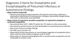
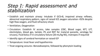
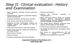
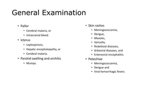
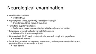
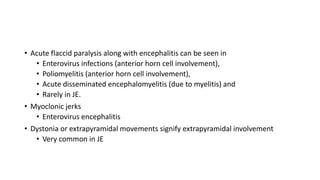
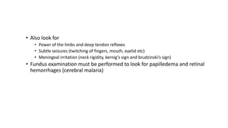
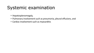
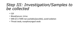
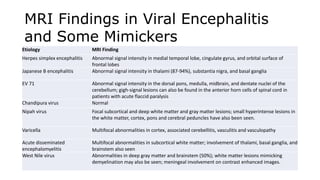
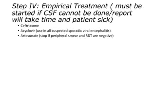
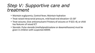
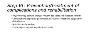
Acute Encephalitis Syndrome
Introduction
• Viral encephalitis is an important cause of mortality and morbidity in children.
• It may be sporadic like herpes simplex encephalitis(HSE), or epidemic such as
Japanese B encephalitis (JE).
Important Definitions
• Encephalopathy
• Encephalopathy describes a clinical syndrome of altered mental status, manifesting as
reduced consciousness or altered behavior.
• Encephalitis
• Encephalitis means inflammation of the brain.
• It is strictly a pathological diagnosis; but surrogate clinical/imaging markers may provide
evidence of inflammation.
• Acute Encephalitis Syndrome
• Clinically, a case of acute encephalitis syndrome is defined as a person of any age, at any time
of year with the acute onset of fever and a change in mental status (including symptoms such
as confusion, disorientation, coma, or inability to talk) AND/OR new onset of seizures
(excluding simple febrile seizures).
• Japanese B encephalitis (JE)
• Laboratory-confirmed JE: A suspected case that has been laboratory-confirmed as JE.
• Probable JE: A suspected case that occurs in close geographic and temporal relationship to a
laboratory-confirmed case of JE, in the context of an outbreak.
• Encephalitis is characterized by brain inflammation as a consequence of
• direct infection of the brain parenchyma,
• a post-infectious process such as acute disseminated encephalomyelitis
(ADEM), or
• a noninfectious condition such as antiN-methyl-D-aspartate receptor
(NMDAR) encephalitis
Diagnostic Criteria for Encephalitis and
Encephalopathy of Presumed Infectious or
Autoimmune Etiology
• Major Criterion (required):
• Patients presenting to medical attention with altered mental status (defined as
decreased or altered level of consciousness, lethargy or personality change) lasting
≥24 h with no alternative cause identified.
• Minor Criteria (2 required for possible encephalitis; ≥3 required for probable or
confirmed encephalitis):
• Documented fever ≥38° C (100.4°F) within the 72 h before or after presentation
• Generalized or partial seizures not fully attributable to a preexisting seizure disorder
• New onset of focal neurologic findings
• CSF WBC count ≥5/cubic mm
• Abnormality of brain parenchyma on neuroimaging suggestive of encephalitis that is
either new from prior studies or appears acute in onset
• Abnormality on electroencephalography that is consistent with encephalitis and not
attributable to another cause.
Evaluation and
Management of a Child
with Acute Encephalitis
Syndrome
Step I: Rapid assessment and
stabilization
• Establish and maintain airway: Intubate if GCS<8, impaired airway reflexes,
abnormal respiratory pattern, signs of raised ICP, oxygen saturation <92% despite
high flow oxygen, and fluid refractory shock
• Ventilation, Oxygenation
• Circulation: Establish IV access, take samples (CBC, Blood sugar, KFT, LFT,
electrolytes, blood gas, lactate, PS and RDT for malarial parasite, serology for
viruses), Fluid bolus if in circulatory failure (20 mL/kg NS), inotropes if required
• Identify signs of cerebral herniation or raised ICP
• Temperature: treat fever and hypothermia
• Treat ongoing seizures- Benzodiazepine, followed by phenytoin loading
Step II: Clinical evaluation: History
and Examination
• Fever, headache, vomiting, seizures, abnormal
posturing
• Altered behavior, cognition, personality changes,
altered consciousness
• Prodromal symptoms- flu-like illness, diarrhea
• Rash, vesicles, past history of chicken pox
• Residence of child: Rural/urban, endemic for
cerebral malaria, any epidemic of AES in
neighborhood
• History of animal contact, insect bite, dog bite
• Drug or toxin exposure- enquire for presence of
any drugs at home
• Recent history of travel
• History of trauma
• Personal or family history of seizure disorder
• Recent immunizations
• History of recurrent episodes of
encephalopathy:
• Other concurrent systemic illness e.g. jaundice
(hepatic failure), pneumonia (hypoxic
encephalopathy), diarrhea (dyselectrolytemia),
dysentery (shigella encephalopathy)
• Past medical illness: Diabetes, congenital heart
disease, chronic kidney or liver disease
• Family history of previous infant/child deaths
• Pre-morbid developmental/ neurological status
of the child
• Risk factors for immunodeficiency- HIV risk
factors, cancer treatment,
steroid/immunosuppressant treatment
Important Points in The History of a
Child With AES
General Examination
• Pallor
• Cerebral malaria, or
• Intracranial bleed.
• Icterus
• Leptospirosis,
• Hepatic encephalopathy, or
• Cerebral malaria.
• Parotid swelling and orchitis
• Mumps.
• Skin rashes
• Meningococcemia,
• Dengue,
• Measles,
• Varicella,
• Rickettsial diseases,
• Arboviral diseases, and
• Enteroviral encephalitis.
• Petechiae
• Meningococcemia,
• Dengue and
• Viral hemorrhagic fevers.
Neurological examination
• Level of consciousness
• Modified GCS
• Pupillary size, shape, symmetry and response to light
• Brainstem and third nerve dysfunction
• Unilateral pupillary dilatation
• Oculomotor nerve compression from ipsilateral uncal herniation
• Progressive symmetrical external ophthalmoplegia
• Bickerstaff brainstem encephalitis
• Oculocephalic (doll’s eye), oculovestibular, corneal, cough and gag reflexes
• Brainstem function
• Trunk, limb position, spontaneous movements, and response to stimulation and
posturing (decerebrate or decorticate)
• Focal deficits
• Acute flaccid paralysis along with encephalitis can be seen in
• Enterovirus infections (anterior horn cell involvement),
• Poliomyelitis (anterior horn cell involvement),
• Acute disseminated encephalomyelitis (due to myelitis) and
• Rarely in JE.
• Myoclonic jerks
• Enterovirus encephalitis
• Dystonia or extrapyramidal movements signify extrapyramidal involvement
• Very common in JE
• Also look for
• Power of the limbs and deep tendon reflexes
• Subtle seizures (twitching of fingers, mouth, eyelid etc)
• Meningeal irritation (neck rigidity, kernig’s sign and brudzinski’s sign)
• Fundus examination must be performed to look for papilledema and retinal
hemorrhages (cerebral malaria)
Systemic examination
• Hepatosplenomegaly,
• Pulmonary involvement such as pneumonia, pleural effusions, and
• Cardiac involvement such as myocarditis
Step III: Investigation/Samples to
be collected
• CSF
• Blood/serum, Urine
• MRI (CT, if MRI not available/possible), avoid sedation
• Throat swab, nasopharyngeal swab
Etiology MRI Finding
Herpes simplex encephalitis Abnormal signal intensity in medial temporal lobe, cingulate gyrus, and orbital surface of
frontal lobes
Japanese B encephalitis Abnormal signal intensity in thalami (87-94%), substantia nigra, and basal ganglia
EV 71 Abnormal signal intensity in the dorsal pons, medulla, midbrain, and dentate nuclei of the
cerebellum; gigh-signal lesions can also be found in the anterior horn cells of spinal cord in
patients with acute flaccid paralysis
Chandipura virus Normal
Nipah virus Focal subcortical and deep white matter and gray matter lesions; small hyperintense lesions in
the white matter, cortex, pons and cerebral peduncles have also been seen.
Varicella Multifocal abnormalities in cortex, associated cerebellitis, vasculitis and vasculopathy
Acute disseminated
encephalomyelitis
Multifocal abnormalities in subcortical white matter; involvement of thalami, basal ganglia, and
brainstem also seen
West Nile virus Abnormalities in deep gray matter and brainstem (50%); white matter lesions mimicking
demyelination may also be seen; meningeal involvement on contrast enhanced images.
MRI Findings in Viral Encephalitis
and Some Mimickers
Step IV: Empirical Treatment ( must be
started if CSF cannot be done/report
will take time and patient sick)
• Ceftriaxone
• Acyclovir (use in all suspected sporadic viral encephalitis)
• Artesunate (stop if peripheral smear and RDT are negative)
Step V: Supportive care and
treatment
• Maintain euglycemia, Control fever, Maintain hydration
• Treat raised intracranial pressure, mild head-end elevation–15-30°
• Treat seizures; Give anticonvulsant if history of seizures or if GCS <8, or child
has features of raised ICT
• Steroids: Pulse steroids (methylprednisolone or dexamethasone) must be
given in children with suspected ADEM.
Step VI: Prevention/treatment of
complications and rehabilitation
• Physiotherapy, posture change, Prevent bed sores and exposure keratitis
• Complications: aspiration pneumonia, nosocomial infections, coagulation
disturbances
• Nutrition: early feeding
• Psychological support to patient and family
Thank you

Acute Encephalitis Syndrome.pptx

  • 1.
  • 2.
    Introduction • Viral encephalitisis an important cause of mortality and morbidity in children. • It may be sporadic like herpes simplex encephalitis(HSE), or epidemic such as Japanese B encephalitis (JE).
  • 3.
    Important Definitions • Encephalopathy •Encephalopathy describes a clinical syndrome of altered mental status, manifesting as reduced consciousness or altered behavior. • Encephalitis • Encephalitis means inflammation of the brain. • It is strictly a pathological diagnosis; but surrogate clinical/imaging markers may provide evidence of inflammation. • Acute Encephalitis Syndrome • Clinically, a case of acute encephalitis syndrome is defined as a person of any age, at any time of year with the acute onset of fever and a change in mental status (including symptoms such as confusion, disorientation, coma, or inability to talk) AND/OR new onset of seizures (excluding simple febrile seizures). • Japanese B encephalitis (JE) • Laboratory-confirmed JE: A suspected case that has been laboratory-confirmed as JE. • Probable JE: A suspected case that occurs in close geographic and temporal relationship to a laboratory-confirmed case of JE, in the context of an outbreak.
  • 4.
    • Encephalitis ischaracterized by brain inflammation as a consequence of • direct infection of the brain parenchyma, • a post-infectious process such as acute disseminated encephalomyelitis (ADEM), or • a noninfectious condition such as antiN-methyl-D-aspartate receptor (NMDAR) encephalitis
  • 5.
    Diagnostic Criteria forEncephalitis and Encephalopathy of Presumed Infectious or Autoimmune Etiology • Major Criterion (required): • Patients presenting to medical attention with altered mental status (defined as decreased or altered level of consciousness, lethargy or personality change) lasting ≥24 h with no alternative cause identified. • Minor Criteria (2 required for possible encephalitis; ≥3 required for probable or confirmed encephalitis): • Documented fever ≥38° C (100.4°F) within the 72 h before or after presentation • Generalized or partial seizures not fully attributable to a preexisting seizure disorder • New onset of focal neurologic findings • CSF WBC count ≥5/cubic mm • Abnormality of brain parenchyma on neuroimaging suggestive of encephalitis that is either new from prior studies or appears acute in onset • Abnormality on electroencephalography that is consistent with encephalitis and not attributable to another cause.
  • 6.
    Evaluation and Management ofa Child with Acute Encephalitis Syndrome
  • 7.
    Step I: Rapidassessment and stabilization • Establish and maintain airway: Intubate if GCS<8, impaired airway reflexes, abnormal respiratory pattern, signs of raised ICP, oxygen saturation <92% despite high flow oxygen, and fluid refractory shock • Ventilation, Oxygenation • Circulation: Establish IV access, take samples (CBC, Blood sugar, KFT, LFT, electrolytes, blood gas, lactate, PS and RDT for malarial parasite, serology for viruses), Fluid bolus if in circulatory failure (20 mL/kg NS), inotropes if required • Identify signs of cerebral herniation or raised ICP • Temperature: treat fever and hypothermia • Treat ongoing seizures- Benzodiazepine, followed by phenytoin loading
  • 8.
    Step II: Clinicalevaluation: History and Examination • Fever, headache, vomiting, seizures, abnormal posturing • Altered behavior, cognition, personality changes, altered consciousness • Prodromal symptoms- flu-like illness, diarrhea • Rash, vesicles, past history of chicken pox • Residence of child: Rural/urban, endemic for cerebral malaria, any epidemic of AES in neighborhood • History of animal contact, insect bite, dog bite • Drug or toxin exposure- enquire for presence of any drugs at home • Recent history of travel • History of trauma • Personal or family history of seizure disorder • Recent immunizations • History of recurrent episodes of encephalopathy: • Other concurrent systemic illness e.g. jaundice (hepatic failure), pneumonia (hypoxic encephalopathy), diarrhea (dyselectrolytemia), dysentery (shigella encephalopathy) • Past medical illness: Diabetes, congenital heart disease, chronic kidney or liver disease • Family history of previous infant/child deaths • Pre-morbid developmental/ neurological status of the child • Risk factors for immunodeficiency- HIV risk factors, cancer treatment, steroid/immunosuppressant treatment Important Points in The History of a Child With AES
  • 9.
    General Examination • Pallor •Cerebral malaria, or • Intracranial bleed. • Icterus • Leptospirosis, • Hepatic encephalopathy, or • Cerebral malaria. • Parotid swelling and orchitis • Mumps. • Skin rashes • Meningococcemia, • Dengue, • Measles, • Varicella, • Rickettsial diseases, • Arboviral diseases, and • Enteroviral encephalitis. • Petechiae • Meningococcemia, • Dengue and • Viral hemorrhagic fevers.
  • 10.
    Neurological examination • Levelof consciousness • Modified GCS • Pupillary size, shape, symmetry and response to light • Brainstem and third nerve dysfunction • Unilateral pupillary dilatation • Oculomotor nerve compression from ipsilateral uncal herniation • Progressive symmetrical external ophthalmoplegia • Bickerstaff brainstem encephalitis • Oculocephalic (doll’s eye), oculovestibular, corneal, cough and gag reflexes • Brainstem function • Trunk, limb position, spontaneous movements, and response to stimulation and posturing (decerebrate or decorticate) • Focal deficits
  • 11.
    • Acute flaccidparalysis along with encephalitis can be seen in • Enterovirus infections (anterior horn cell involvement), • Poliomyelitis (anterior horn cell involvement), • Acute disseminated encephalomyelitis (due to myelitis) and • Rarely in JE. • Myoclonic jerks • Enterovirus encephalitis • Dystonia or extrapyramidal movements signify extrapyramidal involvement • Very common in JE
  • 12.
    • Also lookfor • Power of the limbs and deep tendon reflexes • Subtle seizures (twitching of fingers, mouth, eyelid etc) • Meningeal irritation (neck rigidity, kernig’s sign and brudzinski’s sign) • Fundus examination must be performed to look for papilledema and retinal hemorrhages (cerebral malaria)
  • 13.
    Systemic examination • Hepatosplenomegaly, •Pulmonary involvement such as pneumonia, pleural effusions, and • Cardiac involvement such as myocarditis
  • 14.
    Step III: Investigation/Samplesto be collected • CSF • Blood/serum, Urine • MRI (CT, if MRI not available/possible), avoid sedation • Throat swab, nasopharyngeal swab
  • 15.
    Etiology MRI Finding Herpessimplex encephalitis Abnormal signal intensity in medial temporal lobe, cingulate gyrus, and orbital surface of frontal lobes Japanese B encephalitis Abnormal signal intensity in thalami (87-94%), substantia nigra, and basal ganglia EV 71 Abnormal signal intensity in the dorsal pons, medulla, midbrain, and dentate nuclei of the cerebellum; gigh-signal lesions can also be found in the anterior horn cells of spinal cord in patients with acute flaccid paralysis Chandipura virus Normal Nipah virus Focal subcortical and deep white matter and gray matter lesions; small hyperintense lesions in the white matter, cortex, pons and cerebral peduncles have also been seen. Varicella Multifocal abnormalities in cortex, associated cerebellitis, vasculitis and vasculopathy Acute disseminated encephalomyelitis Multifocal abnormalities in subcortical white matter; involvement of thalami, basal ganglia, and brainstem also seen West Nile virus Abnormalities in deep gray matter and brainstem (50%); white matter lesions mimicking demyelination may also be seen; meningeal involvement on contrast enhanced images. MRI Findings in Viral Encephalitis and Some Mimickers
  • 16.
    Step IV: EmpiricalTreatment ( must be started if CSF cannot be done/report will take time and patient sick) • Ceftriaxone • Acyclovir (use in all suspected sporadic viral encephalitis) • Artesunate (stop if peripheral smear and RDT are negative)
  • 17.
    Step V: Supportivecare and treatment • Maintain euglycemia, Control fever, Maintain hydration • Treat raised intracranial pressure, mild head-end elevation–15-30° • Treat seizures; Give anticonvulsant if history of seizures or if GCS <8, or child has features of raised ICT • Steroids: Pulse steroids (methylprednisolone or dexamethasone) must be given in children with suspected ADEM.
  • 18.
    Step VI: Prevention/treatmentof complications and rehabilitation • Physiotherapy, posture change, Prevent bed sores and exposure keratitis • Complications: aspiration pneumonia, nosocomial infections, coagulation disturbances • Nutrition: early feeding • Psychological support to patient and family
  • 19.