
Recommended
More Related Content
What's hot
What's hot (20)
Similar to ACS ESCGL 2023.pdf
Similar to ACS ESCGL 2023.pdf (20)
More from RajeshPonnada3
More from RajeshPonnada3 (9)
Recently uploaded
Recently uploaded (20)
ACS ESCGL 2023.pdf
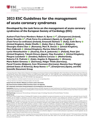
- 1. 2023 ESC Guidelines for the management of acute coronary syndromes Developed by the task force on the management of acute coronary syndromes of the European Society of Cardiology (ESC) Authors/Task Force Members: Robert A. Byrne *† , (Chairperson) (Ireland), Xavier Rossello ‡ , (Task Force Co-ordinator) (Spain), J.J. Coughlan ‡ , (Task Force Co-ordinator) (Ireland), Emanuele Barbato (Italy), Colin Berry (United Kingdom), Alaide Chieffo (Italy), Marc J. Claeys (Belgium), Gheorghe-Andrei Dan (Romania), Marc R. Dweck (United Kingdom), Mary Galbraith (United Kingdom), Martine Gilard (France), Lynne Hinterbuchner (Austria), Ewa A. Jankowska (Poland), Peter Jüni (United Kingdom), Takeshi Kimura (Japan), Vijay Kunadian (United Kingdom), Margret Leosdottir (Sweden), Roberto Lorusso (Netherlands), Roberto F.E. Pedretti (Italy), Angelos G. Rigopoulos (Greece), Maria Rubini Gimenez (Germany), Holger Thiele (Germany), Pascal Vranckx (Belgium), Sven Wassmann (Germany), Nanette Kass Wenger (United States of America), Borja Ibanez *† , (Chairperson) (Spain), and ESC Scientific Document Group * Corresponding authors: Robert A. Byrne, Department of Cardiology and Cardiovascular Research Institute (CVRI) Dublin, Mater Private Network, Dublin, Ireland, and School of Pharmacy and Biomolecular Sciences, RCSI University of Medicine and Health Sciences, Dublin, Ireland. Tel: +353-1-2483190, E-mail: robertabyrne@rcsi.ie; and Borja Ibanez, Clinical Research Department, Centro Nacional de Investigaciones Cardiovasculares Carlos III (CNIC), Madrid, Spain, and Cardiology Department, IIS-Fundación Jiménez Díaz University Hospital, Madrid, Spain, CIBERCV, ISCIII, Madrid, Spain. Tel: +3491 4531200, E-mail: bibanez@cnic.es † The two Chairpersons contributed equally to the document and are joint corresponding authors. ‡ The two Task Force Co-ordinators contributed equally to the document. Author/Task Force Member affiliations are listed in author information. ESC Clinical Practice Guidelines (CPG) Committee: listed in the Appendix. ESC subspecialty communities having participated in the development of this document: Associations: Association of Cardiovascular Nursing & Allied Professions (ACNAP), Association for Acute CardioVascular Care (ACVC), European Association of Cardiovascular Imaging (EACVI), European Association of Preventive Cardiology (EAPC), European Association of Percutaneous Cardiovascular Interventions (EAPCI), European Heart Rhythm Association (EHRA), and Heart Failure Association (HFA). Working Groups: Cardiovascular Pharmacotherapy, Cardiovascular Surgery, E-Cardiology, Myocardial and Pericardial Diseases, Thrombosis. Patient Forum The content of these European Society of Cardiology (ESC) Guidelines has been published for personal and educational use only. No commercial use is authorized. No part of the ESC Guidelines may be translated or reproduced in any form without written permission from the ESC. Permission can be obtained upon submission of a written request to Oxford University Press, the publisher of the European Heart Journal, and the party authorized to handle such permissions on behalf of the ESC (journals.permissions@oup.com). Disclaimer. The ESC Guidelines represent the views of the ESC and were produced after careful consideration of the scientific and medical knowledge and the evidence available at the time of their publication. The ESC is not responsible in the event of any contradiction, discrepancy, and/or ambiguity between the ESC Guidelines and any other official recommendations or guidelines issued by the relevant public health authorities, in particular in relation to good use of healthcare or therapeutic strategies. Health professionals are encouraged to take the ESC Guidelines fully into account when exercising their clinical judgment, as well as in the determination and the implementation of preventive, diagnostic or therapeutic medical strategies; however, the ESC Guidelines do not override, in any way whatsoever, the individual responsibility of health professionals to make appropriate and accurate decisions in consideration of each patient’s health condition and in consultation with that patient and, where appropriate and/or necessary, the patient’s caregiver. Nor do the ESC Guidelines exempt health professionals from taking into full and careful consideration the relevant official updated recommendations or guidelines issued by the competent public health authorities, in order to manage each patient’s case in light of the scientifically accepted data pursuant to their respective ethical and professional obligations. It is also the health professional’s responsibility to verify the applicable rules and regulations relating to drugs and medical devices at the time of prescription. This article is co-published with permission in European Heart Journal and European Heart Journal - Acute Cardiovascular Care. All rights reserved. © The European Society of Cardiology 2023. The articles are identical except for stylistic differences in keeping with each journal’s style. Either citation can be used when citing this article. For permissions, please e-mail: journals.permissions@oup.com European Heart Journal (2023) 00, 1–107 https://doi.org/10.1093/eurheartj/ehad191 ESC GUIDELINES Downloaded from https://academic.oup.com/eurheartj/advance-article/doi/10.1093/eurheartj/ehad191/7243210 by guest on 31 August 2023
- 2. Document Reviewers: Sigrun Halvorsen, (Clinical Practice Guidelines Review Co-ordinator) (Norway), Stefan James, (Clinical Practice Guidelines Review Co-ordinator) (Sweden), Magdy Abdelhamid (Egypt), Victor Aboyans (France), Nina Ajmone Marsan (Netherlands), Sotiris Antoniou (United Kingdom), Riccardo Asteggiano (Italy), Maria Bäck (Sweden), Davide Capodanno (Italy), Ruben Casado-Arroyo (Belgium), Salvatore Cassese (Germany), Jelena Čelutkienė (Lithuania), Maja Cikes (Croatia), Jean-Philippe Collet (France), Gregory Ducrocq (France), Volkmar Falk (Germany), Laurent Fauchier (France), Tobias Geisler (Germany), Diana A. Gorog (United Kingdom), Lene Holmvang (Denmark), Tiny Jaarsma (Sweden), Hywel Wynne Jones (United Kingdom), Lars Køber (Denmark), Konstantinos C. Koskinas (Switzerland), Dipak Kotecha (United Kingdom), Konstantin A. Krychtiuk (Austria), Ulf Landmesser (Germany), George Lazaros (Greece), Basil S. Lewis (Israel), Bertil Lindahl (Sweden), Ales Linhart (Czech Republic), Maja-Lisa Løchen (Norway), Mamas A. Mamas (United Kingdom), John William McEvoy (Ireland), Borislava Mihaylova (United Kingdom), Richard Mindham (United Kingdom), Christian Mueller (Switzerland), Lis Neubeck (United Kingdom), Josef Niebauer (Austria), Jens Cosedis Nielsen (Denmark), Alexander Niessner (Austria), Valeria Paradies (Netherlands), Agnes A. Pasquet (Belgium), Steffen E. Petersen (United Kingdom), Eva Prescott (Denmark), Amina Rakisheva (Kazakhstan), Bianca Rocca (Italy), Giuseppe M.C. Rosano (Italy), Leyla Elif Sade (United States of America / Türkiye), François Schiele (France), Jolanta M. Siller-Matula (Austria), Christian Sticherling (Switzerland), Robert F. Storey (United Kingdom), Matthias Thielmann (Germany), Christiaan Vrints (Belgium), Stephan Windecker (Switzerland), Rune Wiseth (Norway), and Adam Witkowski (Poland) All experts involved in the development of these guidelines have submitted declarations of interest. These have been compiled in a report and simultaneously published in a supplementary document to the guidelines. The report is also available on the ESC website www.escardio.org/Guidelines See the European Heart Journal online for supplementary documents that include background information and evidence tables. Keywords Guidelines • Acute cardiac care • Acute coronary syndrome • Antithrombotic therapy • Fibrinolysis • High- sensitivity troponin • Invasive strategy • MINOCA • Myocardial infarction • Non-ST-elevation myocardial infarction • Patient-centred care • Percutaneous coronary intervention • Recommendations • Reperfusion therapy • Revascularization • Secondary prevention • ST-segment elevation myocardial infarction • Unstable angina Table of contents 1. Preamble ...................................................................................................................... 8 2. Introduction ............................................................................................................... 9 2.1. Definitions | Acute coronary syndromes and myocardial infarction ................................................................................................................... 11 2.2. Epidemiology of acute coronary syndromes .................................... 13 2.3. Number and breakdown of classes of recommendations ......... 13 2.4. What is new ................................................................................................... 14 3. Triage and diagnosis ............................................................................................. 16 3.1. Clinical presentation and physical examination ............................... 16 3.1.1. Clinical presentation ........................................................................... 16 3.1.2. History taking and physical examination ................................... 18 3.2. Diagnostic tools | Electrocardiogram .................................................. 18 3.2.1. Acute coronary syndrome with persistent ST-segment elevation (suspected ST-elevation myocardial infarction) .............. 19 3.2.2. Acute coronary syndrome without persistent ST- segment elevation (non-ST elevation acute coronary syndrome) ........................................................................................................... 19 3.3. Diagnostic tools | Biomarkers ................................................................. 20 3.3.1. High-sensitivity cardiac troponins ................................................. 20 3.3.2. Central laboratory vs. point of care ............................................ 20 3.3.3. Confounders of cardiac troponin concentration .................. 20 3.3.4. Rapid ‘rule-in’ and ‘rule-out’ algorithms ..................................... 20 3.3.4.1. European Society of Cardiology 0 h/1 h and 0 h/2 h algorithms ....................................................................................................... 21 3.3.4.1.1. Rule-out .................................................................................. 21 3.3.4.1.2. Rule-in ..................................................................................... 21 3.3.4.1.3. Observe .................................................................................. 21 3.3.4.2. Practical guidance on how to implement the European Society of Cardiology 0 h/1 h algorithm ...................... 23 3.3.5. Other biomarkers ............................................................................... 23 3.4. Diagnostic tools | Non-invasive imaging ............................................. 23 3.4.1. Echocardiography ................................................................................ 23 3.4.2. Computed tomography .................................................................... 23 3.4.3. Cardiac magnetic resonance imaging with or without stress testing ...................................................................................................... 23 3.5. Differential diagnosis for acute chest pain ........................................ 24 4. Initial measures for patients presenting with suspected acute coronary syndrome | Initial treatment ............................................................. 24 4.1. Pre-hospital logistics of care .................................................................... 24 4.1.1. Time to treatment .............................................................................. 24 2 ESC Guidelines Downloaded from https://academic.oup.com/eurheartj/advance-article/doi/10.1093/eurheartj/ehad191/7243210 by guest on 31 August 2023
- 3. 4.1.2. Healthcare systems and system delays ....................................... 24 4.1.3. Emergency medical services ............................................................ 24 4.1.4. General practitioners ......................................................................... 25 4.1.5. Organization of ST-elevation myocardial infarction treatment in networks ................................................................................... 25 4.2. Emergency care ............................................................................................. 25 4.2.1. Initial diagnosis and monitoring ..................................................... 25 4.2.2. Acute pharmacotherapy ................................................................... 25 4.2.2.1. Oxygen ............................................................................................ 25 4.2.2.2. Nitrates ........................................................................................... 25 4.2.2.3. Pain relief ........................................................................................ 25 4.2.2.4. Intravenous beta-blockers ....................................................... 25 5. Acute-phase management of patients with acute coronary syndrome ....................................................................................................................... 26 5.1. Selection of invasive strategy and reperfusion therapy ............... 26 5.2. Acute coronary syndrome managed with invasive strategy ...... 26 5.2.1. Primary percutaneous coronary intervention strategy for ST-elevation myocardial infarction ........................................................... 26 5.2.1.1. Invasive strategy in ST-elevation myocardial infarction late presenters .............................................................................................. 28 5.2.2. Immediate invasive strategy for non-ST elevation acute coronary syndrome ......................................................................................... 28 5.2.3. Routine vs. selective invasive strategy ......................................... 28 5.2.3.1. Early vs. delayed invasive strategy for non-ST elevation acute coronary syndrome ................................................... 28 5.2.4. Summary of invasive strategies for patients with non-ST elevation acute coronary syndrome ........................................................ 28 5.3. Fibrinolysis and pharmaco-invasive strategy in patients with ST-elevation myocardial infarction ................................................................ 30 5.3.1. Benefit and indication of fibrinolysis ............................................ 30 5.3.1.1. Pre-hospital fibrinolysis ............................................................. 30 5.3.1.2. Angiography and percutaneous coronary intervention after fibrinolysis (pharmaco-invasive strategy) ..... 30 5.3.1.2.1. Comparison of fibrinolytic agents ............................... 30 5.3.1.2.2. Hazards of fibrinolysis and contraindications ......... 30 5.4. Patients not undergoing reperfusion ................................................... 30 5.4.1. Patients who are not candidates for invasive coronary angiography ......................................................................................................... 30 5.4.2. Patients with coronary artery disease not amenable to revascularization ............................................................................................... 30 6. Antithrombotic therapy ..................................................................................... 31 6.1. Antiplatelet therapy in the acute phase ............................................. 33 6.1.1. Oral antiplatelet therapy .................................................................. 33 6.1.2. Timing of loading dose of oral antiplatelet therapy .............. 34 6.1.2.1. Pre-treatment in patients with suspected ST- elevation myocardial infarction ............................................................. 34 6.1.2.2. Pre-treatment in patients with non-ST-elevation acute coronary syndrome ....................................................................... 34 6.1.2.3. Summary of pre-treatment strategies ................................ 34 6.1.3. Intravenous antiplatelet drugs ........................................................ 34 6.2. Anticoagulant treatment in the acute phase .................................... 35 6.2.1. Anticoagulation in patients with ST-elevation myocardial infarction undergoing primary percutaneous coronary intervention ........................................................................................................ 35 6.2.2. Anticoagulation in patients with non-ST-elevation acute coronary syndrome undergoing angiography and percutaneous coronary intervention if indicated ............................................................ 35 6.3. Maintenance antithrombotic therapy after revascularization ... 36 6.3.1. Shortening dual antiplatelet therapy ............................................ 37 6.3.2. De-escalation from potent P2Y12 inhibitor to clopidogrel 37 6.3.3. Summary of alternative antiplatelet strategies to reduce bleeding risk in the first 12 months after acute coronary syndrome ............................................................................................................. 38 6.4. Long-term treatment .................................................................................. 40 6.4.1. Prolonging antithrombotic therapy beyond 12 months ..... 40 6.5. Antiplatelet therapy in patients requiring oral anticoagulation 41 6.5.1. Acute coronary syndrome patients requiring anticoagulation .................................................................................................. 41 6.5.2. Patients requiring vitamin K antagonists or undergoing coronary artery bypass surgery ................................................................. 43 6.6. Antithrombotic therapy as an adjunct to fibrinolysis ................... 43 6.7. Antithrombotic therapy in patients not undergoing reperfusion ............................................................................................................... 43 7. Acute coronary syndrome with unstable presentation ....................... 43 7.1. Out-of-hospital cardiac arrest in acute coronary syndrome .... 44 7.1.1. Systems of care ..................................................................................... 44 7.2. Cardiogenic shock complicating acute coronary syndrome ...... 44 8. Management of acute coronary syndrome during hospitalization .. 45 8.1. Coronary care unit/intensive cardiac care unit ............................... 45 8.1.1. Monitoring .............................................................................................. 45 8.1.2. Ambulation ............................................................................................. 45 8.1.3. Length of stay in the intensive cardiac care unit .................... 46 8.2. In-hospital care .............................................................................................. 46 8.2.1. Length of hospital stay ...................................................................... 46 8.2.2. Risk assessment .................................................................................... 46 8.2.2.1. Clinical risk assessment ............................................................. 46 8.2.2.2. Imaging risk assessment ............................................................ 46 8.2.2.3. Biomarkers for risk assessment ............................................ 46 8.2.2.4. Bleeding risk assessment .......................................................... 46 8.2.2.5. Integrating ischaemic and bleeding risks ............................ 46 9. Technical aspects of invasive strategies ....................................................... 47 9.1. Percutaneous coronary intervention ................................................... 47 9.1.1. Vascular access ..................................................................................... 47 9.1.2. Intravascular imaging/physiology of the infarct-related artery ..................................................................................................................... 47 9.1.2.1. Intravascular imaging .................................................................. 47 9.1.2.2. Intravascular physiology ............................................................ 48 9.1.3. Timing of revascularization with percutaneous coronary intervention ........................................................................................................ 48 9.1.4. Balloons and stents ............................................................................. 49 9.1.5. Embolic protection and microvascular salvage strategies .. 49 9.1.5.1. Thrombus aspiration ................................................................. 49 9.1.5.2. Interventions to protect the microcirculation ............... 49 9.2. Coronary artery bypass grafting ............................................................ 49 9.2.1. Indication and timing of coronary artery bypass grafting in acute coronary syndrome patients .......................................................... 49 9.2.2. Technical considerations specific to acute coronary syndrome patients ........................................................................................... 49 9.3. Spontaneous coronary artery dissection ........................................... 49 9.3.1. Intravascular imaging .......................................................................... 50 9.3.2. Revascularization .................................................................................. 50 10. Management of patients with multivessel disease ................................ 50 10.1. Management of multivessel disease in acute coronary syndrome complicated by cardiogenic shock ........................................... 50 ESC Guidelines 3 Downloaded from https://academic.oup.com/eurheartj/advance-article/doi/10.1093/eurheartj/ehad191/7243210 by guest on 31 August 2023
- 4. 10.2. Patients with multivessel coronary artery disease undergoing primary percutaneous coronary intervention .................. 51 10.3. Timing of non-infarct-related artery revascularization in acute coronary syndrome ................................................................................. 52 10.3.1. Patients presenting with ST-elevation myocardial infarction and multivessel coronary artery disease ........................... 52 10.3.2. Patients presenting with non-ST-elevation acute coronary syndrome and multivessel coronary artery disease ...... 52 10.4. Evaluation of non-infarct-related artery stenosis severity (angiography vs. physiology) ............................................................................. 52 10.5. Hybrid revascularization ......................................................................... 53 11. Myocardial infarction with non-obstructive coronary arteries ...... 53 12. Special situations ................................................................................................. 56 12.1. Type 2 myocardial infarction and acute myocardial injury ...... 56 12.2. Complications ............................................................................................. 56 12.2.1. Heart failure ........................................................................................ 56 12.2.2. Mechanical complications .............................................................. 57 12.2.3. Left ventricular thrombus .............................................................. 57 12.2.4. Post-acute coronary syndrome pericarditis .......................... 57 12.2.5. Arrhythmias ......................................................................................... 57 12.2.5.1. Atrial fibrillation ......................................................................... 57 12.2.5.2. Ventricular arrhythmias ......................................................... 58 12.2.6. Bleeding ................................................................................................. 58 12.2.6.1. Management of bleeding ....................................................... 58 12.3. Comorbid conditions ............................................................................... 59 12.3.1. Patients at high bleeding risk and with blood disorders (anaemia and thrombocytopaenia) .......................................................... 59 12.3.2. Chronic kidney disease ................................................................... 60 12.3.3. Diabetes mellitus ............................................................................... 60 12.3.4. Older adults with frailty and multimorbidity ........................ 60 12.3.4.1. The older person ...................................................................... 60 12.3.4.2. Frailty and multimorbidity ..................................................... 60 12.3.5. Pregnancy ............................................................................................. 61 12.3.6. Drug abuse ........................................................................................... 61 12.3.7. Patients with cancer ......................................................................... 61 12.3.8. Coronavirus disease (COVID-19) ............................................. 61 13. Long-term treatment ........................................................................................ 62 13.1. Cardiac rehabilitation ............................................................................... 64 13.1.1. Comprehensive cardiac rehabilitation ...................................... 64 13.1.2. Digital health ....................................................................................... 64 13.1.3. Adherence and persistence .......................................................... 64 13.2. Lifestyle management ............................................................................... 64 13.2.1. Tobacco ................................................................................................ 64 13.2.2. Nutrition and alcohol ...................................................................... 64 13.2.3. Physical activity and exercise ....................................................... 65 13.2.4. Psychological considerations ........................................................ 65 13.2.5. Resumption of activities ................................................................. 65 13.3. Pharmacological treatment .................................................................... 65 13.3.1. Antithrombotic therapy ................................................................. 65 13.3.2. Lipid-lowering therapy .................................................................... 65 13.3.3. Beta-blockers ...................................................................................... 66 13.3.4. Nitrates and calcium channel blockers .................................... 67 13.3.5. Renin–angiotensin–aldosterone system inhibitors .............. 67 13.3.6. Medications for diabetes ................................................................ 67 13.3.6.1. Sodium–glucose co-transporter 2 inhibitors ................ 67 13.3.6.2. Glucagon-like peptide-1 receptor agonists ................... 68 13.3.7. Proton pump inhibitors .................................................................. 68 13.3.8. Vaccination ........................................................................................... 68 13.3.9. Anti-inflammatory drugs ................................................................ 68 13.3.10. Hormone replacement therapy ............................................... 68 14. Patient perspectives .......................................................................................... 69 14.1. Patient-centred care ................................................................................. 69 14.2. Shared decision-making ........................................................................... 70 14.3. Informed consent ...................................................................................... 70 14.4. Research participation and consent in the acute setting .......... 71 14.5. Patient satisfaction and expectations ................................................ 71 14.6. Patient-reported outcome measures and patient-reported experience measures ........................................................................................... 72 14.7. Preparation for discharge ....................................................................... 72 15. Key messages ....................................................................................................... 72 16. Gaps in evidence ................................................................................................. 74 17. Sex differences .................................................................................................... 76 18. ‘What to do’ and ‘What not to do’ messages from the Guidelines 77 19. Quality indicators ............................................................................................... 82 20. Supplementary data ........................................................................................... 82 21. Data availability statement .............................................................................. 82 22. Author information ........................................................................................... 82 23. Appendix ................................................................................................................ 82 24. References ............................................................................................................. 83 Tables of Recommendations Recommendation Table 1 — Recommendations for clinical and diagnostic tools for patients with suspected acute coronary syndrome ....................................................................................................................... 19 Recommendation Table 2 — Recommendations for non-invasive imaging in the initial assessment of patients with suspected acute coronary syndrome ................................................................................................... 23 Recommendation Table 3 — Recommendations for the initial management of patients with acute coronary syndrome ........................ 26 Recommendation Table 4 — Recommendations for reperfusion therapy and timing of invasive strategy ............................................................ 31 Recommendation Table 5 — Recommendations for antiplatelet and anticoagulant therapy in acute coronary syndrome ................................... 39 Recommendation Table 6 — Recommendations for alternative antithrombotic therapy regimens ................................................ 40 Recommendation Table 7 — Recommendations for fibrinolytic therapy ............................................................................................................................ 43 Recommendation Table 8 — Recommendations for cardiac arrest and out-of-hospital cardiac arrest ...................................................................... 44 Recommendation Table 9 — Recommendations for cardiogenic shock ............................................................................................................................... 45 Recommendation Table 10 — Recommendations for in-hospital management ................................................................................................................. 47 Recommendation Table 11 — Recommendations for technical aspects of invasive strategies ................................................................................. 50 Recommendation Table 12 — Recommendations for management of patients with multivessel disease ................................................................... 53 Recommendation Table 13 — Recommendations for myocardial infarction with non-obstructive coronary arteries ...................................... 56 Recommendation Table 14 — Recommendations for acute coronary syndrome complications ..................................................................... 58 Recommendation Table 15 — Recommendations for acute coronary syndrome comorbid conditions ...................................................... 62 Recommendation Table 16 — Recommendations for long-term management ................................................................................................................. 68 4 ESC Guidelines Downloaded from https://academic.oup.com/eurheartj/advance-article/doi/10.1093/eurheartj/ehad191/7243210 by guest on 31 August 2023
- 5. Recommendation Table 17 — Recommendations for patient perspectives in acute coronary syndrome care ............................................ 72 List of tables Table 1 Classes of recommendations .................................................................. 9 Table 2 Levels of evidence ........................................................................................ 9 Table 3 Definitions of terms related to invasive strategy and reperfusion therapy commonly used in this document ............................ 12 Table 4 New recommendations ......................................................................... 14 Table 5 Revised recommendations .................................................................... 15 Table 6 Dose regimen of antiplatelet and anticoagulant drugs in acute coronary syndrome patients .................................................................... 32 Table 7 Suggested strategies to reduce bleeding risk related to percutaneous coronary intervention ................................................................. 41 Table 8 Gaps in evidence ....................................................................................... 74 Table 9 ‘What to do’ and ‘What not to do’ .................................................. 77 List of figures Figure 1 Central illustration ................................................................................... 10 Figure 2 The spectrum of clinical presentations, electrocardiographic findings, and high-sensitivity cardiac troponin levels in patients with acute coronary syndrome ...................................................................................... 11 Figure 3 Classification of patients presenting with suspected acute coronary syndrome: from a working to a final diagnosis ......................... 13 Figure 4 An overview of the initial triage, management and investigation of patients who present with signs and symptoms potentially consistent with acute coronary syndrome .............................. 17 Figure 5 The A.C.S. assessment for the initial evaluation of patients with suspected acute coronary syndrome ...................................................... 18 Figure 6 The 0 h/1 h or 0 h/2 h rule-out and rule-in algorithms using high-sensitivity cardiac troponin assays in patients presenting to the emergency department with suspected NSTEMI and without an indication for immediate invasive angiography .............................................. 22 Figure 7 Modes of presentation and pathways to invasive management and myocardial revascularization in patients presenting with STEMI ................................................................................................................... 27 Figure 8 Selection of invasive strategy and reperfusion therapy in patients presenting with NSTE-ACS ................................................................. 29 Figure 9 Antithrombotic treatments in acute coronary syndrome: pharmacological targets ........................................................................................... 33 Figure 10 Recommended default antithrombotic therapy regimens in acute coronary syndrome patients without an indication for oral anticoagulation ............................................................................................................ 36 Figure 11 Alternative antiplatelet strategies to reduce bleeding risk in the first 12 months after an ACS ....................................................................... 38 Figure 12 Antithrombotic regimens in patients with acute coronary syndrome and an indication for oral anticoagulation ...................................... 42 Figure 13 A practical algorithm to guide intravascular imaging in acute coronary syndrome patients .................................................................... 48 Figure 14 Algorithm for the management of acute coronary syndrome patients with multivessel coronary artery disease ................. 51 Figure 15 Underlying causes for patients with a working diagnosis of myocardial infarction with non-obstructive coronary arteries .............. 54 Figure 16 Evaluation of patients with a working diagnosis of MINOCA 55 Figure 17 Long-term management after acute coronary syndrome ... 63 Figure 18 Lipid-lowering therapy in ACS patients ....................................... 66 Figure 19 A person-centred approach to the ACS journey ................... 70 Figure 20 Acute coronary syndrome patient expectations ..................... 71 Abbreviations and acronyms AβYSS Beta Blocker Interruption After Uncomplicated Myocardial Infarction ACCOAST A Comparison of Prasugrel at the Time of Percutaneous Coronary Intervention or as Pretreatment at the Time of Diagnosis in Patients with Non-ST Elevation Myocardial Infarction ACE Angiotensin-converting enzyme ACS Acute coronary syndrome AF Atrial fibrillation AFIRE Atrial Fibrillation and Ischemic Events With Rivaroxaban in Patients With Stable Coronary Artery Disease AMI Acute myocardial infarction ARB Angiotensin receptor blocker ARC-HBR Academic Research Consortium for High Bleeding Risk ARNI Angiotensin receptor/neprilysin inhibitor ASCVD Atherosclerotic cardiovascular disease ASSENT 3 ASsessment of the Safety and Efficacy of a New Thrombolytic 3 ATLANTIC Administration of Ticagrelor in the Cath Lab or in the Ambulance for New ST Elevation Myocardial Infarction to Open the Coronary Artery AUGUSTUS An Open-Label, 2 × 2 Factorial, Randomized Controlled, Clinical Trial to Evaluate the Safety of Apixaban Versus Vitamin K Antagonist and Aspirin Versus Aspirin Placebo in Patients With Atrial Fibrillation and Acute Coronary Syndrome or Percutaneous Coronary Intervention AV Atrioventricular BARC Bleeding Academic Research Consortium b.i.d. Bis in die (twice a day) BBB Bundle branch block BEACON Better Evaluation of Acute Chest Pain with Coronary Computed Tomography Angiography BETAMI BEtablocker Treatment After Acute Myocardial Infarction in Patients Without Reduced Left Ventricular Systolic Function BMS Bare metal stent BNP Brain natriuretic peptide CABG Coronary artery bypass grafting CAD Coronary artery disease CAPITAL-RCT Carvedilol Post-Intervention Long-Term Administration in Large-scale Randomized Controlled Trial CAPRICORN CArvedilol Post-infaRct survIval COntRolled evaluatioN CCS Chronic coronary syndrome ESC Guidelines 5 Downloaded from https://academic.oup.com/eurheartj/advance-article/doi/10.1093/eurheartj/ehad191/7243210 by guest on 31 August 2023
- 6. CCTA Coronary computed tomography angiography CCU Coronary care unit CHA2DS2-VASc Congestive heart failure, Hypertension, Age, Diabetes, Stroke or TIA-Vascular disease CHAMPION PCI Cangrelor versus Standard Therapy to Achieve Optimal Management of Platelet Inhibition CHAMPION PHOENIX A Clinical Trial Comparing Cangrelor to Clopidogrel Standard Therapy in Subjects Who Require Percutaneous Coronary Intervention CHAMPION PLATFORM Cangrelor Versus Standard Therapy to Achieve Optimal Management of Platelet Inhibition CKD Chronic kidney disease CMR Cardiac magnetic resonance CI Confidence interval COACT Coronary Angiography after Cardiac Arrest COLCOT Colchicine Cardiovascular Outcomes Trial COMFORTABLE- AMI Comparison of Biolimus Eluted From an Erodible Stent Coating With Bare Metal Stents in Acute ST-Elevation Myocardial Infarction COMPARE-ACUTE Comparison Between FFR Guided Revascularization Versus Conventional Strategy in Acute STEMI Patients With MVD COMPASS Cardiovascular Outcomes for People Using Anticoagulation Strategies COMPLETE Complete vs. Culprit-only Revascularization to Treat Multivessel Disease After Early PCI for STEMI COVID-19 Coronavirus disease 2019 CR Cardiac rehabilitation CRT Cardiac resynchronization therapy— defibrillator/pacemaker CS Cardiogenic shock CT Computed tomography CV Cardiovascular CVD Cardiovascular disease CvLPRIT Complete versus Lesion-only Primary PCI Trial cTn Cardiac troponin CULPRIT-SHOCK Culprit Lesion Only PCI versus Multivessel PCI in Cardiogenic Shock DANAMI-3– PRIMULTI Third Danish Study of Optimal Acute Treatment of Patients with ST-Segment Elevation Myocardial Infarction—Primary PCI in Multivessel Disease DANBLOCK Danish Trial of Beta Blocker Treatment After Myocardial Infarction Without Reduced Ejection Fraction DAPT Dual antiplatelet therapy DAT Dual antithrombotic therapy DCB Drug-coated balloon DES Drug-eluting stent(s) DM Diabetes mellitus ECG Electrocardiography/gram ECMO Extracorporeal membrane oxygenation eGFR Estimated glomerular filtration rate ED Emergency department EMS Emergency medical service(s) EPHESUS Eplerenone Post-AMI Heart failure Efficacy and SUrvival Study ESC European Society of Cardiology EXAMINATION Everolimus-Eluting Stents Versus Bare-Metal Stents in ST Segment Elevation Myocardial Infarction ExTRACT-TIMI 25 Enoxaparin and Thrombolysis Reperfusion for Acute myocardial infarction Treatment Thrombolysis In Myocardial Infarction—Study 25 FAME Fractional Flow Reserve versus Angiography for Multivessel Evaluation FAMOUS-NSTEMI Fractional flow reserve (FFR) versus angiography in guiding management to optimise outcomes in non-ST segment elevation myocardial infarction FAST-MI French Registry of Acute ST-elevation and non-ST-elevation Myocardial Infarction FFR Fractional flow reserve FLOWER-MI Flow Evaluation to Guide Revascularization in Multivessel ST-Elevation Myocardial Infarction FMC First medical contact GLP-1RA Glucagon-like peptide-1 receptor agonist GP Glycoprotein GRACE Global Registry of Acute Coronary Events HBR High bleeding risk HCR Hybrid coronary revascularization HF Heart failure HFrEF Heart failure with reduced ejection fraction HOST-REDUCE-P- OLYTECH-ACS Harmonizing Optimal Strategy for Treatment of Coronary Artery Diseases Trial— Comparison of REDUCTION of PrasugrEl Dose & POLYmer TECHnology in ACS Patients HR Hazard ratio HR-QoL Health-related quality of life hs-cTn High-sensitivity cardiac troponin IABP Intra-aortic balloon counter pulsation/pumping IABP-SHOCK II Intraaortic Balloon Pump in Cardiogenic Shock II ICA Invasive coronary angiography ICCU Intensive cardiac care unit ICD Implantable cardioverter defibrillator ICU Intensive care unit IMPROVE-IT Improved Reduction of Outcomes: Vytorin Efficacy International Trial INR International normalized ratio IRA Infarct-related artery ISAR-REACT 5 Intracoronary stenting and Antithrombotic regimen Rapid Early Action for Coronary Treatment ISIS-4 Fourth International Study of Infarct Survival i.v. Intravenous IVUS Intravascular ultrasound LAD Left anterior descending LBBB Left bundle branch block LD Loading dose LDL-C Low-density lipoprotein-cholesterol LIMA Left internal mammary artery LMWH Low-molecular-weight heparin LoDoCo2 Low-dose Colchicine trial-2 LV Left ventricular(cle) LVAD Left ventricular assist device LVEF Left ventricular ejection fraction MACE Major adverse cardiovascular events 6 ESC Guidelines Downloaded from https://academic.oup.com/eurheartj/advance-article/doi/10.1093/eurheartj/ehad191/7243210 by guest on 31 August 2023
- 7. MASTER DAPT Management of High Bleeding Risk Patients Post Bioresorbable Polymer Coated Stent Implantation With an Abbreviated Versus Prolonged DAPT Regimen MATRIX Minimizing Adverse Haemorrhagic Events by Transradial Access Site and Systemic Implementation of angioX MCS Mechanical circulatory support MD Maintenance dose MI Myocardial infarction MINOCA Myocardial infarction with non-obstructive coronary arteries MRA Mineralocorticoid receptor antagonist MVD Multivessel disease MVO Microvascular obstruction NOAC Non-vitamin K antagonist oral anticoagulant NORSTENT Norwegian Coronary Stent Trial NPV Negative predictive value NRT Nicotine replacement therapy NSTE Non-ST elevation NSTE-ACS Non-ST elevation acute coronary syndrome NSTEMI Non-ST-elevation myocardial infarction NT-pro BNP N-terminal pro B-type natriuretic peptide NYHA New York Heart Association o.d. Once a day OAC Oral anticoagulant/ation OASIS-5 Fifth Organization to Assess Strategies in Acute Ischemic Syndromes OASIS-6 The Safety and Efficacy of Fondaparinux Versus Control Therapy in Patients With ST Segment Elevation Acute Myocardial Infarction OAT Occluded Artery Trial OCT Optical coherence tomography ODYSSEY OUTCOMES Evaluation of Cardiovascular Outcomes After an Acute Coronary Syndrome During Treatment With Alirocumab OHCA Out-of-hospital cardiac arrest OR Odds ratio PARADISE-MI Prospective ARNI vs ACE Inhibitor Trial to Determine Superiority in Reducing Heart Failure Events After MI PCI Percutaneous coronary intervention PCSK9 Proprotein convertase subtilisin/kexin type 9 PE Pulmonary embolism PEGASUS-TIMI 54 PrEvention with TicaGrelor of SecondAry Thrombotic Events in High-RiSk Patients with Prior AcUte Coronary Syndrome— Thrombolysis In Myocardial Infarction PEPCAD NSTEMI Bare Metal Stent Versus Drug Coated Balloon With Provisional Stenting in Non-ST-Elevation Myocardial Infarction PLATO PLATelet inhibition and patient Outcomes POC Point of care POPular Genetics Cost-effectiveness of CYP2C19 Genotype Guided Treatment With Antiplatelet Drugs in Patients With ST-segment-elevation Myocardial Infarction Undergoing Immediate PCI With Stent Implantation: Optimization of Treatment PPCI Primary percutaneous coronary intervention PPI Proton pump inhibitor PPV Positive predictive value PRAMI Preventive Angioplasty in Myocardial Infarction PREM Patient-reported experience measure PROM Patient-reported outcome measure QI Quality indicator RAAS Renin–angiotensin–aldosterone system RAPID-CTCA Rapid Assessment of Potential Ischaemic heart Disease with CTCA RCT Randomized controlled trial REALITY Restrictive and Liberal Transfusion Strategies in Patients With Acute Myocardial Infarction REBOOT-CNIC TREatment With Beta-blockers After myOcardial Infarction withOut Reduced Ejection fracTion REDUCE-SWEDE- HEART Evaluation of Decreased Usage of Betablockers After Myocardial Infarction in the SWEDEHEART Registry REMINDER Double-Blind, Randomized, Placebo-Controlled Trial Evaluating The Safety And Efficacy Of Early Treatment With Eplerenone In Patients With Acute Myocardial Infarction REVELATION REVascularization With PaclitaxEL-Coated Balloon Angioplasty Versus Drug-Eluting Stenting in Acute Myocardial InfarcTION RIVAL RadIal Vs femorAL access for coronary intervention ROMICAT II Multicenter Study to Rule Out Myocardial Infarction by Cardiac Computed Tomography ROSC Return of spontaneous circulation RR Relative risk RV Right ventricular SAPT Single antiplatelet therapy SBP Systolic blood pressure s.c. Subcutaneous SCAD Spontaneous coronary artery dissection SHOCK Should We Emergently Revascularize Occluded Coronaries for Cardiogenic Shock SGLT2 Sodium–glucose co-transporter 2 SMART-DECISION Long-term Beta-blocker Therapy After Acute Myocardial Infarction SPECT Single-photon emission computerized tomography STE ST elevation STEMI ST-elevation myocardial infarction STOPDAPT-2-ACS ShorT and OPtimal Duration of Dual AntiPlatelet Therapy-2 Study for the Patients With ACS STREAM Strategic Reperfusion Early After Myocardial Infarction SWEDEHEART Swedish Web-System for Enhancement and Development of Evidence-Based Care in Heart Disease Evaluated According to Recommended Therapies TALOS-AMI TicAgrelor Versus CLOpidogrel in Stabilized Patients With Acute Myocardial Infarction TAT Triple antithrombotic therapy ESC Guidelines 7 Downloaded from https://academic.oup.com/eurheartj/advance-article/doi/10.1093/eurheartj/ehad191/7243210 by guest on 31 August 2023
- 8. TICO Ticagrelor Monotherapy After 3 Months in the Patients Treated With New Generation Sirolimus Stent for Acute Coronary Syndrome TIMI Thrombolysis In Myocardial Infarction TLR Target lesion revascularization TOMAHAWK Immediate Unselected Coronary Angiography Versus Delayed Triage in Survivors of Out-of-hospital Cardiac Arrest Without ST-segment Elevation TOPIC Timing of Platelet Inhibition After Acute Coronary Syndrome TOTAL Trial of routine aspiration ThrOmbecTomy with PCI vs. PCI ALone in patients with STEMI TRITON-TIMI 38 TRial to Assess Improvement in Therapeutic Outcomes by Optimizing Platelet InhibitioN with Prasugrel Thrombolysis In Myocardial Infarction 38 TROPICAL-ACS Testing Responsiveness to Platelet Inhibition on Chronic Antiplatelet Treatment For Acute Coronary Syndromes TTE Transthoracic echocardiography TWILIGHT Ticagrelor With Aspirin or Alone in High-Risk Patients After Coronary Intervention UA Unstable angina UFH Unfractionated heparin VA-ECMO Veno-arterial extracorporeal membrane oxygenation VALIANT VALsartan In Acute myocardial iNfarcTion VF Ventricular fibrillation VKA Vitamin K antagonist VT Ventricular tachycardia 1. Preamble Guidelines evaluate and summarize available evidence with the aim of as- sisting health professionals in proposing the best diagnostic or therapeut- ic approach for an individual patient with a given condition. Guidelines are intended for use by health professionals and the European Society of Cardiology (ESC) makes its Guidelines freely available. ESC Guidelines do not override the individual responsibility of health professionals to make appropriate and accurate decisions in consider- ation of each patient’s health condition and in consultation with that pa- tient or the patient’s caregiver where appropriate and/or necessary. It is also the health professional’s responsibility to verify the rules and reg- ulations applicable in each country to drugs and devices at the time of prescription, and, where appropriate, to respect the ethical rules of their profession. ESC Guidelines represent the official position of the ESC on a given topic and are regularly updated. ESC Policies and Procedures for for- mulating and issuing ESC Guidelines can be found on the ESC website (https://www.escardio.org/Guidelines). The Members of this Task Force were selected by the ESC to represent professionals involved with the medical care of patients with this pathology. The selection procedure aimed to include members from across the whole of the ESC region and from rele- vant ESC Subspecialty Communities. Consideration was given to diversity and inclusion, notably with respect to gender and country of origin. The Task Force performed a critical evaluation of diag- nostic and therapeutic approaches, including assessment of the risk-benefit ratio. The strength of every recommendation and the level of evidence supporting them were weighed and scored ac- cording to predefined scales as outlined below. The Task Force fol- lowed ESC voting procedures, and all approved recommendations were subject to a vote and achieved at least 75% agreement among voting members. The experts of the writing and reviewing panels provided declaration of interest forms for all relationships that might be perceived as real or potential sources of conflicts of interest. Their declarations of interest were reviewed according to the ESC declaration of interest rules and can be found on the ESC website (http://www.escardio.org/ Guidelines) and have been compiled in a report published in a supple- mentary document with the guidelines. The Task Force received its en- tire financial support from the ESC without any involvement from the healthcare industry. The ESC Clinical Practice Guidelines (CPG) Committee supervises and co-ordinates the preparation of new guidelines and is responsible for the approval process. ESC Guidelines undergo extensive review by the CPG Committee and external experts, including members from across the whole of the ESC region and from relevant ESC Subspecialty Communities and National Cardiac Societies. After appro- priate revisions, the guidelines are signed off by all the experts involved in the Task Force. The finalized document is signed off by the CPG Committee for publication in the European Heart Journal. The guidelines were developed after careful consideration of the scientific and medical knowledge and the evidence available at the time of their writing. Tables of evidence summarizing the findings of studies informing development of the guidelines are included. The ESC warns readers that the technical language may be misinterpreted and declines any responsibility in this respect. Off-label use of medication may be presented in this guideline if a sufficient level of evidence shows that it can be considered medically ap- propriate for a given condition. However, the final decisions concerning an individual patient must be made by the responsible health profes- sional giving special consideration to: • The specific situation of the patient. Unless otherwise provided for by national regulations, off-label use of medication should be limited to situations where it is in the patient’s interest with regard to the quality, safety, and efficacy of care, and only after the patient has been informed and has provided consent. • Country-specific health regulations, indications by governmental drug regulatory agencies, and the ethical rules to which health profes- sionals are subject, where applicable. 8 ESC Guidelines Downloaded from https://academic.oup.com/eurheartj/advance-article/doi/10.1093/eurheartj/ehad191/7243210 by guest on 31 August 2023
- 9. 2. Introduction The major aspects of the management of patients with acute coronary syndromes described in this European Society of Cardiology (ESC) Guideline are summarized in Figure 1. Table 2 Levels of evidence Level of evidence A Level of evidence B Level of evidence C Data derived from multiple randomized clinical trials or meta-analyses. Data derived from a single randomized clinical trial or large non-randomized studies. Consensus of opinion of the experts and/or small studies, retrospective studies, registries. ©ESC 2023 © ESC 2023 Table 1 Classes of recommendations ©ESC 2023 Classes of recommendations Class I Evidence and/or general agreement that a given treatment or procedure is beneficial, useful, effective. Conflicting evidence and/or a divergence of opinion about the usefulness/ efficacy of the given treatment or procedure. Is recommended or is indicated Wording to use Definition Class III Evidence or general agreement that the given treatment or procedure is not useful/effective, and in some cases may be harmful. Is not recommended Class IIb Usefulness/efficacy is less well established by evidence/opinion. May be considered Class IIa Weight of evidence/opinion is in favour of usefulness/efficacy. Should be considered Class II © ESC 2023 ESC Guidelines 9 Downloaded from https://academic.oup.com/eurheartj/advance-article/doi/10.1093/eurheartj/ehad191/7243210 by guest on 31 August 2023
- 10. Aim for complete revascularization 2 3 4 1 5 ACS encompasses a spectrum Think secondary prevention Abnormal ECG? Clinical context? Stable patient? Think ‘A.C.S.’ at initial assessment Think antithrombotic therapy Primary PCI Fibrinolysis (If timely primary PCI not feasible) Immediate angiography ± PCI AND OR OR OR Early (<24 h) angiography should be considered Aspirin UFH LMWH Bivalirudin Fondaparinux P2Y12 inhibitor STEMI OR Very high-risk NSTE-ACS High-risk NSTE-ACS Antiplatelet therapy + Anticoagulant therapy PCI Antithrombotic therapy Lipid lowering therapy Smoking cessation Cardiac rehabilitation Risk factor management Psychosocial considerations CABG Based on clinical status, co-morbidities, and disease complexity OR Consider adjunctive tests to guide revascularization Think invasive management Think revascularization Intravascular imaging Intravascular physiology Unstable angina NSTEMI STEMI Figure 1 Central illustration. ACS, acute coronary syndrome; CABG, coronary artery bypass grafting; ECG, electrocardiogram; LMWH, low molecular- weight heparin; NSTE-ACS, non-ST-elevation acute coronary syndrome; PCI, percutaneous coronary intervention; PPCI, primary percutaneous coronary intervention; STEMI, ST-elevation myocardial infarction; UFH, unfractionated heparin. Patients with acute coronary syndrome (ACS) can initially present with a wide variety of clinical signs and symptoms and it is important that there is a high degree of awareness of this amongst both the general public and healthcare providers. If ACS is suspected, think ‘A.C.S.’ for the initial triage and assessment. This involves performing an electrocardiogram (ECG) to assess for Abnormalities or evidence of ischaemia, taking a targeted clinical history to assess the clinical Context of the presentation, and carrying out a targeted clinical examination to assess for clinical and haemodynamic Stability. Based on the initial assessment, the healthcare provider can decide whether immediate invasive management is required. Patients with ST-elevation myocardial infarction (STEMI) require primary percutaneous coronary intervention (PPCI) (or fibrinoly- sis if PPCI within 120 min is not feasible); patients with non-ST-elevation ACS (NSTE-ACS) with very high-risk features require immediate angiography ± PCI if indicated; patients with NSTE-ACS and high-risk features should undergo inpatient angiography (angiography within 24 h should be considered). A com- bination of antiplatelet and anticoagulant therapy is indicated acutely for patients with ACS. The majority of patients with ACS will eventually undergo re- vascularization, most commonly with PCI. Once the final diagnosis of ACS has been established, it is important to implement measures to prevent recurrent events and to optimize cardiovascular risk. This consists of medical therapy, lifestyle changes and cardiac rehabilitation, as well as consideration of psycho- social factors. 10 ESC Guidelines Downloaded from https://academic.oup.com/eurheartj/advance-article/doi/10.1093/eurheartj/ehad191/7243210 by guest on 31 August 2023
- 11. 2.1. Definitions | Acute coronary syndromes and myocardial infarction Acute coronary syndromes (ACS) encompass a spectrum of condi- tions that include patients presenting with recent changes in clinical symptoms or signs, with or without changes on 12-lead electrocardio- gram (ECG) and with or without acute elevations in cardiac tropo- nin (cTn) concentrations (Figure 2). Patients presenting with suspected ACS may eventually receive a diagnosis of acute myocar- dial infarction (AMI) or unstable angina (UA). The diagnosis of myocardial infarction (MI) is associated with cTn release and is made based on the fourth universal definition of MI.1 UA is defined as myocardial ischaemia at rest or on minimal exertion in the ab- sence of acute cardiomyocyte injury/necrosis. It is characterized by specific clinical findings of prolonged (>20 min) angina at rest; new onset of severe angina; angina that is increasing in frequency, longer in duration, or lower in threshold; or angina that occurs after a re- cent episode of MI. ACS are associated with a broad range of clinical presentations, from patients who are symptom free at presentation to patients with ongoing chest discomfort/symptoms and patients Increasing chest pain/symptoms Oligo/ asymptomatic Persistent chest pain/symptoms Cardiogenic shock/ acute heart failure Cardiac arrest Normal ST segment depression ST segment elevation Malignant arrhythmia Clinical presentation ECG findings hs-cTn levels The ACS spectrum Final diagnosis Working diagnosis Non-elevated Unstable angina NSTE-ACS NSTEMI Rise and fall STEMI STEMI Figure 2 The spectrum of clinical presentations, electrocardiographic findings, and high-sensitivity cardiac troponin levels in patients with acute coronary syndrome. ACS, acute coronary syndrome; ECG, electrocardiogram; hs-cTn, high-sensitivity cardiac troponin; NSTE-ACS, non-ST-elevation acute coronary syndrome; NSTEMI, non-ST-elevation myocardial infarction; STEMI, ST-elevation myocardial infarction. ESC Guidelines 11 Downloaded from https://academic.oup.com/eurheartj/advance-article/doi/10.1093/eurheartj/ehad191/7243210 by guest on 31 August 2023
- 12. with cardiac arrest, electrical/haemodynamic instability, or cardio- genic shock (CS) (Figure 2). Patients presenting with suspected ACS are typically classified based on ECG at presentation for the purposes of initial management. After this, patients can be further classified based on the presence or absence of cardiac troponin elevation (once these results are available), as de- monstrated in Figures 2 and 3. These features (ECG changes and cardiac troponin elevation) are important in the initial triage and diagnosis of patients with ACS, helping to risk stratify patients and guide the initial management strategy. However, after the acute management and sta- bilization phase, most aspects of the subsequent management strategy are common to all patients with ACS (regardless of the initial ECG pattern or the presence/absence of cardiac troponin elevation at presentation) and can therefore be considered under a common pathway. A glossary of the terms related to invasive strategies and reperfusion therapy commonly used in this document, and their associated definitions, is provided in Table 3. While they are closely related, it is important to recognize that ACS is not the same as MI.1 AMI is defined as cardiomyocyte necrosis in the clinical setting of acute myocardial ischaemia. This includes MI due to atherothrombotic events (Type 1 MI) and also other potential causes of myocardial ischaemia and myocyte necrosis (Type 2–5 MI) (Supplementary data online, Table S1). Myocardial injury is another distinct entity, used to describe troponin release due to mechanisms other than myocardial ischaemia and not meeting the criteria for MI outlined in Supplementary data online, Table S1. Myocardial injury can be acute or chronic depending on whether there is evidence of dynamic change in the elevated troponins on serial testing. Some causes of myocardial injury include myocarditis, sepsis, takotsubo car- diomyopathy, heart valve disease, cardiac arrhythmias, and heart fail- ure (HF). The focus of this guideline is largely centred on the management of patients who will eventually receive a diagnosis of Type 1 MI. However, at every stage of the management of patients presenting with ACS, physicians must carefully consider other differential diag- noses in their clinical assessment because they are common, asso- ciated with different underlying pathological mechanisms, have different prognoses, and frequently require different treatment ap- proaches. More information is provided in the Supplementary data online. In general, detailed information regarding the results of individual trials will not be provided in the main guideline. However, where appropriate, this information is provided in the Supplementary data online evidence tables. Table 3 Definitions of terms related to invasive strat- egy and reperfusion therapy commonly used in this document Term Definition First medical contact (FMC) The time point when the patient is initially assessed by a physician, paramedic, nurse, or other trained emergency medical services worker who can obtain and interpret the ECG and deliver initial interventions (e.g. defibrillation). FMC can be either in the pre-hospital setting or upon patient arrival at the hospital (e.g. the emergency department) STEMI diagnosis The time at which a patient with ischaemic symptoms is interpreted as presenting with ACS and ST-segment elevation (or ST-segment elevation equivalent) Primary PCIa Emergent PCI with balloon, stent, or other approved device, performed on the IRA without previous fibrinolytic treatment Primary PCI strategya Emergency coronary angiography and PCI of the IRA if indicated Rescue PCIa Emergency PCI performed as soon as possible in cases of failed fibrinolytic treatment Routine early PCI strategy after fibrinolysisa Coronary angiography, with PCI of the IRA if indicated, performed between 2 h and 24 h after successful fibrinolysis Pharmaco-invasive strategya Fibrinolysis combined with rescue PCI (in cases of failed fibrinolysis) or routine early PCI strategy (in cases of successful fibrinolysis) Immediate invasive strategy Emergency coronary angiography (i.e. as soon as possible) and PCI/CABG of the IRA if indicated Early invasive strategy Early coronary angiography (<24 h from diagnosis of ACS) and PCI/CABG of the IRA if indicated Selective invasive strategy Coronary angiography ± PCI/CABG based on clinical assessment and/or non-invasive testing © ESC 2023 ACS, acute coronary syndrome; CABG, coronary artery bypass grafting; ECG, electrocardiogram; IRA, infarct-related artery; PCI, percutaneous coronary intervention; STE-ACS, ST-segment-elevation acute coronary syndrome. a CABG may also be indicated instead of PCI in certain circumstances. 12 ESC Guidelines Downloaded from https://academic.oup.com/eurheartj/advance-article/doi/10.1093/eurheartj/ehad191/7243210 by guest on 31 August 2023
- 13. 2.2. Epidemiology of acute coronary syndromes Cardiovascular disease (CVD) is the most common cause of mortality and morbidity worldwide, with a substantial portion of this burden borne by low- and middle-income countries.2,3 ACS is often the first clinical manifestation of CVD. In 2019, there were an estimated 5.8 mil- lion new cases of ischaemic heart disease in the 57 ESC member coun- tries.3 The median age-standardized incidence estimate per 100 000 people was 293.3 (interquartile ratio 195.8–529.5). CVD remains the most common cause of death within ESC member countries, account- ing for just under 2.2 million deaths in females and just over 1.9 million deaths in males in the most recent year of available data. Ischaemic heart disease is the most common cause of CVD death, accounting for 38% of all CVD deaths in females and 44% in males.3 2.3. Number and breakdown of classes of recommendations The total number of recommendations in this guideline is 193. A sum- mary of the recommendations according to Class of Recommendation and Level of Evidence (LoE) is also provided. As per Class of Recommendation, there were 106 Class I, 70 Class II, and 17 Class III recommendations. As per LoE, there were 56 LoE A, 64 LoE B, and 73 LoE C recommendations. STEMI NSTE-ACS STEMI NSTEMI Unstable angina Non-ACS diagnosis hs-cTn levels ± Angiography ± Imaging Final diagnosisb Working diagnosisa Further investigations Clinical presentation If a patient has signs/symptoms suggestive of ACS, perform an ECG within 10 min of FMC ECG Figure 3 Classification of patients presenting with suspected acute coronary syndrome: from a working to a final diagnosis. ACS, acute coronary syndrome; ECG, electrocardiogram; FMC, first medical contact; hs-cTn, high-sensitivity cardiac troponin; MI, myocardial infarction; NSTE-ACS, non-ST-elevation acute coronary syndrome; NSTEMI, non-ST-elevation myocardial infarction: STEMI, ST-elevation myocardial infarction. a The working ACS diagnosis can be clas- sified as STEMI or NSTE-ACS on the basis of available clinical information and ECG findings. This allows for initial triage and assessment. b The final diagnosis is based on symptoms, ECG and troponin for the diagnosis of MI as well as the results of other tests (i.e. imaging and/or angiography) to facilitate understanding of the mechanism and subclassification of the type of MI. Patients initially assigned a working diagnosis of STEMI or NSTE-ACS may eventually receive a final non-ACS diagnosis. ESC Guidelines 13 Downloaded from https://academic.oup.com/eurheartj/advance-article/doi/10.1093/eurheartj/ehad191/7243210 by guest on 31 August 2023
- 14. 2.4. What is new Table 4 New recommendations Recommendations Classa Levelb Recommendations for antiplatelet and anticoagulant therapy in acute coronary syndrome If patients presenting with ACS stop DAPT to undergo coronary artery bypass grafting, it is recommended they resume DAPT after surgery for at least 12 months. I C In older ACS patients, especially if HBR, clopidogrel as the P2Y12 receptor inhibitor may be considered. IIb B Recommendations for alternative antithrombotic therapy regimens In patients who are event-free after 3–6 months of DAPT and who are not high ischaemic risk, single antiplatelet therapy (preferably with a P2Y12 receptor inhibitor) should be considered. IIa A P2Y12 inhibitor monotherapy may be considered as an alternative to aspirin monotherapy for long-term treatment. IIb A In HBR patients, aspirin or P2Y12 receptor inhibitor monotherapy after 1 month of DAPT may be considered. IIb B In patients requiring OAC, withdrawing antiplatelet therapy at 6 months while continuing OAC may be considered. IIb B De-escalation of antiplatelet therapy in the first 30 days after an ACS event is not recommended. III B Recommendations for cardiac arrest and out-of-hospital cardiac arrest Evaluation of neurological prognosis (no earlier than 72 h after admission) is recommended in all comatose survivors after cardiac arrest. I C Transport of patients with out-of-hospital cardiac arrest to a cardiac arrest centre according to local protocol should be considered. IIa C Recommendations for technical aspects of invasive strategies In patients with spontaneous coronary artery dissection, PCI is recommended only for patients with symptoms and signs of ongoing myocardial ischaemia, a large area of myocardium in jeopardy, and reduced antegrade flow. I C Intravascular imaging should be considered to guide PCI. IIa A Intravascular imaging (preferably optical coherence tomography) may be considered in patients with ambiguous culprit lesions. IIb C Recommendations for multivessel disease in ACS patients presenting in cardiogenic shock Staged PCI of non-IRA should be considered. IIa C Recommendations for multivessel disease in haemodynamically stable STEMI patients undergoing primary PCI It is recommended that PCI of the non-IRA is based on angiographic severity. I B Invasive epicardial functional assessment of non-culprit segments of the IRA is not recommended during the index procedure. III C Recommendations for acute coronary syndrome complications Implantation of a permanent pacemaker is recommended when high-degree AV block does not resolve within a waiting period of at least 5 days after MI. I C Cardiac magnetic resonance imaging should be considered in patients with equivocal echocardiographic images or in cases of high clinical suspicion of LV thrombus. IIa C Following an acute anterior MI, a contrast echocardiogram may be considered for the detection of LV thrombus if the apex is not well visualized on echocardiography. IIb C In selected patients with high-degree AV block in the context of an anterior wall MI and acute heart failure, early device implantation (cardiac resynchronization therapy—defibrillator/pacemaker) may be considered. IIb C In patients with recurrent life-threatening ventricular arrhythmias, sedation or general anaesthesia to reduce sympathetic drive may be considered. IIb C Recommendations for acute coronary syndrome comorbid conditions It is recommended to base the choice of long-term glucose-lowering treatment on the presence of comorbidities, including heart failure, chronic kidney disease, and obesity. I A For frail older patients with comorbidities, a holistic approach is recommended to individualize interventional and pharmacological treatments after careful evaluation of the risks and benefits. I B An invasive strategy is recommended in cancer patients presenting with high-risk ACS with expected survival ≥6 months. I B A temporary interruption of cancer therapy is recommended in patients in whom the cancer therapy is suspected to be a contributing cause of ACS. I C A conservative non-invasive strategy should be considered in ACS patients with poor cancer prognosis (i.e. with expected life survival <6 months) and/or very high bleeding risk. IIa C Aspirin is not recommended in cancer patients with a platelet count <10 000/μL. III C Continued 14 ESC Guidelines Downloaded from https://academic.oup.com/eurheartj/advance-article/doi/10.1093/eurheartj/ehad191/7243210 by guest on 31 August 2023
- 15. Clopidogrel is not recommended in cancer patients with a platelet count <30 000/μL. III C In ACS patients with cancer and <50 000/μL platelet count, prasugrel or ticagrelor are not recommended. III C Recommendations for long-term management It is recommended to intensify lipid-lowering therapy during the index ACS hospitalization for patients who were on lipid-lowering therapy before admission. I C Low-dose colchicine (0.5 mg once a day) may be considered, particularly if other risk factors are insufficiently controlled or if recurrent cardiovascular disease events occur under optimal therapy. IIb A Combination therapy with a high-dose statin plus ezetimibe may be considered during index hospitalization. IIb B Recommendations for patient perspectives in acute coronary syndrome care Patient-centred care is recommended by assessing and adhering to individual patient preferences, needs and beliefs, ensuring that patient values are used to inform all clinical decisions. I B It is recommended to include ACS patients in decision-making (as much as their condition allows) and to inform them about the risk of adverse events, radiation exposure, and alternative options. Decision aids should be used to facilitate the discussion. I B It is recommended to assess symptoms using methods that help patients to describe their experience. I C Use of the ‘teach back’ technique for decision support during the securing of informed consent should be considered. IIa B Patient discharge information should be provided in both written and verbal formats prior to discharge. Adequate preparation and education for patient discharge using the teach back technique and/or motivational interviewing, giving information in chunks, and checking for understanding, should be considered. IIa B Assessment of mental well-being using a validated tool and onward psychological referral when appropriate should be considered. IIa B © ESC 2023 ACS, acute coronary syndrome; AV, atrioventricular; DAPT, dual antiplatelet therapy; HBR, high bleeding risk; IRA, infarct-related artery; LV, left ventricular(cle); MI, myocardial infarction; OAC, oral anticoagulant/ation; PCI, percutaneous coronary intervention; STEMI, ST-elevation myocardial infarction. a Class of recommendation. b Level of evidence. Table 5 Revised recommendations Recommendations in 2017 and 2020 versions Classa LoEb Recommendations in 2023 version Classa LoEb Recommendations for imaging for patients with suspected NSTE-ACS In patients with no recurrence of chest pain, normal ECG findings, and normal levels of cardiac troponin (preferably high sensitivity), but still with suspected ACS, a non-invasive stress test (preferably with imaging) for inducible ischaemia or CCTA is recommended before deciding on an invasive approach. I B In patients with suspected ACS, non-elevated (or uncertain) hs-cTn, no ECG changes and no recurrence of pain, incorporating CCTA or a non-invasive stress imaging test as part of the initial workup should be considered. IIa A Recommendations for timing of invasive strategy in NSTE-ACS An early invasive strategy within 24 h is recommended in patients with any of the following high-risk criteria: • Diagnosis of NSTEMI suggested by the diagnostic algorithm recommended in Section 3 • Dynamic or presumably new contiguous ST/T-segment changes suggesting ongoing ischaemia • Transient ST-segment elevation • GRACE risk score >140. I A An early invasive strategy within 24 h should be considered in patients with at least one of the following high-risk criteria: • Confirmed diagnosis of NSTEMI based on current recommended ESC hs-cTn algorithms • Dynamic ST-segment or T wave changes • Transient ST-segment elevation • GRACE risk score >140. IIa A Recommendations for antiplatelet and anticoagulant therapy in STEMI A potent P2Y12 inhibitor (prasugrel or ticagrelor), or clopidogrel if these are not available or are contraindicated, is recommended before (or at latest at the time of) PCI, and maintained over 12 months, unless there are contraindications such as excessive risk of bleeding. I A Pre-treatment with a P2Y12 receptor inhibitor may be considered in patients undergoing a primary PCI strategy. IIb B Recommendations for long-term antithrombotic therapy After stent implantation in patients undergoing a strategy of DAPT, stopping aspirin after 3–6 months should be considered, depending on the balance between the ischaemic and bleeding risks. IIa A In patients who are event-free after 3–6 months of DAPT and who are not high ischaemic risk, SAPT (preferably with a P2Y12 receptor inhibitor) should be considered. IIa A Continued ESC Guidelines 15 Downloaded from https://academic.oup.com/eurheartj/advance-article/doi/10.1093/eurheartj/ehad191/7243210 by guest on 31 August 2023
- 16. New/revised concepts • ACS should be considered a spectrum, which encompasses both non-ST-elevation (NSTE)-ACS and ST-elevation MI (STEMI). • A section on the management of ACS in patients with cancer is provided. • A section on patient perspectives is provided. 3. Triage and diagnosis 3.1. Clinical presentation and physical examination 3.1.1. Clinical presentation Acute chest discomfort—which may be described as pain, pressure, tightness, heaviness, or burning—is the leading presenting symptom prompting consideration of the clinical diagnosis of ACS and the initi- ation of testing aligned with specific diagnostic algorithms (Figure 4). Chest pain descriptors should be classified as cardiac, possibly car- diac, and likely non-cardiac. Further information on the suggested use of these terms is provided in the Supplementary data online. The use of the descriptor ‘atypical’ should be avoided. Chest pain-equivalent symptoms include dyspnoea, epigastric pain, and pain in the left or right arm or neck/jaw. Misdiagnosis or delayed diagnosis is sometimes due to an incomplete history or difficulty in eliciting symptoms from the patient. In order to understand the complexity of ACS-related symptomatology, careful history taking and comprehensive interaction with the patient are crucial and may help to facilitate an early and accurate diagnosis. Further information is provided in the Supplementary data online, in- cluding Figure S1, which outlines some of the most common symptoms of ACS in women and men. Recommendations for cardiac arrest and out-of-hospital cardiac arrest Delayed as opposed to immediate angiography should be considered among haemodynamically stable patients without ST-segment elevation successfully resuscitated after out-of-hospital cardiac arrest. IIa B Routine immediate angiography after resuscitated cardiac arrest is not recommended in haemodynamically stable patients without persistent ST-segment elevation (or equivalents). III A Targeted temperature management (also called therapeutic hypothermia), aiming for a constant temperature between 32 and 36 C for at least 24 h, is indicated in patients who remain unconscious after resuscitation from cardiac arrest (of presumed cardiac cause). I B Temperature control (i.e. continuous monitoring of core temperature and active prevention of fever [i.e. >37.7°C]) is recommended after either out-of-hospital or in-hospital cardiac arrest for adults who remain unresponsive after return of spontaneous circulation. I B Recommendations for in-hospital management When echocardiography is suboptimal/inconclusive, an alternative imaging method (CMR preferably) should be considered. IIa C When echocardiography is suboptimal/inconclusive, CMR imaging may be considered. IIb C Recommendations for management of multivessel disease in haemodynamically stable STEMI patients undergoing primary PCI Routine revascularization of non-IRA lesions should be considered in STEMI patients with multivessel disease before hospital discharge. IIa A Complete revascularization is recommended either during the index PCI procedure or within 45 days. I A Recommendations for acute coronary syndrome comorbid conditions Glucose-lowering therapy should be considered in ACS patients with blood glucose >10 mmol/L (>180 mg/dL), with the target adapted to comorbidities, while episodes of hypoglycaemia should be avoided. IIa B Glucose-lowering therapy should be considered in patients with ACS with persistent hyperglycaemia, while episodes of hypoglycaemia should be avoided. IIa C © ESC 2023 ACS, acute coronary syndrome; CCTA, coronary computed tomography angiography; CMR, cardiac magnetic resonance; DAPT, dual antiplatelet therapy; ECG, electrocardiography/gram; ESC European Society of Cardiology; GRACE, Global Registry of Acute Coronary Events; hs-cTn, high-sensitivity cardiac troponin; IRA, infarct-related artery; NSTE-ACS, non-ST-elevation acute coronary syndrome; NSTEMI, non-ST-elevation myocardial infarction; PCI, percutaneous coronary intervention; SAPT, single antiplatelet therapy; STEMI, ST-elevation myocardial infarction. a Class of recommendation. b Level of evidence. 16 ESC Guidelines Downloaded from https://academic.oup.com/eurheartj/advance-article/doi/10.1093/eurheartj/ehad191/7243210 by guest on 31 August 2023
- 17. It is important that awareness of the symptoms associated with ACS is high among the general population, in particular red flag symptoms such as prolonged chest pain (>15 min) and/or recurrent pain within 1 h, which should prompt patients or other members of the public to seek urgent medical help. Continuous education, pro- motion, and advocacy efforts are important to make sure that this information is as widely available as possible to the general population. ACS presentation Initial A.C.S. assessment ECG Physical examination Clinical history Vital signs hs-cTna levels Non-immediate angiography Echo Intravascular imaging hs-cTna levels Non-invasive imaging ECG monitoring PCI CABG Long-term medical therapy Lifestyle measures Smoking cessation STEMI NSTE-ACS with very high-risk featuresb NSTE-ACS without very high-risk featuresb Immediate angiography ± PPCI or fibrinolysis if timely PPCI not feasible PPCI ATT Fibrinolysis Immediate angiography ± PCI Consider angiography within 24 h for NSTE-ACS with high risk features Working diagnosis Early invasive angiography according to patient risk Further management Further investigations PCI ATT PCI ATT Figure 4 An overview of the initial triage, management and investigation of patients who present with signs and symptoms potentially consistent with acute coronary syndrome. ACS, acute coronary syndrome; ATT, antithrombotic therapy; CABG, coronary artery bypass grafting; ECG, electrocardiogram; hs-cTn, high-sensitivity cardiac troponin; NSTE-ACS, non-ST-elevation acute coronary syndrome; PPCI, primary percutaneous coronary intervention; STEMI, ST-elevation myocardial infarction. The ‘A.C.S.’ assessment is detailed in Figure 5. a Results of hs-cTn measurements are not required for the initial stratifi- cation of ACS and the initial emergency management (i.e. for patients with a working diagnosis of STEMI or very high-risk NSTE-ACS) should not be delayed based on this. b For patients with NSTE-ACS with very high-risk features, immediate angiography is recommended. For patients with NSTE-ACS with high- risk features, early invasive angiography (i.e. <24 h) should be considered and inpatient invasive angiography is recommended. See Recommendation Table 4 for details. ESC Guidelines 17 Downloaded from https://academic.oup.com/eurheartj/advance-article/doi/10.1093/eurheartj/ehad191/7243210 by guest on 31 August 2023
- 18. 3.1.2. History taking and physical examination Patients with suspected ACS present in a broad range of clinical scen- arios, including in the community, at the emergency department (ED), or in the inpatient setting. It is crucial to take a focused medical history and accurately characterize the presenting symptoms in order to manage the patient via the appropriate care pathway as soon as possible. Prompt assessment of vital signs is recommended at first medical contact (FMC), at the same time as acquisition of an initial ECG (Figure 5). In patients presenting with suspected ACS, physical examin- ation is recommended and is useful both to eliminate differential diag- noses and to identify very high-risk and high-risk ACS features. This may be particularly relevant for patients presenting with cardiac arrest, signs of CS, and/or haemodynamic or electrical instability.4 Focused physical examination should include checking for the presence of all major pulses, measurement of blood pressure in both arms, auscultation of the heart and lungs, and assessing for signs of HF or circulatory compromise. 3.2. Diagnostic tools | Electrocardiogram The resting 12-lead ECG is the first-line diagnostic tool in the assess- ment of patients with suspected ACS. It is recommended that an ECG is obtained immediately upon FMC and interpreted by a qualified emergency medical technician or physician within 10 min.4,5 It should be repeated as necessary, especially if symptoms have waned at FMC. Based on the initial ECG, patients with suspected ACS can be differen- tiated into two working diagnoses: • Patients with acute chest pain (or chest pain-equivalent signs/symptoms) and persistent ST-segment elevation (or ST-segment elevation equivalents) on ECG (working diagnosis: ST-segment elevation MI: STEMI). The vast major- ity of these patients will sustain myocardial necrosis and troponin ele- vation, fulfilling the criteria for an MI, but MI will not be the final diagnosis in all patients with a working diagnosis of STEMI. • Patients with acute chest pain (or chest pain-equivalent signs/symptoms) but without persistent ST-segment A C S Abnormal ECG? Clinical context? Stable patient? Perform an ECG to assess for evidence of ischaemia or other abnormalities Consider the clinical context and available investigations Perform an exam to assess if the patient is clinically and vitally stable Figure 5 The A.C.S. assessment for the initial evaluation of patients with suspected acute coronary syndrome. ECG, electrocardiogram. This figure sum- marizes the initial ‘A.C.S. assessment’ that can be performed for a patient presenting with suspected ACS. ‘A’ stands for ‘Abnormal ECG?’: an ECG should be performed within 10 min of FMC and assessed for evidence of abnormalities or ischaemia. ‘C’ stands for ‘Clinical Context?’: it is important to consider the clinical context of the patient’s presentation and the results of any investigations that are available. This should also include a targeted history with the aim of determining the patient’s symptoms and elucidating any other relevant background information. ‘S’ stands for ‘Stable Patient?’: the patient should be quickly assessed to determine if they are clinically stable—this should include assessment of the clinical vital signs, including heart rate, blood pressure, and oxygen saturations, if possible, as well as checking for potential signs of CS. 18 ESC Guidelines Downloaded from https://academic.oup.com/eurheartj/advance-article/doi/10.1093/eurheartj/ehad191/7243210 by guest on 31 August 2023
- 19. elevation (or ST-segment elevation equivalents) on ECG (working diagnosis: non-ST-elevation [NSTE]-ACS). These patients may exhibit other ECG alterations, including transient ST-segment elevation, persistent or transient ST-segment depres- sion, and T wave abnormalities, including hyperacute T waves, T wave inversion, biphasic T waves, flat T waves, and pseudo- normalization of T waves. Alternatively, the ECG may be normal. The majority of patients in this category who subsequently display a typical rise and fall in cardiac troponin levels (i.e. fulfilling MI criteria as per the fourth universal definition of MI) will receive a final diagno- sis of non-ST-elevation MI (NSTEMI). In other patients, the troponin level will remain below the 99th centile and they will receive a final diagnosis of UA, although with high-sensitivity troponin assays this diagnosis has become less common. It is also important to recognize that NSTEMI or UA will not be the final diagnosis in all patients with an initial working diagnosis of NSTE-ACS. 3.2.1. Acute coronary syndrome with persistent ST-segment elevation (suspected ST-elevation myocardial infarction) The priority for these patients is the implementation of reperfusion ther- apy as soon as possible (see Section 5). In the appropriate clinical context, ST-segment elevation (measured at the J-point) is considered suggestive of ongoing coronary artery acute occlusion in the following cases: New ST elevation at the J-point in at least two contiguous leads: • ≥2.5 mm in men <40 years, ≥2 mm in men ≥40 years, or ≥1.5 mm in women regardless of age in leads V2–V3 • and/or ≥1 mm in the other leads (in the absence of left ventricular [LV] hypertrophy or left bundle branch block [LBBB]). In patients with suspected inferior STEMI, it is recommended to re- cord right precordial leads (V3R and V4R) in order to assess for ST-segment elevation.6 Posterior leads (V7–V9) can also be recorded to investigate for posterior STEMI, particularly in patients with ongoing symptoms and an inconclusive standard 12-lead ECG. The diagnosis of ongoing acute coronary artery occlusion on ECG can sometimes be challenging, and some cases may warrant prompt management and triage for immediate reperfusion therapy despite the absence of ST-segment elevation. It is also important to recognize that while the most sensitive sign for ongoing acute coronary artery occlusion is ST-segment elevation, there are other ECG find- ings that can be suggestive of ongoing coronary artery occlusion (or severe ischaemia). If these findings are present, prompt triage for immediate reperfusion therapy is indicated (see Supplementary data online, Figure S2). ST-segment depression in leads V1–V3 (especially when the terminal T wave is positive) and/or ST-segment elevation in V7–V9 are highly suggestive of posterior coronary artery occlusion (often the left cir- cumflex artery).1,7 ST-segment elevation in V3R and V4R is highly sug- gestive of ongoing RV ischaemia.8 ST depression ≥1 mm in ≥6 surface leads (inferolateral ST depression), coupled with ST-segment elevation in aVR and/or V1, suggests multivessel ischaemia or left main coronary artery obstruction, particularly if the patient presents with haemo- dynamic compromise.9–11 Bundle branch block (BBB). In patients with a high clinical sus- picion of ongoing myocardial ischaemia, the presence of LBBB, right bundle branch block (RBBB), or a paced rhythm precludes an accurate assessment of the presence or absence of ST-segment elevation. Therefore, patients presenting with these ECG patterns in combination with signs/symptoms that are highly suspicious for ongoing myocardial ischaemia should be managed similarly to those with clear ST-segment elevation, regardless of whether the BBB is previously known (see Supplementary data online).4 3.2.2. Acute coronary syndrome without persistent ST-segment elevation (non-ST elevation acute coronary syndrome) While the ECG in the setting of NSTE-ACS may be normal in more than one-third of patients, characteristic ECG abnormalities are fre- quently present and increase the diagnostic probability of ACS.12–16 These ECG abnormalities include ST depression and T wave changes (especially biphasic T waves or prominent negative T waves [Wellens’ sign, related to severe proximal left anterior descending ar- tery stenosis]), (see Supplementary data online, Figure S3). Recommendation Table 1 — Recommendations for clinical and diagnostic tools for patients with suspected acute coronary syndrome Recommendations Classa Levelb It is recommended to base the diagnosis and initial short-term risk stratification of ACS on a combination of clinical history, symptoms, vital signs, other physical findings, ECG, and hs-cTn.1,17,18 I B ECG Twelve-lead ECG recording and interpretation is recommended as soon as possible at the point of FMC, with a target of <10 min.5,19 I B Continuous ECG monitoring and the availability of defibrillator capacity is recommended as soon as possible in all patients with suspected STEMI, in suspected ACS with other ECG changes or ongoing chest pain, and once the diagnosis of MI is made.20,21 I B The use of additional ECG leads (V3R, V4R, and V7– V9) is recommended in cases of inferior STEMI or if total vessel occlusion is suspected and standard leads are inconclusive.22–24 I B An additional 12-lead ECG is recommended in cases with recurrent symptoms or diagnostic uncertainty. I C Continued ESC Guidelines 19 Downloaded from https://academic.oup.com/eurheartj/advance-article/doi/10.1093/eurheartj/ehad191/7243210 by guest on 31 August 2023
- 20. 3.3. Diagnostic tools | Biomarkers 3.3.1. High-sensitivity cardiac troponins After excluding clinical and ECG signs suggestive of STEMI or very high- risk NSTE-ACS, biomarkers play a complementary role in the diagnosis, risk stratification, and management of patients with suspected ACS. Measurement of a biomarker of cardiomyocyte injury, preferably high- sensitivity cardiac troponin (hs-cTn), is recommended in all patients with suspected ACS.15,17,25–27,53,54 If the clinical presentation is com- patible with myocardial ischaemia, then a rise and/or fall in cTn above the 99th percentile of healthy individuals points to a diagnosis of MI as per the criteria in the fourth universal definition of MI.1 In patients with MI, levels of cTn rise rapidly (i.e. usually within 1 h if using high- sensitivity assays) after symptom onset and remain elevated for a vari- able period of time (usually several days).1,15,26,53,55–58 Advances in technology have led to a refinement in cTn assays and have improved their accuracy in detecting and quantifying cardiomyo- cyte injury.1,12–15,18,26,34,35,53,55–60 Data from large multicentre studies have consistently shown that hs-cTn assays increase diagnostic accuracy for MI at the time of presentation in comparison to conventional assays, especially in patients presenting early after chest pain onset, enabling more rapid ‘rule-in’ and ‘rule-out’ of MI.1,12–15,26,34,35,53,55–58 Overall, hs-cTn T and hs-cTn I subunit assays appear to provide comparable diagnostic accuracy in the early diagnosis of MI.28,32,61,62 The use of the terms ‘normal’ and ‘abnormal’ to describe hs-cTn levels should be avoided; instead, the terms ‘non-elevated’ and ‘elevated’ should be used to refer to hs-cTn levels below and above the 99th percentile. Some of the clinical implications of hs-cTn assays are detailed in Supplementary data online, Table S2. It is also important to consider that there are other clinical conditions apart from Type 1 MI in which elevations in cTn can be observed (see Supplementary data online, Section 3.3.1 and Table S3). 3.3.2. Central laboratory vs. point of care The vast majority of cTn assays that run on automated platforms in the central laboratory are sensitive (i.e. allow for the detection of cTn in ∼20–50% of healthy individuals) or high-sensitivity (i.e. allow for the de- tection of cTn in ∼50–95% of healthy individuals) assays. High-sensitivity assays are recommended over lower-sensitivity assays, as they provide higher diagnostic accuracy at an identical low cost.1,12,15,25–27,57,63 The majority of currently used point-of-care (POC) tests cannot be considered high-sensitivity assays.64 The advantage of POC tests is a shorter turnaround time. However, this is counterbalanced by lower sensitivity, lower diagnostic accuracy, and lower negative predictive va- lue (NPV). A randomized trial in low-risk chest pain patients with sus- pected NSTE-ACS and onset of symptoms ≥2 h before ambulance presentation reported that the use of a pre-hospital rule-out strategy (with a single POC conventional troponin T test) resulted in a signifi- cant reduction of 30-day healthcare costs and a comparable major ad- verse cardiovascular event (MACE) rate in comparison to an ED rule-out strategy (with evaluation as per standard local practice).65 Overall, automated assays have been more thoroughly evaluated than POC tests and are currently preferred.1,12–15,26,34,35,53,55–58 However, this is a rapidly developing field and it will be important to re-evaluate this preference when more extensively validated high- sensitivity POC tests are clinically available.66–68 3.3.3. Confounders of cardiac troponin concentration In patients presenting with suspected NSTE-ACS, four clinical variables affect hs-cTn concentrations beyond the presence or absence of MI. These variables are: age (concentrations in healthy very young vs. ‘healthy’ very old individuals differ by up to 300%); renal dysfunction (differences between otherwise healthy patients with very high vs. very low estimated glomerular filtration rate [eGFR] of up to 300%); time from chest pain onset (>300%); and, to a lesser extent, sex (≈40%).28,34,35,69–76 Despite the potential baseline differences in hs-cTn values based on these four variables, absolute changes in hs-cTn levels are still of diagnostic and prognostic value. Current data on the use of sex-specific hs-cTn values in the diagnosis of MI have been controversial and failed to demonstrate a clear clinical bene- fit.74,75,77–80 Therefore, until automated tools (i.e. risk assessment cal- culators) incorporating the effect of all four clinical variables (age, eGFR, time from chest pain onset, and sex) are available, the use of uni- form cut-off concentrations should remain the standard of care for the early diagnosis of MI.28,30,31,34,35,73,81,82 3.3.4. Rapid ‘rule-in’ and ‘rule-out’ algorithms Due to their higher sensitivity and diagnostic accuracy for the detection of MI at presentation, the time interval to the second cTn assessment can be shortened with the use of hs-cTn assays. This substantially re- duces the delay to diagnosis, translating into shorter stays in the ED, lower costs, and less diagnostic uncertainty for patients.15,83–88 It is re- commended to use the 0 h/1 h algorithm (best option) or the 0 h/2 h Blood sampling It is recommended to measure cardiac troponins with high-sensitivity assays immediately after presentation and to obtain the results within 60 min of blood sampling.15,25–27 I B It is recommended to use an ESC algorithmic approach with serial hs-cTn measurements (0 h/1 h or 0 h/2 h) to rule in and rule out NSTEMI.28–44 I B Additional testing after 3 h is recommended if the first two hs-cTn measurements of the 0 h/1 h algorithm are inconclusive and no alternative diagnoses explaining the condition have been made.45,46 I B The use of established risk scores (e.g. GRACE risk score) for prognosis estimation should be considered.47–49 IIa B Triage for emergency reperfusion strategy It is recommended that patients with suspected STEMI are immediately triaged for an emergency reperfusion strategy.50–52 I A © ESC 2023 ACS, acute coronary syndrome; ECG, electrocardiogram; ESC, European Society of Cardiology; FMC, first medical contact; GRACE, Global Registry of Acute Coronary Events; hs-cTn, high-sensitivity cardiac troponin; MI, myocardial infarction; NSTEMI, non-ST-elevation myocardial infarction; STEMI, ST-elevation myocardial infarction. a Class of recommendation. b Level of evidence. 20 ESC Guidelines Downloaded from https://academic.oup.com/eurheartj/advance-article/doi/10.1093/eurheartj/ehad191/7243210 by guest on 31 August 2023