2. Document Reviewers: Sigrun Halvorsen, (Clinical Practice Guidelines Review Co-ordinator) (Norway),
Stefan James, (Clinical Practice Guidelines Review Co-ordinator) (Sweden), Magdy Abdelhamid (Egypt),
Victor Aboyans (France), Nina Ajmone Marsan (Netherlands), Sotiris Antoniou (United Kingdom),
Riccardo Asteggiano (Italy), Maria Bäck (Sweden), Davide Capodanno (Italy), Ruben Casado-Arroyo (Belgium),
Salvatore Cassese (Germany), Jelena Čelutkienė (Lithuania), Maja Cikes (Croatia), Jean-Philippe Collet (France),
Gregory Ducrocq (France), Volkmar Falk (Germany), Laurent Fauchier (France), Tobias Geisler (Germany), Diana
A. Gorog (United Kingdom), Lene Holmvang (Denmark), Tiny Jaarsma (Sweden), Hywel Wynne Jones (United
Kingdom), Lars Køber (Denmark), Konstantinos C. Koskinas (Switzerland), Dipak Kotecha (United Kingdom),
Konstantin A. Krychtiuk (Austria), Ulf Landmesser (Germany), George Lazaros (Greece), Basil S. Lewis (Israel),
Bertil Lindahl (Sweden), Ales Linhart (Czech Republic), Maja-Lisa Løchen (Norway), Mamas A. Mamas (United
Kingdom), John William McEvoy (Ireland), Borislava Mihaylova (United Kingdom), Richard Mindham (United
Kingdom), Christian Mueller (Switzerland), Lis Neubeck (United Kingdom), Josef Niebauer (Austria), Jens
Cosedis Nielsen (Denmark), Alexander Niessner (Austria), Valeria Paradies (Netherlands), Agnes A. Pasquet
(Belgium), Steffen E. Petersen (United Kingdom), Eva Prescott (Denmark), Amina Rakisheva (Kazakhstan),
Bianca Rocca (Italy), Giuseppe M.C. Rosano (Italy), Leyla Elif Sade (United States of America / Türkiye),
François Schiele (France), Jolanta M. Siller-Matula (Austria), Christian Sticherling (Switzerland), Robert F. Storey
(United Kingdom), Matthias Thielmann (Germany), Christiaan Vrints (Belgium), Stephan Windecker
(Switzerland), Rune Wiseth (Norway), and Adam Witkowski (Poland)
All experts involved in the development of these guidelines have submitted declarations of interest. These have
been compiled in a report and simultaneously published in a supplementary document to the guidelines. The
report is also available on the ESC website www.escardio.org/Guidelines
See the European Heart Journal online for supplementary documents that include background information and
evidence tables.
Keywords Guidelines • Acute cardiac care • Acute coronary syndrome • Antithrombotic therapy • Fibrinolysis • High-
sensitivity troponin • Invasive strategy • MINOCA • Myocardial infarction • Non-ST-elevation myocardial infarction
• Patient-centred care • Percutaneous coronary intervention • Recommendations • Reperfusion therapy •
Revascularization • Secondary prevention • ST-segment elevation myocardial infarction • Unstable angina
Table of contents
1. Preamble .............................................................................................................. 3727
2. Introduction ....................................................................................................... 3728
2.1. Definitions | Acute coronary syndromes and myocardial
infarction ............................................................................................................. 3730
2.2. Epidemiology of acute coronary syndromes .............................. 3732
2.3. Number and breakdown of classes of recommendations ... 3732
2.4. What is new ............................................................................................. 3733
3. Triage and diagnosis ........................................................................................ 3735
3.1. Clinical presentation and physical examination ......................... 3735
3.1.1. Clinical presentation ...................................................................... 3735
3.1.2. History taking and physical examination .............................. 3737
3.2. Diagnostic tools | Electrocardiogram ............................................ 3737
3.2.1. Acute coronary syndrome with persistent ST-segment
elevation (suspected ST-elevation myocardial infarction) ......... 3738
3.2.2. Acute coronary syndrome without persistent ST-
segment elevation (non-ST elevation acute coronary
syndrome) ...................................................................................................... 3738
3.3. Diagnostic tools | Biomarkers ........................................................... 3739
3.3.1. High-sensitivity cardiac troponins ............................................ 3739
3.3.2. Central laboratory vs. point of care ....................................... 3739
3.3.3. Confounders of cardiac troponin concentration ............. 3739
3.3.4. Rapid ‘rule-in’ and ‘rule-out’ algorithms ................................ 3739
3.3.4.1. European Society of Cardiology 0 h/1 h and 0 h/2 h
algorithms .................................................................................................. 3740
3.3.4.1.1. Rule-out ............................................................................. 3740
3.3.4.1.2. Rule-in ................................................................................ 3740
3.3.4.1.3. Observe ............................................................................. 3740
3.3.4.2. Practical guidance on how to implement the
European Society of Cardiology 0 h/1 h algorithm ................. 3742
3.3.5. Other biomarkers .......................................................................... 3742
3.4. Diagnostic tools | Non-invasive imaging ....................................... 3742
3.4.1. Echocardiography ........................................................................... 3742
3.4.2. Computed tomography ............................................................... 3742
3.4.3. Cardiac magnetic resonance imaging with or without
stress testing ................................................................................................. 3742
3.5. Differential diagnosis for acute chest pain .................................. 3743
4. Initial measures for patients presenting with suspected acute
coronary syndrome | Initial treatment ........................................................ 3743
4.1. Pre-hospital logistics of care .............................................................. 3743
4.1.1. Time to treatment ......................................................................... 3743
ESC Guidelines 3721
Downloaded
from
https://academic.oup.com/eurheartj/article/44/38/3720/7243210
by
guest
on
17
April
2024
3. 4.1.2. Healthcare systems and system delays .................................. 3743
4.1.3. Emergency medical services ....................................................... 3743
4.1.4. General practitioners .................................................................... 3744
4.1.5. Organization of ST-elevation myocardial infarction
treatment in networks .............................................................................. 3744
4.2. Emergency care ....................................................................................... 3744
4.2.1. Initial diagnosis and monitoring ................................................ 3744
4.2.2. Acute pharmacotherapy .............................................................. 3744
4.2.2.1. Oxygen ....................................................................................... 3744
4.2.2.2. Nitrates ...................................................................................... 3744
4.2.2.3. Pain relief ................................................................................... 3744
4.2.2.4. Intravenous beta-blockers .................................................. 3744
5. Acute-phase management of patients with acute coronary
syndrome .................................................................................................................. 3745
5.1. Selection of invasive strategy and reperfusion therapy ......... 3745
5.2. Acute coronary syndrome managed with invasive strategy 3745
5.2.1. Primary percutaneous coronary intervention strategy for
ST-elevation myocardial infarction ...................................................... 3745
5.2.1.1. Invasive strategy in ST-elevation myocardial infarction
late presenters ......................................................................................... 3747
5.2.2. Immediate invasive strategy for non-ST elevation acute
coronary syndrome .................................................................................... 3747
5.2.3. Routine vs. selective invasive strategy .................................... 3747
5.2.3.1. Early vs. delayed invasive strategy for non-ST
elevation acute coronary syndrome .............................................. 3747
5.2.4. Summary of invasive strategies for patients with non-ST
elevation acute coronary syndrome ................................................... 3747
5.3. Fibrinolysis and pharmaco-invasive strategy in patients with
ST-elevation myocardial infarction .......................................................... 3749
5.3.1. Benefit and indication of fibrinolysis ....................................... 3749
5.3.1.1. Pre-hospital fibrinolysis ........................................................ 3749
5.3.1.2. Angiography and percutaneous coronary
intervention after fibrinolysis (pharmaco-invasive strategy) 3749
5.3.1.2.1. Comparison of fibrinolytic agents .......................... 3749
5.3.1.2.2. Hazards of fibrinolysis and contraindications .... 3749
5.4. Patients not undergoing reperfusion ............................................. 3749
5.4.1. Patients who are not candidates for invasive coronary
angiography .................................................................................................... 3749
5.4.2. Patients with coronary artery disease not amenable to
revascularization .......................................................................................... 3749
6. Antithrombotic therapy ................................................................................ 3750
6.1. Antiplatelet therapy in the acute phase ....................................... 3752
6.1.1. Oral antiplatelet therapy ............................................................. 3752
6.1.2. Timing of loading dose of oral antiplatelet therapy ......... 3753
6.1.2.1. Pre-treatment in patients with suspected ST-
elevation myocardial infarction ........................................................ 3753
6.1.2.2. Pre-treatment in patients with non-ST-elevation
acute coronary syndrome .................................................................. 3753
6.1.2.3. Summary of pre-treatment strategies ........................... 3753
6.1.3. Intravenous antiplatelet drugs ................................................... 3753
6.2. Anticoagulant treatment in the acute phase .............................. 3754
6.2.1. Anticoagulation in patients with ST-elevation myocardial
infarction undergoing primary percutaneous coronary
intervention ................................................................................................... 3754
6.2.2. Anticoagulation in patients with non-ST-elevation acute
coronary syndrome undergoing angiography and percutaneous
coronary intervention if indicated ....................................................... 3754
6.3. Maintenance antithrombotic therapy after revascularization 3755
6.3.1. Shortening dual antiplatelet therapy ....................................... 3756
6.3.2. De-escalation from potent P2Y12 inhibitor to
clopidogrel ..................................................................................................... 3756
6.3.3. Summary of alternative antiplatelet strategies to reduce
bleeding risk in the first 12 months after acute coronary
syndrome ........................................................................................................ 3757
6.4. Long-term treatment ............................................................................ 3759
6.4.1. Prolonging antithrombotic therapy beyond 12 months 3759
6.5. Antiplatelet therapy in patients requiring oral
anticoagulation ................................................................................................. 3760
6.5.1. Acute coronary syndrome patients requiring
anticoagulation ............................................................................................. 3760
6.5.2. Patients requiring vitamin K antagonists or undergoing
coronary artery bypass surgery ............................................................ 3762
6.6. Antithrombotic therapy as an adjunct to fibrinolysis ............. 3762
6.7. Antithrombotic therapy in patients not undergoing
reperfusion ......................................................................................................... 3762
7. Acute coronary syndrome with unstable presentation .................. 3762
7.1. Out-of-hospital cardiac arrest in acute coronary syndrome 3763
7.1.1. Systems of care ................................................................................ 3763
7.2. Cardiogenic shock complicating acute coronary syndrome 3763
8. Management of acute coronary syndrome during hospitalization 3764
8.1. Coronary care unit/intensive cardiac care unit ......................... 3764
8.1.1. Monitoring ......................................................................................... 3764
8.1.2. Ambulation ........................................................................................ 3764
8.1.3. Length of stay in the intensive cardiac care unit ............... 3765
8.2. In-hospital care ........................................................................................ 3765
8.2.1. Length of hospital stay ................................................................. 3765
8.2.2. Risk assessment ............................................................................... 3765
8.2.2.1. Clinical risk assessment ........................................................ 3765
8.2.2.2. Imaging risk assessment ....................................................... 3765
8.2.2.3. Biomarkers for risk assessment ....................................... 3765
8.2.2.4. Bleeding risk assessment ..................................................... 3765
8.2.2.5. Integrating ischaemic and bleeding risks ....................... 3765
9. Technical aspects of invasive strategies .................................................. 3766
9.1. Percutaneous coronary intervention ............................................. 3766
9.1.1. Vascular access ................................................................................ 3766
9.1.2. Intravascular imaging/physiology of the infarct-related
artery ................................................................................................................ 3766
9.1.2.1. Intravascular imaging ............................................................. 3766
9.1.2.2. Intravascular physiology ....................................................... 3767
9.1.3. Timing of revascularization with percutaneous coronary
intervention ................................................................................................... 3767
9.1.4. Balloons and stents ........................................................................ 3768
9.1.5. Embolic protection and microvascular salvage strategies 3768
9.1.5.1. Thrombus aspiration ............................................................ 3768
9.1.5.2. Interventions to protect the microcirculation .......... 3768
9.2. Coronary artery bypass grafting ...................................................... 3768
9.2.1. Indication and timing of coronary artery bypass grafting in
acute coronary syndrome patients ..................................................... 3768
9.2.2. Technical considerations specific to acute coronary
syndrome patients ...................................................................................... 3768
9.3. Spontaneous coronary artery dissection ..................................... 3768
9.3.1. Intravascular imaging ..................................................................... 3769
9.3.2. Revascularization ............................................................................. 3769
10. Management of patients with multivessel disease ........................... 3769
10.1. Management of multivessel disease in acute coronary
syndrome complicated by cardiogenic shock ..................................... 3769
3722 ESC Guidelines
Downloaded
from
https://academic.oup.com/eurheartj/article/44/38/3720/7243210
by
guest
on
17
April
2024
4. 10.2. Patients with multivessel coronary artery disease
undergoing primary percutaneous coronary intervention ............ 3770
10.3. Timing of non-infarct-related artery revascularization in
acute coronary syndrome ........................................................................... 3771
10.3.1. Patients presenting with ST-elevation myocardial
infarction and multivessel coronary artery disease ...................... 3771
10.3.2. Patients presenting with non-ST-elevation acute
coronary syndrome and multivessel coronary artery disease . 3771
10.4. Evaluation of non-infarct-related artery stenosis severity
(angiography vs. physiology) ....................................................................... 3771
10.5. Hybrid revascularization ................................................................... 3772
11. Myocardial infarction with non-obstructive coronary arteries . 3772
12. Special situations ............................................................................................ 3775
12.1. Type 2 myocardial infarction and acute myocardial injury 3775
12.2. Complications ....................................................................................... 3775
12.2.1. Heart failure ................................................................................... 3775
12.2.2. Mechanical complications ......................................................... 3776
12.2.3. Left ventricular thrombus ......................................................... 3776
12.2.4. Post-acute coronary syndrome pericarditis ..................... 3776
12.2.5. Arrhythmias .................................................................................... 3776
12.2.5.1. Atrial fibrillation .................................................................... 3776
12.2.5.2. Ventricular arrhythmias .................................................... 3777
12.2.6. Bleeding ............................................................................................ 3777
12.2.6.1. Management of bleeding .................................................. 3777
12.3. Comorbid conditions ......................................................................... 3778
12.3.1. Patients at high bleeding risk and with blood disorders
(anaemia and thrombocytopaenia) ..................................................... 3778
12.3.2. Chronic kidney disease .............................................................. 3779
12.3.3. Diabetes mellitus .......................................................................... 3779
12.3.4. Older adults with frailty and multimorbidity ................... 3779
12.3.4.1. The older person ................................................................. 3779
12.3.4.2. Frailty and multimorbidity ................................................ 3779
12.3.5. Pregnancy ........................................................................................ 3780
12.3.6. Drug abuse ...................................................................................... 3780
12.3.7. Patients with cancer .................................................................... 3780
12.3.8. Coronavirus disease (COVID-19) ........................................ 3780
13. Long-term treatment ................................................................................... 3781
13.1. Cardiac rehabilitation ......................................................................... 3783
13.1.1. Comprehensive cardiac rehabilitation ................................. 3783
13.1.2. Digital health .................................................................................. 3783
13.1.3. Adherence and persistence ..................................................... 3783
13.2. Lifestyle management ......................................................................... 3783
13.2.1. Tobacco ........................................................................................... 3783
13.2.2. Nutrition and alcohol ................................................................. 3783
13.2.3. Physical activity and exercise .................................................. 3784
13.2.4. Psychological considerations ................................................... 3784
13.2.5. Resumption of activities ............................................................ 3784
13.3. Pharmacological treatment .............................................................. 3784
13.3.1. Antithrombotic therapy ............................................................ 3784
13.3.2. Lipid-lowering therapy ............................................................... 3784
13.3.3. Beta-blockers ................................................................................. 3785
13.3.4. Nitrates and calcium channel blockers ............................... 3786
13.3.5. Renin–angiotensin–aldosterone system inhibitors ......... 3786
13.3.6. Medications for diabetes ........................................................... 3786
13.3.6.1. Sodium–glucose co-transporter 2 inhibitors ........... 3786
13.3.6.2. Glucagon-like peptide-1 receptor agonists .............. 3787
13.3.7. Proton pump inhibitors ............................................................. 3787
13.3.8. Vaccination ...................................................................................... 3787
13.3.9. Anti-inflammatory drugs ........................................................... 3787
13.3.10. Hormone replacement therapy .......................................... 3787
14. Patient perspectives ..................................................................................... 3788
14.1. Patient-centred care ........................................................................... 3788
14.2. Shared decision-making ..................................................................... 3789
14.3. Informed consent ................................................................................ 3789
14.4. Research participation and consent in the acute setting .... 3790
14.5. Patient satisfaction and expectations .......................................... 3790
14.6. Patient-reported outcome measures and patient-reported
experience measures ..................................................................................... 3791
14.7. Preparation for discharge ................................................................. 3791
15. Key messages .................................................................................................. 3791
16. Gaps in evidence ............................................................................................ 3793
17. Sex differences ............................................................................................... 3795
18. ‘What to do’ and ‘What not to do’ messages from the
Guidelines ................................................................................................................. 3796
19. Quality indicators .......................................................................................... 3801
20. Supplementary data ...................................................................................... 3801
21. Data availability statement ......................................................................... 3801
22. Author information ...................................................................................... 3801
23. Appendix ........................................................................................................... 3801
24. References ........................................................................................................ 3802
Tables of Recommendations
Recommendation Table 1 — Recommendations for clinical and
diagnostic tools for patients with suspected acute coronary
syndrome .................................................................................................................. 3738
Recommendation Table 2 — Recommendations for non-invasive
imaging in the initial assessment of patients with suspected acute
coronary syndrome .............................................................................................. 3742
Recommendation Table 3 — Recommendations for the initial
management of patients with acute coronary syndrome ................... 3745
Recommendation Table 4 — Recommendations for reperfusion
therapy and timing of invasive strategy ....................................................... 3750
Recommendation Table 5 — Recommendations for antiplatelet and
anticoagulant therapy in acute coronary syndrome .............................. 3758
Recommendation Table 6 — Recommendations for
alternative antithrombotic therapy regimens ........................................... 3759
Recommendation Table 7 — Recommendations for fibrinolytic
therapy ....................................................................................................................... 3762
Recommendation Table 8 — Recommendations for cardiac arrest
and out-of-hospital cardiac arrest ................................................................. 3763
Recommendation Table 9 — Recommendations for cardiogenic
shock .......................................................................................................................... 3764
Recommendation Table 10 — Recommendations for in-hospital
management ............................................................................................................ 3766
Recommendation Table 11 — Recommendations for technical
aspects of invasive strategies ............................................................................ 3769
Recommendation Table 12 — Recommendations for management
of patients with multivessel disease .............................................................. 3772
Recommendation Table 13 — Recommendations for myocardial
infarction with non-obstructive coronary arteries ................................. 3775
Recommendation Table 14 — Recommendations for acute
coronary syndrome complications ................................................................ 3777
Recommendation Table 15 — Recommendations for acute
coronary syndrome comorbid conditions ................................................. 3781
ESC Guidelines 3723
Downloaded
from
https://academic.oup.com/eurheartj/article/44/38/3720/7243210
by
guest
on
17
April
2024
5. Recommendation Table 16 — Recommendations for long-term
management ............................................................................................................ 3787
Recommendation Table 17 — Recommendations for patient
perspectives in acute coronary syndrome care ....................................... 3791
List of tables
Table 1 Classes of recommendations .......................................................... 3728
Table 2 Levels of evidence ................................................................................ 3728
Table 3 Definitions of terms related to invasive strategy and
reperfusion therapy commonly used in this document ....................... 3731
Table 4 New recommendations .................................................................... 3733
Table 5 Revised recommendations ............................................................... 3734
Table 6 Dose regimen of antiplatelet and anticoagulant drugs in
acute coronary syndrome patients ............................................................... 3751
Table 7 Suggested strategies to reduce bleeding risk related to
percutaneous coronary intervention ............................................................ 3760
Table 8 Gaps in evidence .................................................................................. 3793
Table 9 ‘What to do’ and ‘What not to do’ ............................................. 3796
List of figures
Figure 1 Central illustration .............................................................................. 3729
Figure 2 The spectrum of clinical presentations, electrocardiographic
findings, and high-sensitivity cardiac troponin levels in patients with
acute coronary syndrome ................................................................................. 3730
Figure 3 Classification of patients presenting with suspected acute
coronary syndrome: from a working to a final diagnosis .................... 3732
Figure 4 An overview of the initial triage, management and
investigation of patients who present with signs and symptoms
potentially consistent with acute coronary syndrome ......................... 3736
Figure 5 The A.C.S. assessment for the initial evaluation of patients
with suspected acute coronary syndrome ................................................. 3737
Figure 6 The 0 h/1 h or 0 h/2 h rule-out and rule-in algorithms using
high-sensitivity cardiac troponin assays in patients presenting to the
emergency department with suspected NSTEMI and without an
indication for immediate invasive angiography ......................................... 3741
Figure 7 Modes of presentation and pathways to invasive
management and myocardial revascularization in patients presenting
with STEMI .............................................................................................................. 3746
Figure 8 Selection of invasive strategy and reperfusion therapy in
patients presenting with NSTE-ACS ............................................................ 3748
Figure 9 Antithrombotic treatments in acute coronary syndrome:
pharmacological targets ...................................................................................... 3752
Figure 10 Recommended default antithrombotic therapy regimens
in acute coronary syndrome patients without an indication for oral
anticoagulation ....................................................................................................... 3755
Figure 11 Alternative antiplatelet strategies to reduce bleeding risk in
the first 12 months after an ACS .................................................................. 3757
Figure 12 Antithrombotic regimens in patients with acute coronary
syndrome and an indication for oral anticoagulation ................................ 3761
Figure 13 A practical algorithm to guide intravascular imaging in
acute coronary syndrome patients ............................................................... 3767
Figure 14 Algorithm for the management of acute coronary
syndrome patients with multivessel coronary artery disease ............ 3770
Figure 15 Underlying causes for patients with a working diagnosis of
myocardial infarction with non-obstructive coronary arteries ......... 3773
Figure 16 Evaluation of patients with a working diagnosis of
MINOCA .................................................................................................................. 3774
Figure 17 Long-term management after acute coronary syndrome 3782
Figure 18 Lipid-lowering therapy in ACS patients .................................. 3785
Figure 19 A person-centred approach to the ACS journey .............. 3789
Figure 20 Acute coronary syndrome patient expectations ................ 3790
Abbreviations and acronyms
AβYSS Beta Blocker Interruption After
Uncomplicated Myocardial Infarction
ACCOAST A Comparison of Prasugrel at the Time of
Percutaneous Coronary Intervention or as
Pretreatment at the Time of Diagnosis in
Patients with Non-ST Elevation Myocardial
Infarction
ACE Angiotensin-converting enzyme
ACS Acute coronary syndrome
AF Atrial fibrillation
AFIRE Atrial Fibrillation and Ischemic Events With
Rivaroxaban in Patients With Stable Coronary
Artery Disease
AMI Acute myocardial infarction
ARB Angiotensin receptor blocker
ARC-HBR Academic Research Consortium for High
Bleeding Risk
ARNI Angiotensin receptor/neprilysin inhibitor
ASCVD Atherosclerotic cardiovascular disease
ASSENT 3 ASsessment of the Safety and Efficacy of a New
Thrombolytic 3
ATLANTIC Administration of Ticagrelor in the Cath Lab or
in the Ambulance for New ST Elevation
Myocardial Infarction to Open the Coronary
Artery
AUGUSTUS An Open-Label, 2 × 2 Factorial, Randomized
Controlled, Clinical Trial to Evaluate the Safety
of Apixaban Versus Vitamin K Antagonist and
Aspirin Versus Aspirin Placebo in Patients With
Atrial Fibrillation and Acute Coronary
Syndrome or Percutaneous Coronary
Intervention
AV Atrioventricular
BARC Bleeding Academic Research Consortium
b.i.d. Bis in die (twice a day)
BBB Bundle branch block
BEACON Better Evaluation of Acute Chest Pain with
Coronary Computed Tomography Angiography
BETAMI BEtablocker Treatment After Acute
Myocardial Infarction in Patients Without
Reduced Left Ventricular Systolic Function
BMS Bare metal stent
BNP Brain natriuretic peptide
CABG Coronary artery bypass grafting
CAD Coronary artery disease
CAPITAL-RCT Carvedilol Post-Intervention Long-Term
Administration in Large-scale Randomized
Controlled Trial
CAPRICORN CArvedilol Post-infaRct survIval COntRolled
evaluatioN
CCS Chronic coronary syndrome
3724 ESC Guidelines
Downloaded
from
https://academic.oup.com/eurheartj/article/44/38/3720/7243210
by
guest
on
17
April
2024
6. CCTA Coronary computed tomography angiography
CCU Coronary care unit
CHA2DS2-VASc Congestive heart failure, Hypertension, Age,
Diabetes, Stroke or TIA-Vascular disease
CHAMPION PCI Cangrelor versus Standard Therapy to
Achieve Optimal Management of Platelet
Inhibition
CHAMPION
PHOENIX
A Clinical Trial Comparing Cangrelor to
Clopidogrel Standard Therapy in Subjects
Who Require Percutaneous Coronary
Intervention
CHAMPION
PLATFORM
Cangrelor Versus Standard Therapy to
Achieve Optimal Management of Platelet
Inhibition
CKD Chronic kidney disease
CMR Cardiac magnetic resonance
CI Confidence interval
COACT Coronary Angiography after Cardiac Arrest
COLCOT Colchicine Cardiovascular Outcomes Trial
COMFORTABLE-
AMI
Comparison of Biolimus Eluted From an Erodible
Stent Coating With Bare Metal Stents in Acute
ST-Elevation Myocardial Infarction
COMPARE-ACUTE Comparison Between FFR Guided
Revascularization Versus Conventional
Strategy in Acute STEMI Patients With MVD
COMPASS Cardiovascular Outcomes for People Using
Anticoagulation Strategies
COMPLETE Complete vs. Culprit-only Revascularization to
Treat Multivessel Disease After Early PCI for
STEMI
COVID-19 Coronavirus disease 2019
CR Cardiac rehabilitation
CRT Cardiac resynchronization therapy—
defibrillator/pacemaker
CS Cardiogenic shock
CT Computed tomography
CV Cardiovascular
CVD Cardiovascular disease
CvLPRIT Complete versus Lesion-only Primary PCI Trial
cTn Cardiac troponin
CULPRIT-SHOCK Culprit Lesion Only PCI versus Multivessel PCI
in Cardiogenic Shock
DANAMI-3–
PRIMULTI
Third Danish Study of Optimal Acute
Treatment of Patients with ST-Segment
Elevation Myocardial Infarction—Primary PCI
in Multivessel Disease
DANBLOCK Danish Trial of Beta Blocker Treatment After
Myocardial Infarction Without Reduced
Ejection Fraction
DAPT Dual antiplatelet therapy
DAT Dual antithrombotic therapy
DCB Drug-coated balloon
DES Drug-eluting stent(s)
DM Diabetes mellitus
ECG Electrocardiography/gram
ECMO Extracorporeal membrane oxygenation
eGFR Estimated glomerular filtration rate
ED Emergency department
EMS Emergency medical service(s)
EPHESUS Eplerenone Post-AMI Heart failure Efficacy and
SUrvival Study
ESC European Society of Cardiology
EXAMINATION Everolimus-Eluting Stents Versus Bare-Metal
Stents in ST Segment Elevation Myocardial
Infarction
ExTRACT-TIMI 25 Enoxaparin and Thrombolysis Reperfusion for
Acute myocardial infarction Treatment
Thrombolysis In Myocardial Infarction—Study 25
FAME Fractional Flow Reserve versus Angiography
for Multivessel Evaluation
FAMOUS-NSTEMI Fractional flow reserve (FFR) versus
angiography in guiding management to
optimise outcomes in non-ST segment
elevation myocardial infarction
FAST-MI French Registry of Acute ST-elevation and
non-ST-elevation Myocardial Infarction
FFR Fractional flow reserve
FLOWER-MI Flow Evaluation to Guide Revascularization in
Multivessel ST-Elevation Myocardial Infarction
FMC First medical contact
GLP-1RA Glucagon-like peptide-1 receptor agonist
GP Glycoprotein
GRACE Global Registry of Acute Coronary Events
HBR High bleeding risk
HCR Hybrid coronary revascularization
HF Heart failure
HFrEF Heart failure with reduced ejection fraction
HOST-REDUCE-P
OLYTECH-ACS
Harmonizing Optimal Strategy for Treatment
of Coronary Artery Diseases Trial—
Comparison of REDUCTION of PrasugrEl
Dose & POLYmer TECHnology in ACS
Patients
HR Hazard ratio
HR-QoL Health-related quality of life
hs-cTn High-sensitivity cardiac troponin
IABP Intra-aortic balloon counter pulsation/pumping
IABP-SHOCK II Intraaortic Balloon Pump in Cardiogenic Shock II
ICA Invasive coronary angiography
ICCU Intensive cardiac care unit
ICD Implantable cardioverter defibrillator
ICU Intensive care unit
IMPROVE-IT Improved Reduction of Outcomes: Vytorin
Efficacy International Trial
INR International normalized ratio
IRA Infarct-related artery
ISAR-REACT 5 Intracoronary stenting and Antithrombotic
regimen Rapid Early Action for Coronary
Treatment
ISIS-4 Fourth International Study of Infarct Survival
i.v. Intravenous
IVUS Intravascular ultrasound
LAD Left anterior descending
LBBB Left bundle branch block
LD Loading dose
LDL-C Low-density lipoprotein-cholesterol
LIMA Left internal mammary artery
LMWH Low-molecular-weight heparin
LoDoCo2 Low-dose Colchicine trial-2
LV Left ventricular(cle)
LVAD Left ventricular assist device
LVEF Left ventricular ejection fraction
MACE Major adverse cardiovascular events
ESC Guidelines 3725
Downloaded
from
https://academic.oup.com/eurheartj/article/44/38/3720/7243210
by
guest
on
17
April
2024
7. MASTER DAPT Management of High Bleeding Risk Patients
Post Bioresorbable Polymer Coated Stent
Implantation With an Abbreviated Versus
Prolonged DAPT Regimen
MATRIX Minimizing Adverse Haemorrhagic Events by
Transradial Access Site and Systemic
Implementation of angioX
MCS Mechanical circulatory support
MD Maintenance dose
MI Myocardial infarction
MINOCA Myocardial infarction with non-obstructive
coronary arteries
MRA Mineralocorticoid receptor antagonist
MVD Multivessel disease
MVO Microvascular obstruction
NOAC Non-vitamin K antagonist oral anticoagulant
NORSTENT Norwegian Coronary Stent Trial
NPV Negative predictive value
NRT Nicotine replacement therapy
NSTE Non-ST elevation
NSTE-ACS Non-ST elevation acute coronary syndrome
NSTEMI Non-ST-elevation myocardial infarction
NT-pro BNP N-terminal pro B-type natriuretic peptide
NYHA New York Heart Association
o.d. Once a day
OAC Oral anticoagulant/ation
OASIS-5 Fifth Organization to Assess Strategies in Acute
Ischemic Syndromes
OASIS-6 The Safety and Efficacy of Fondaparinux Versus
Control Therapy in Patients With ST Segment
Elevation Acute Myocardial Infarction
OAT Occluded Artery Trial
OCT Optical coherence tomography
ODYSSEY
OUTCOMES
Evaluation of Cardiovascular Outcomes After
an Acute Coronary Syndrome During
Treatment With Alirocumab
OHCA Out-of-hospital cardiac arrest
OR Odds ratio
PARADISE-MI Prospective ARNI vs ACE Inhibitor Trial to
Determine Superiority in Reducing Heart
Failure Events After MI
PCI Percutaneous coronary intervention
PCSK9 Proprotein convertase subtilisin/kexin type 9
PE Pulmonary embolism
PEGASUS-TIMI 54 PrEvention with TicaGrelor of SecondAry
Thrombotic Events in High-RiSk Patients with
Prior AcUte Coronary Syndrome—
Thrombolysis In Myocardial Infarction
PEPCAD NSTEMI Bare Metal Stent Versus Drug Coated Balloon
With Provisional Stenting in Non-ST-Elevation
Myocardial Infarction
PLATO PLATelet inhibition and patient Outcomes
POC Point of care
POPular Genetics Cost-effectiveness of CYP2C19 Genotype
Guided Treatment With Antiplatelet Drugs in
Patients With ST-segment-elevation Myocardial
Infarction Undergoing Immediate PCI With
Stent Implantation: Optimization of Treatment
PPCI Primary percutaneous coronary intervention
PPI Proton pump inhibitor
PPV Positive predictive value
PRAMI Preventive Angioplasty in Myocardial Infarction
PREM Patient-reported experience measure
PROM Patient-reported outcome measure
QI Quality indicator
RAAS Renin–angiotensin–aldosterone system
RAPID-CTCA Rapid Assessment of Potential Ischaemic heart
Disease with CTCA
RCT Randomized controlled trial
REALITY Restrictive and Liberal Transfusion
Strategies in Patients With Acute Myocardial
Infarction
REBOOT-CNIC TREatment With Beta-blockers After
myOcardial Infarction withOut Reduced
Ejection fracTion
REDUCE-SWEDE
HEART
Evaluation of Decreased Usage of Betablockers
After Myocardial Infarction in the
SWEDEHEART Registry
REMINDER Double-Blind, Randomized,
Placebo-Controlled Trial Evaluating The Safety
And Efficacy Of Early Treatment With
Eplerenone In Patients With Acute Myocardial
Infarction
REVELATION REVascularization With PaclitaxEL-Coated
Balloon Angioplasty Versus Drug-Eluting
Stenting in Acute Myocardial InfarcTION
RIVAL RadIal Vs femorAL access for coronary
intervention
ROMICAT II Multicenter Study to Rule Out Myocardial
Infarction by Cardiac Computed Tomography
ROSC Return of spontaneous circulation
RR Relative risk
RV Right ventricular
SAPT Single antiplatelet therapy
SBP Systolic blood pressure
s.c. Subcutaneous
SCAD Spontaneous coronary artery dissection
SHOCK Should We Emergently Revascularize
Occluded Coronaries for Cardiogenic Shock
SGLT2 Sodium–glucose co-transporter 2
SMART-DECISION Long-term Beta-blocker Therapy After Acute
Myocardial Infarction
SPECT Single-photon emission computerized
tomography
STE ST elevation
STEMI ST-elevation myocardial infarction
STOPDAPT-2-ACS ShorT and OPtimal Duration of Dual
AntiPlatelet Therapy-2 Study for the Patients
With ACS
STREAM Strategic Reperfusion Early After Myocardial
Infarction
SWEDEHEART Swedish Web-System for Enhancement and
Development of Evidence-Based Care in Heart
Disease Evaluated According to
Recommended Therapies
TALOS-AMI TicAgrelor Versus CLOpidogrel in Stabilized
Patients With Acute Myocardial Infarction
TAT Triple antithrombotic therapy
3726 ESC Guidelines
Downloaded
from
https://academic.oup.com/eurheartj/article/44/38/3720/7243210
by
guest
on
17
April
2024
8. TICO Ticagrelor Monotherapy After 3 Months in the
Patients Treated With New Generation
Sirolimus Stent for Acute Coronary Syndrome
TIMI Thrombolysis In Myocardial Infarction
TLR Target lesion revascularization
TOMAHAWK Immediate Unselected Coronary Angiography
Versus Delayed Triage in Survivors of
Out-of-hospital Cardiac Arrest Without
ST-segment Elevation
TOPIC Timing of Platelet Inhibition After Acute
Coronary Syndrome
TOTAL Trial of routine aspiration ThrOmbecTomy
with PCI vs. PCI ALone in patients with STEMI
TRITON-TIMI 38 TRial to Assess Improvement in Therapeutic
Outcomes by Optimizing Platelet InhibitioN
with Prasugrel Thrombolysis In Myocardial
Infarction 38
TROPICAL-ACS Testing Responsiveness to Platelet Inhibition
on Chronic Antiplatelet Treatment For Acute
Coronary Syndromes
TTE Transthoracic echocardiography
TWILIGHT Ticagrelor With Aspirin or Alone in High-Risk
Patients After Coronary Intervention
UA Unstable angina
UFH Unfractionated heparin
VA-ECMO Veno-arterial extracorporeal membrane
oxygenation
VALIANT VALsartan In Acute myocardial iNfarcTion
VF Ventricular fibrillation
VKA Vitamin K antagonist
VT Ventricular tachycardia
1. Preamble
Guidelines evaluate and summarize available evidence with the aim of as
sisting health professionals in proposing the best diagnostic or therapeut
ic approach for an individual patient with a given condition. Guidelines are
intended for use by health professionals and the European Society of
Cardiology (ESC) makes its Guidelines freely available.
ESC Guidelines do not override the individual responsibility of health
professionals to make appropriate and accurate decisions in consider
ation of each patient’s health condition and in consultation with that pa
tient or the patient’s caregiver where appropriate and/or necessary. It is
also the health professional’s responsibility to verify the rules and reg
ulations applicable in each country to drugs and devices at the time of
prescription, and, where appropriate, to respect the ethical rules of
their profession.
ESC Guidelines represent the official position of the ESC on a given
topic and are regularly updated. ESC Policies and Procedures for for
mulating and issuing ESC Guidelines can be found on the ESC website
(https://www.escardio.org/Guidelines).
The Members of this Task Force were selected by the ESC to
represent professionals involved with the medical care of patients
with this pathology. The selection procedure aimed to include
members from across the whole of the ESC region and from rele
vant ESC Subspecialty Communities. Consideration was given to
diversity and inclusion, notably with respect to gender and country
of origin. The Task Force performed a critical evaluation of diag
nostic and therapeutic approaches, including assessment of the
risk-benefit ratio. The strength of every recommendation and the
level of evidence supporting them were weighed and scored ac
cording to predefined scales as outlined below. The Task Force fol
lowed ESC voting procedures, and all approved recommendations
were subject to a vote and achieved at least 75% agreement among
voting members.
The experts of the writing and reviewing panels provided declaration
of interest forms for all relationships that might be perceived as real or
potential sources of conflicts of interest. Their declarations of interest
were reviewed according to the ESC declaration of interest rules and
can be found on the ESC website (http://www.escardio.org/
Guidelines) and have been compiled in a report published in a supple
mentary document with the guidelines. The Task Force received its en
tire financial support from the ESC without any involvement from the
healthcare industry.
The ESC Clinical Practice Guidelines (CPG) Committee supervises
and co-ordinates the preparation of new guidelines and is responsible
for the approval process. ESC Guidelines undergo extensive review
by the CPG Committee and external experts, including members
from across the whole of the ESC region and from relevant ESC
Subspecialty Communities and National Cardiac Societies. After appro
priate revisions, the guidelines are signed off by all the experts involved
in the Task Force. The finalized document is signed off by the CPG
Committee for publication in the European Heart Journal. The guidelines
were developed after careful consideration of the scientific and medical
knowledge and the evidence available at the time of their writing. Tables
of evidence summarizing the findings of studies informing development
of the guidelines are included. The ESC warns readers that the technical
language may be misinterpreted and declines any responsibility in this
respect.
Off-label use of medication may be presented in this guideline if a
sufficient level of evidence shows that it can be considered medically ap
propriate for a given condition. However, the final decisions concerning
an individual patient must be made by the responsible health profes
sional giving special consideration to:
• The specific situation of the patient. Unless otherwise provided for
by national regulations, off-label use of medication should be limited
to situations where it is in the patient’s interest with regard to the
quality, safety, and efficacy of care, and only after the patient has
been informed and has provided consent.
• Country-specific health regulations, indications by governmental
drug regulatory agencies, and the ethical rules to which health profes
sionals are subject, where applicable.
ESC Guidelines 3727
Downloaded
from
https://academic.oup.com/eurheartj/article/44/38/3720/7243210
by
guest
on
17
April
2024
10. Aim for complete
revascularization
2
3
4
1
5
ACS encompasses a spectrum
Think secondary prevention
Abnormal
ECG?
Clinical
context?
Stable
patient?
Think ‘A.C.S.’ at initial assessment
Think antithrombotic therapy
Primary PCI Fibrinolysis
(If timely primary PCI not feasible)
Immediate angiography ± PCI
AND
OR OR OR
Early (<24 h) angiography
should be considered
Aspirin UFH LMWH Bivalirudin Fondaparinux
P2Y12 inhibitor
STEMI
OR
Very high-risk NSTE-ACS High-risk NSTE-ACS
Antiplatelet therapy
+
Anticoagulant therapy
PCI
Antithrombotic
therapy
Lipid lowering
therapy
Smoking
cessation
Cardiac
rehabilitation
Risk factor
management
Psychosocial
considerations
CABG
Based on clinical status, co-morbidities,
and disease complexity
OR
Consider adjunctive tests
to guide revascularization
Think invasive management
Think revascularization
Intravascular imaging Intravascular physiology
Unstable angina NSTEMI STEMI
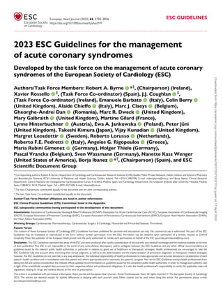
Figure 1 Central illustration. ACS, acute coronary syndrome; CABG, coronary artery bypass grafting; ECG, electrocardiogram; LMWH, low molecular-
weight heparin; NSTE-ACS, non-ST-elevation acute coronary syndrome; PCI, percutaneous coronary intervention; PPCI, primary percutaneous coronary
intervention; STEMI, ST-elevation myocardial infarction; UFH, unfractionated heparin. Patients with acute coronary syndrome (ACS) can initially present with
a wide variety of clinical signs and symptoms and it is important that there is a high degree of awareness of this amongst both the general public and healthcare
providers. If ACS is suspected, think ‘A.C.S.’ for the initial triage and assessment. This involves performing an electrocardiogram (ECG) to assess for
Abnormalities or evidence of ischaemia, taking a targeted clinical history to assess the clinical Context of the presentation, and carrying out a targeted clinical
examination to assess for clinical and haemodynamic Stability. Based on the initial assessment, the healthcare provider can decide whether immediate invasive
management is required. Patients with ST-elevation myocardial infarction (STEMI) require primary percutaneous coronary intervention (PPCI) (or fibrinoly
sis if PPCI within 120 min is not feasible); patients with non-ST-elevation ACS (NSTE-ACS) with very high-risk features require immediate angiography ± PCI
if indicated; patients with NSTE-ACS and high-risk features should undergo inpatient angiography (angiography within 24 h should be considered). A com
bination of antiplatelet and anticoagulant therapy is indicated acutely for patients with ACS. The majority of patients with ACS will eventually undergo re
vascularization, most commonly with PCI. Once the final diagnosis of ACS has been established, it is important to implement measures to prevent recurrent
events and to optimize cardiovascular risk. This consists of medical therapy, lifestyle changes and cardiac rehabilitation, as well as consideration of psycho
social factors.
ESC Guidelines 3729
Downloaded
from
https://academic.oup.com/eurheartj/article/44/38/3720/7243210
by
guest
on
17
April
2024
11. 2.1. Definitions | Acute coronary
syndromes and myocardial infarction
Acute coronary syndromes (ACS) encompass a spectrum of condi
tions that include patients presenting with recent changes in clinical
symptoms or signs, with or without changes on 12-lead electrocardio
gram (ECG) and with or without acute elevations in cardiac tropo
nin (cTn) concentrations (Figure 2). Patients presenting with
suspected ACS may eventually receive a diagnosis of acute myocar
dial infarction (AMI) or unstable angina (UA). The diagnosis of
myocardial infarction (MI) is associated with cTn release and is
made based on the fourth universal definition of MI.1
UA is defined
as myocardial ischaemia at rest or on minimal exertion in the ab
sence of acute cardiomyocyte injury/necrosis. It is characterized by
specific clinical findings of prolonged (>20 min) angina at rest; new
onset of severe angina; angina that is increasing in frequency, longer
in duration, or lower in threshold; or angina that occurs after a re
cent episode of MI. ACS are associated with a broad range of clinical
presentations, from patients who are symptom free at presentation
to patients with ongoing chest discomfort/symptoms and patients
Increasing chest
pain/symptoms
Oligo/
asymptomatic
Persistent chest
pain/symptoms
Cardiogenic shock/
acute heart failure
Cardiac
arrest
Normal ST segment
depression
ST segment
elevation
Malignant
arrhythmia
Clinical
presentation
ECG
findings
hs-cTn
levels
The ACS spectrum
Final
diagnosis
Working
diagnosis
Non-elevated
Unstable
angina
NSTE-ACS
NSTEMI
Rise and fall
STEMI
STEMI
Figure 2 The spectrum of clinical presentations, electrocardiographic findings, and high-sensitivity cardiac troponin levels in patients with acute coronary
syndrome. ACS, acute coronary syndrome; ECG, electrocardiogram; hs-cTn, high-sensitivity cardiac troponin; NSTE-ACS, non-ST-elevation acute coronary
syndrome; NSTEMI, non-ST-elevation myocardial infarction; STEMI, ST-elevation myocardial infarction.
3730 ESC Guidelines
Downloaded
from
https://academic.oup.com/eurheartj/article/44/38/3720/7243210
by
guest
on
17
April
2024
13. 2.2. Epidemiology of acute coronary
syndromes
Cardiovascular disease (CVD) is the most common cause of mortality
and morbidity worldwide, with a substantial portion of this burden
borne by low- and middle-income countries.2,3
ACS is often the first
clinical manifestation of CVD. In 2019, there were an estimated 5.8 mil
lion new cases of ischaemic heart disease in the 57 ESC member coun
tries.3
The median age-standardized incidence estimate per 100 000
people was 293.3 (interquartile ratio 195.8–529.5). CVD remains the
most common cause of death within ESC member countries, account
ing for just under 2.2 million deaths in females and just over 1.9 million
deaths in males in the most recent year of available data. Ischaemic
heart disease is the most common cause of CVD death, accounting
for 38% of all CVD deaths in females and 44% in males.3
2.3. Number and breakdown of classes of
recommendations
The total number of recommendations in this guideline is 193. A sum
mary of the recommendations according to Class of Recommendation
and Level of Evidence (LoE) is also provided. As per Class of
Recommendation, there were 106 Class I, 70 Class II, and 17 Class III
recommendations. As per LoE, there were 56 LoE A, 64 LoE B, and
73 LoE C recommendations.
STEMI
NSTE-ACS
STEMI
NSTEMI
Unstable angina
Non-ACS diagnosis
hs-cTn levels
± Angiography
± Imaging
Final diagnosisb
Working diagnosisa Further investigations
Clinical presentation
If a patient has
signs/symptoms
suggestive of ACS,
perform an ECG
within 10 min of FMC
ECG
Figure 3 Classification of patients presenting with suspected acute coronary syndrome: from a working to a final diagnosis. ACS, acute coronary syndrome;
ECG, electrocardiogram; FMC, first medical contact; hs-cTn, high-sensitivity cardiac troponin; MI, myocardial infarction; NSTE-ACS, non-ST-elevation acute
coronary syndrome; NSTEMI, non-ST-elevation myocardial infarction: STEMI, ST-elevation myocardial infarction. a
The working ACS diagnosis can be clas
sified as STEMI or NSTE-ACS on the basis of available clinical information and ECG findings. This allows for initial triage and assessment. b
The final diagnosis is
based on symptoms, ECG and troponin for the diagnosis of MI as well as the results of other tests (i.e. imaging and/or angiography) to facilitate understanding
of the mechanism and subclassification of the type of MI. Patients initially assigned a working diagnosis of STEMI or NSTE-ACS may eventually receive a final
non-ACS diagnosis.
3732 ESC Guidelines
Downloaded
from
https://academic.oup.com/eurheartj/article/44/38/3720/7243210
by
guest
on
17
April
2024
14. 2.4. What is new
Table 4 New recommendations
Recommendations Classa
Levelb
Recommendations for antiplatelet and anticoagulant therapy in acute coronary syndrome
If patients presenting with ACS stop DAPT to undergo coronary artery bypass grafting, it is recommended they resume DAPT after surgery
for at least 12 months.
I C
In older ACS patients, especially if HBR, clopidogrel as the P2Y12 receptor inhibitor may be considered. IIb B
Recommendations for alternative antithrombotic therapy regimens
In patients who are event-free after 3–6 months of DAPT and who are not high ischaemic risk, single antiplatelet therapy (preferably with a
P2Y12 receptor inhibitor) should be considered.
IIa A
P2Y12 inhibitor monotherapy may be considered as an alternative to aspirin monotherapy for long-term treatment. IIb A
In HBR patients, aspirin or P2Y12 receptor inhibitor monotherapy after 1 month of DAPT may be considered. IIb B
In patients requiring OAC, withdrawing antiplatelet therapy at 6 months while continuing OAC may be considered. IIb B
De-escalation of antiplatelet therapy in the first 30 days after an ACS event is not recommended. III B
Recommendations for cardiac arrest and out-of-hospital cardiac arrest
Evaluation of neurological prognosis (no earlier than 72 h after admission) is recommended in all comatose survivors after cardiac arrest. I C
Transport of patients with out-of-hospital cardiac arrest to a cardiac arrest centre according to local protocol should be considered. IIa C
Recommendations for technical aspects of invasive strategies
In patients with spontaneous coronary artery dissection, PCI is recommended only for patients with symptoms and signs of ongoing
myocardial ischaemia, a large area of myocardium in jeopardy, and reduced antegrade flow.
I C
Intravascular imaging should be considered to guide PCI. IIa A
Intravascular imaging (preferably optical coherence tomography) may be considered in patients with ambiguous culprit lesions. IIb C
Recommendations for multivessel disease in ACS patients presenting in cardiogenic shock
Staged PCI of non-IRA should be considered. IIa C
Recommendations for multivessel disease in haemodynamically stable STEMI patients undergoing primary PCI
It is recommended that PCI of the non-IRA is based on angiographic severity. I B
Invasive epicardial functional assessment of non-culprit segments of the IRA is not recommended during the index procedure. III C
Recommendations for acute coronary syndrome complications
Implantation of a permanent pacemaker is recommended when high-degree AV block does not resolve within a waiting period of at least 5
days after MI.
I C
Cardiac magnetic resonance imaging should be considered in patients with equivocal echocardiographic images or in cases of high clinical
suspicion of LV thrombus.
IIa C
Following an acute anterior MI, a contrast echocardiogram may be considered for the detection of LV thrombus if the apex is not well
visualized on echocardiography.
IIb C
In selected patients with high-degree AV block in the context of an anterior wall MI and acute heart failure, early device implantation (cardiac
resynchronization therapy—defibrillator/pacemaker) may be considered.
IIb C
In patients with recurrent life-threatening ventricular arrhythmias, sedation or general anaesthesia to reduce sympathetic drive may be
considered.
IIb C
Recommendations for acute coronary syndrome comorbid conditions
It is recommended to base the choice of long-term glucose-lowering treatment on the presence of comorbidities, including heart failure,
chronic kidney disease, and obesity.
I A
For frail older patients with comorbidities, a holistic approach is recommended to individualize interventional and pharmacological
treatments after careful evaluation of the risks and benefits.
I B
An invasive strategy is recommended in cancer patients presenting with high-risk ACS with expected survival ≥6 months. I B
A temporary interruption of cancer therapy is recommended in patients in whom the cancer therapy is suspected to be a contributing cause
of ACS.
I C
A conservative non-invasive strategy should be considered in ACS patients with poor cancer prognosis (i.e. with expected life survival <6
months) and/or very high bleeding risk.
IIa C
Aspirin is not recommended in cancer patients with a platelet count <10 000/μL. III C
Continued
ESC Guidelines 3733
Downloaded
from
https://academic.oup.com/eurheartj/article/44/38/3720/7243210
by
guest
on
17
April
2024
17. It is important that awareness of the symptoms associated with
ACS is high among the general population, in particular red flag
symptoms such as prolonged chest pain (>15 min) and/or recurrent
pain within 1 h, which should prompt patients or other members of
the public to seek urgent medical help. Continuous education, pro
motion, and advocacy efforts are important to make sure that this
information is as widely available as possible to the general
population.
ACS
presentation
Initial A.C.S.
assessment
ECG Physical examination Clinical history Vital signs hs-cTna levels
Non-immediate
angiography
Echo
Intravascular
imaging
hs-cTna
levels
Non-invasive
imaging
ECG
monitoring
PCI CABG
Long-term
medical therapy
Lifestyle
measures
Smoking
cessation
STEMI
NSTE-ACS
with very high-risk featuresb
NSTE-ACS
without very high-risk featuresb
Immediate angiography ±
PPCI or fibrinolysis if timely
PPCI not feasible
PPCI ATT Fibrinolysis
Immediate angiography
± PCI
Consider angiography
within 24 h for NSTE-ACS
with high risk features
Working
diagnosis
Early invasive
angiography
according to
patient risk
Further
management
Further
investigations
PCI ATT PCI ATT
Figure 4 An overview of the initial triage, management and investigation of patients who present with signs and symptoms potentially consistent with acute
coronary syndrome. ACS, acute coronary syndrome; ATT, antithrombotic therapy; CABG, coronary artery bypass grafting; ECG, electrocardiogram; hs-cTn,
high-sensitivity cardiac troponin; NSTE-ACS, non-ST-elevation acute coronary syndrome; PPCI, primary percutaneous coronary intervention; STEMI,
ST-elevation myocardial infarction. The ‘A.C.S.’ assessment is detailed in Figure 5. a
Results of hs-cTn measurements are not required for the initial stratifi
cation of ACS and the initial emergency management (i.e. for patients with a working diagnosis of STEMI or very high-risk NSTE-ACS) should not be delayed
based on this. b
For patients with NSTE-ACS with very high-risk features, immediate angiography is recommended. For patients with NSTE-ACS with high-
risk features, early invasive angiography (i.e. <24 h) should be considered and inpatient invasive angiography is recommended. See Recommendation Table 4
for details.
3736 ESC Guidelines
Downloaded
from
https://academic.oup.com/eurheartj/article/44/38/3720/7243210
by
guest
on
17
April
2024
18. 3.1.2. History taking and physical examination
Patients with suspected ACS present in a broad range of clinical scen
arios, including in the community, at the emergency department (ED),
or in the inpatient setting. It is crucial to take a focused medical history
and accurately characterize the presenting symptoms in order to
manage the patient via the appropriate care pathway as soon as
possible.
Prompt assessment of vital signs is recommended at first medical
contact (FMC), at the same time as acquisition of an initial ECG
(Figure 5). In patients presenting with suspected ACS, physical examin
ation is recommended and is useful both to eliminate differential diag
noses and to identify very high-risk and high-risk ACS features. This may
be particularly relevant for patients presenting with cardiac arrest, signs
of CS, and/or haemodynamic or electrical instability.4
Focused physical
examination should include checking for the presence of all major
pulses, measurement of blood pressure in both arms, auscultation of
the heart and lungs, and assessing for signs of HF or circulatory
compromise.
3.2. Diagnostic tools | Electrocardiogram
The resting 12-lead ECG is the first-line diagnostic tool in the assess
ment of patients with suspected ACS. It is recommended that an
ECG is obtained immediately upon FMC and interpreted by a qualified
emergency medical technician or physician within 10 min.4,5
It should
be repeated as necessary, especially if symptoms have waned at FMC.
Based on the initial ECG, patients with suspected ACS can be differen
tiated into two working diagnoses:
• Patients with acute chest pain (or chest pain-equivalent
signs/symptoms) and persistent ST-segment elevation
(or ST-segment elevation equivalents) on ECG (working
diagnosis: ST-segment elevation MI: STEMI). The vast major
ity of these patients will sustain myocardial necrosis and troponin ele
vation, fulfilling the criteria for an MI, but MI will not be the final
diagnosis in all patients with a working diagnosis of STEMI.
• Patients with acute chest pain (or chest pain-equivalent
signs/symptoms) but without persistent ST-segment
A C S
Abnormal
ECG?
Clinical
context?
Stable
patient?
Perform an ECG to assess
for evidence of ischaemia
or other abnormalities
Consider the clinical
context and available
investigations
Perform an exam to assess
if the patient is clinically
and vitally stable
Figure 5 The A.C.S. assessment for the initial evaluation of patients with suspected acute coronary syndrome. ECG, electrocardiogram. This figure sum
marizes the initial ‘A.C.S. assessment’ that can be performed for a patient presenting with suspected ACS. ‘A’ stands for ‘Abnormal ECG?’: an ECG should be
performed within 10 min of FMC and assessed for evidence of abnormalities or ischaemia. ‘C’ stands for ‘Clinical Context?’: it is important to consider the
clinical context of the patient’s presentation and the results of any investigations that are available. This should also include a targeted history with the aim of
determining the patient’s symptoms and elucidating any other relevant background information. ‘S’ stands for ‘Stable Patient?’: the patient should be quickly
assessed to determine if they are clinically stable—this should include assessment of the clinical vital signs, including heart rate, blood pressure, and oxygen
saturations, if possible, as well as checking for potential signs of CS.
ESC Guidelines 3737
Downloaded
from
https://academic.oup.com/eurheartj/article/44/38/3720/7243210
by
guest
on
17
April
2024
19. elevation (or ST-segment elevation equivalents) on ECG
(working diagnosis: non-ST-elevation [NSTE]-ACS).
These patients may exhibit other ECG alterations, including transient
ST-segment elevation, persistent or transient ST-segment depres
sion, and T wave abnormalities, including hyperacute T waves, T
wave inversion, biphasic T waves, flat T waves, and pseudo-
normalization of T waves. Alternatively, the ECG may be normal.
The majority of patients in this category who subsequently display
a typical rise and fall in cardiac troponin levels (i.e. fulfilling MI criteria
as per the fourth universal definition of MI) will receive a final diagno
sis of non-ST-elevation MI (NSTEMI). In other patients, the troponin
level will remain below the 99th centile and they will receive a final
diagnosis of UA, although with high-sensitivity troponin assays this
diagnosis has become less common. It is also important to recognize
that NSTEMI or UA will not be the final diagnosis in all patients with
an initial working diagnosis of NSTE-ACS.
3.2.1. Acute coronary syndrome with persistent
ST-segment elevation (suspected ST-elevation
myocardial infarction)
The priority for these patients is the implementation of reperfusion ther
apy as soon as possible (see Section 5). In the appropriate clinical context,
ST-segment elevation (measured at the J-point) is considered suggestive
of ongoing coronary artery acute occlusion in the following cases:
New ST elevation at the J-point in at least two contiguous leads:
• ≥2.5 mm in men <40 years, ≥2 mm in men ≥40 years, or ≥1.5 mm
in women regardless of age in leads V2–V3
• and/or ≥1 mm in the other leads (in the absence of left ventricular
[LV] hypertrophy or left bundle branch block [LBBB]).
In patients with suspected inferior STEMI, it is recommended to re
cord right precordial leads (V3R and V4R) in order to assess for
ST-segment elevation.6
Posterior leads (V7–V9) can also be recorded
to investigate for posterior STEMI, particularly in patients with ongoing
symptoms and an inconclusive standard 12-lead ECG.
The diagnosis of ongoing acute coronary artery occlusion on ECG
can sometimes be challenging, and some cases may warrant prompt
management and triage for immediate reperfusion therapy despite
the absence of ST-segment elevation. It is also important to recognize
that while the most sensitive sign for ongoing acute coronary
artery occlusion is ST-segment elevation, there are other ECG find
ings that can be suggestive of ongoing coronary artery occlusion (or
severe ischaemia). If these findings are present, prompt triage for
immediate reperfusion therapy is indicated (see Supplementary data
online, Figure S2).
ST-segment depression in leads V1–V3 (especially when the terminal
T wave is positive) and/or ST-segment elevation in V7–V9 are highly
suggestive of posterior coronary artery occlusion (often the left cir
cumflex artery).1,7
ST-segment elevation in V3R and V4R is highly sug
gestive of ongoing RV ischaemia.8
ST depression ≥1 mm in ≥6 surface
leads (inferolateral ST depression), coupled with ST-segment elevation
in aVR and/or V1, suggests multivessel ischaemia or left main coronary
artery obstruction, particularly if the patient presents with haemo
dynamic compromise.9–11
Bundle branch block (BBB). In patients with a high clinical sus
picion of ongoing myocardial ischaemia, the presence of LBBB, right
bundle branch block (RBBB), or a paced rhythm precludes an accurate
assessment of the presence or absence of ST-segment elevation.
Therefore, patients presenting with these ECG patterns in combination
with signs/symptoms that are highly suspicious for ongoing myocardial
ischaemia should be managed similarly to those with clear ST-segment
elevation, regardless of whether the BBB is previously known (see
Supplementary data online).4
3.2.2. Acute coronary syndrome without persistent
ST-segment elevation (non-ST elevation acute
coronary syndrome)
While the ECG in the setting of NSTE-ACS may be normal in more
than one-third of patients, characteristic ECG abnormalities are fre
quently present and increase the diagnostic probability of ACS.12–16
These ECG abnormalities include ST depression and T wave changes
(especially biphasic T waves or prominent negative T waves
[Wellens’ sign, related to severe proximal left anterior descending ar
tery stenosis]), (see Supplementary data online, Figure S3).
Recommendation Table 1 — Recommendations for
clinical and diagnostic tools for patients with suspected
acute coronary syndrome
Recommendations Classa
Levelb
It is recommended to base the diagnosis and initial
short-term risk stratification of ACS on a
combination of clinical history, symptoms, vital signs,
other physical findings, ECG, and hs-cTn.1,17,18
I B
ECG
Twelve-lead ECG recording and interpretation is
recommended as soon as possible at the point of
FMC, with a target of <10 min.5,19
I B
Continuous ECG monitoring and the availability of
defibrillator capacity is recommended as soon as
possible in all patients with suspected STEMI, in
suspected ACS with other ECG changes or ongoing
chest pain, and once the diagnosis of MI is made.20,21
I B
The use of additional ECG leads (V3R, V4R, and V7–
V9) is recommended in cases of inferior STEMI or if
total vessel occlusion is suspected and standard leads
are inconclusive.22–24
I B
An additional 12-lead ECG is recommended in cases
with recurrent symptoms or diagnostic uncertainty.
I C
Continued
3738 ESC Guidelines
Downloaded
from
https://academic.oup.com/eurheartj/article/44/38/3720/7243210
by
guest
on
17
April
2024