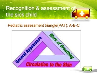
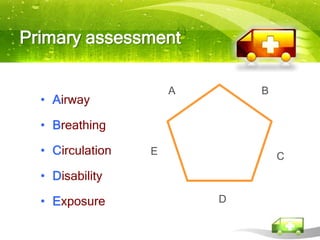
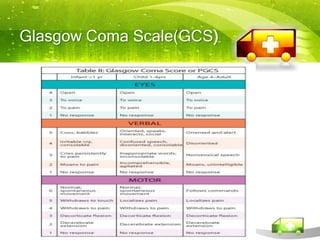
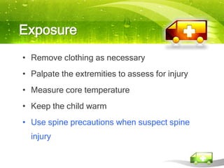
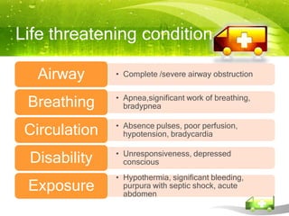
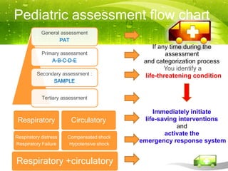
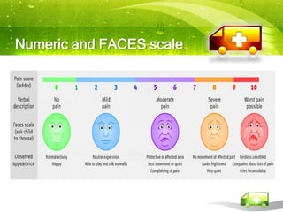
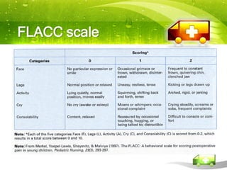
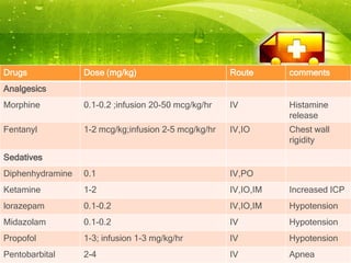
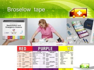
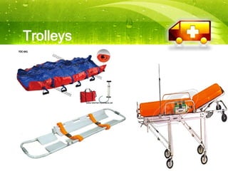
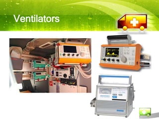
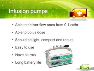
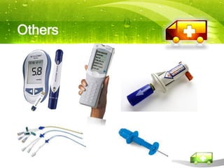
Pediatric Transport


 Darin Aranwutikul, MD.
Goal

Early stabilization and initiation of advanced care
  at the referring institution, with continuation of
  critical care therapies and monitoring en route.
Recognition & assessment of
the sick child
Pediatric assessment triangle(PAT): A-B-C
Appearance :
   TICLS Mnemonic
Tone            Refers to child’s muscle tone
Interactivity   Refers to degree of interaction the child has with his/her
                environment or those attempting to interact with the child


Consolability   Refers to the child’s response to parents or caregivers


Look /gaze      Identifies whether the child tracks things appropriately
                with his/her eyes or has a nonfocused gaze.


Speech/cry      Refers to how the child vocalizes
Primary assessment

                      A       B
  • Airway

  • Breathing

  • Circulation   E               C
  • Disability

  • Exposure              D
Airway
• Patency
• Need simple management
      positioning
      head tilt-chin lift
      Use airway adjuncts ( oral airway)
• Require advanced intervention
      ET intubation
      cricothyroidotomy
      CPAP
Breathing


 • Respiratory rate

 • Respiratory effort

 • Airway and lung sounds

 • Pulse oximetry
Normal respiratory rates
by age
              Age         Breaths per minutes

Infant (<1 year)               30 to 60

Todler( 1-3 yrs)               24 to 40

Preschooler ( 4-5 yrs)         22 to 34

School age ( 6-12 yrs)         18 to 30

Adolescent ( 13-18 yrs)        12 to 16
Respiratory rate


     • Apnea

     • Tachypnea

     • Bradypnea

      Bradypnea or irregular respiratory rate in an
accutely ill infant or child often signals impending arrest
Abnormal lung and airway sounds


   • Stridor upper airway obstruction

   • Wheezinglower airway obstruction

   • Grunting lung tissue disease

   • Crackles  lung tissue disease
Pulse oximetry


   • Above 94% in room air

   • Additional intervention is required if O2 sat<90%
     in child receiving 100% oxygen .

   • Be careful to interpret pulse oximetry in
     conjunction with clinical assessment and other
     signs.
Circulation

• Evaluate cardiovascular • Evaluate end-organ
  function                       function
     heart rate and rhythm         brain perfusion
     pulses                        skin perfusion
     capillary refill time         renal perfusion
     blood pressure and pulse
      pressure
Normal heart rates in children

         Age           Awake rate          Mean         Sleep rate

    NB to 3 mo           85-205              140          80-160

     3 mo to 2 y         100-190             130          75-160

     2 y to 10 y         60-140              80            60-90

        >10 y            60-100              75            50-90

 Typical physiologic response to a fall in cardiac output is tachycardia.
Blood pressure
Definition of hypotension


             Age            Systolic BP (mmHg)
       Term neonates                  <60

            Infants                   <70

       Children 1-10 yr
       5th BP percentile    <70 + (age in years x 2)


       Children > 10 yr               <90
Systemic perfusion
• Peripheral Pulses
  – Present/Absent
  – Strength
• Skin Perfusion
  – Capillary refill time
  – Temperature
  – Color
  – Mottling
Systemic perfusion
• CNS Perfusion
  – Level of
    consciousness


• Renal Perfusion
  – Urine 1-2 cc/kg/hr
Disability

  • Establish the child's level of consciousness

  • Standard evaluations are

       AVPU pediatric response scale

       Glasgow Coma Scale (GCS)

       pupillary responses
AVPU pediatric response scale
Glasgow Coma Scale(GCS)
    -
Pupillary response
Exposure
• Remove clothing as necessary
• Palpate the extremities to assess for injury
• Measure core temperature
• Keep the child warm
• Use spine precautions when suspect spine
  injury
Life threatening condition

   Airway      • Complete /severe airway obstruction


 Breathing     • Apnea,significant work of breathing,
                 bradypnea


 Circulation   • Absence pulses, poor perfusion,
                 hypotension, bradycardia


 Disability    • Unresponsiveness, depressed
                 conscious
               • Hypothermia, significant bleeding,
 Exposure        purpura with septic shock, acute
                 abdomen
Secondary assessment
• Signs and Symptoms

• Allergies

• Medications

• Past medical history

• Last meal

• Events leading to presentation
Pediatric assessment flow chart
            General assessment
                   PAT
                                               If any time during the
            Primary assessment                      assessment
                A-B-C-D-E                   and categorization process
                                                    You identify a
          Secondary assessment :            life-threatening condition
                SAMPLE


            Tertiary assessment

                                               Immediately initiate
 Respiratory            Circulatory         life-saving interventions
                                                       and
                                                   activate the
Respiratory distress   Compensated shock
Respiratory Failure    Hypotensive shock
                                           emergency response system


Respiratory +circulatory
Breathing is everything to a child
• The common denominator for unexpected deaths
  in children is hypoxia.

• Do not increase the child's level of anxiety
Not only the child
• Needs of parents or caregivers must
  be addressed.
• Be calm and confident.
• Written information and involve them
  in plan of care.
Children don’t have less pain than adult
Numeric and FACES scale
FLACC scale
Drugs             Dose (mg/kg)                        Route      comments
Analgesics
Morphine          0.1-0.2 ;infusion 20-50 mcg/kg/hr   IV         Histamine
                                                                 release
Fentanyl          1-2 mcg/kg;infusion 2-5 mcg/kg/hr   IV,IO      Chest wall
                                                                 rigidity
Sedatives
Diphenhydramine   0.1                                 IV,PO
Ketamine          1-2                                 IV,IO,IM   Increased ICP
lorazepam         0.1-0.2                             IV,IO,IM   Hypotension
Midazolam         0.1-0.2                             IV         Hypotension
Propofol          1-3; infusion 1-3 mg/kg/hr          IV         Hypotension
Pentobarbital     2-4                                 IV         Apnea
YOU CANNOT REMEMBER
NORMAL WEIGHTS, RESPIRATORY
RATES, BLOOD PRESSURES, HEART
RATES, AND CALCULATE DRUG DOSES
IN YOUR HEAD SO DON'T TRY
Broselow tape
HOW TO STABILIZE THE CHILD
Stabilization of the respiratory
system

  • Well oxygenated and ventilating prior to transfer

  • Consider the need for intubation and mechanical
    ventilation.

  • Confirm ETT placement and secure the tube.

  • Obtain blood gases while ventilating on the transport
    ventilator before leaving

  • Consider the need for sedation and paralysis
Stabilization of the
cardiovascular system

•   Hemodynamically stable before departure.

• Treat compensated shock before departure.

• Invasive arterial blood pressure monitoring in
    patients with inotropic support.

•   At least 2 good, working points of IV access.

• Ensure availability of emergency or special drugs
Medications to Maintain Cardiac Output and for
  Postresuscitation Stabilization
  Medication              Dose Range                                Comment
                 0.75–1 mg/kg IV/IO over 5
Inamrinone       minutes; may repeat × 2
                                                      Inodilator
                 then 5-10 mcg/kg/min
Dobutamine       2–20 mcg/kg/min IV/IO
                                                      Inotrope; vasodilator
                 2–20 mcg/kg/min IV/IO                Inotrope; chronotrope; renal and
Dopamine
                                                      splanchnic vasodilator in low doses;
                                                      pressor in high doses
Epinephrine      0.1–1 mcg/kg/min IV/IO               Inotrope; chronotrope; vasodilator in
                                                      low doses; pressor in higher doses
                 Loading dose: 50 mcg/kg IV/IO
Milrinone        over 10–60 min                       Inodilator
                 then 0.25-0.75 mcg/kg/min
Norepinephrine   0.1–2 mcg/kg/min                     Vasopressor

Sodium           Initial: 0.5–1 mcg/kg/min; titrate   Vasodilator
                 to effect up to 8 mcg/kg/min
nitroprusside                                         Prepare only in D5W
Stabilization of the central nervous
system

 • Minimize secondary brain injury due to
   hypotension and hypoxia

 • Appropriate treatment of prolonged seizures

 • Adequate sedation
Stabilization of the gastrointestinal
system

  • Placement of a nasogastric tube and left
    on free drainage.

  • Stop feeding and aspirate the stomach
    before transfer.
Stabilization of the renal system

    • Consider urethral catheterisation in
     children
      – with shock

      – who are paralysed and sedated

      – who have received diuretics or mannitol
Transport team assessment and
initial stabilization

 • Rapid assessment

 • Urgent therapy and manage life-threatening
   conditions is priority

 • Have patient as stable as possible before
   loading into the transport vehicle.
EQUIPMENT USED IN PEDIATRIC
TRANSPORT
General features of all
equipment
•   Self-contained, lightweight and portable
•   Durable and robust
•   Long battery life and short recharge time
•   Clear displays
•   Suitable for all ages
•   Visible and audible alarms
•   Data storage and download capability
•   Secure
Batteries
• Use external sources of power when
 available.

• Choose equipment that is not solely
 reliant on internal rechargeable batteries.

• Do not rely on leaving them charging all
 the time.
Trolleys
Ventilators
Humidification

  Heat and moisture exchangers (HMEs):
Temperature maintenance
Infusion pumps

 • Able to deliver flow rates from 0.1 cc/hr

 • Able to bolus dose

 • Should be light, compact and robust

 • Easy to use

 • Have alarms

 • Long battery life
Suction equipment

  • Portable suction units with battery power
  • Foot pump suction units
Defibrillators
• Portable defibrillator or AED
Monitoring
Others
Reference

 • American Academy of Pediatrics. Guidelines for Air and Ground
    Transport of neonatal and pediatric patients, 3rd edition.

 • David G. Jaimovich . Handbook of Pediatric and Neonatal
    transport medicine, 2nd edition.

 • Peter Barry.Paediatric and Neonatal critical care transport, BMJ
    2003

 • American Academy of Pediatrics. Pediatric Advanced life Support
    provider manual 2006

Acem 2011 pediatric transport darin

  • 1.
    Pediatric Transport DarinAranwutikul, MD.
  • 2.
    Goal Early stabilization andinitiation of advanced care at the referring institution, with continuation of critical care therapies and monitoring en route.
  • 3.
    Recognition & assessmentof the sick child Pediatric assessment triangle(PAT): A-B-C
  • 4.
    Appearance : TICLS Mnemonic Tone Refers to child’s muscle tone Interactivity Refers to degree of interaction the child has with his/her environment or those attempting to interact with the child Consolability Refers to the child’s response to parents or caregivers Look /gaze Identifies whether the child tracks things appropriately with his/her eyes or has a nonfocused gaze. Speech/cry Refers to how the child vocalizes
  • 5.
    Primary assessment A B • Airway • Breathing • Circulation E C • Disability • Exposure D
  • 6.
    Airway • Patency • Needsimple management  positioning  head tilt-chin lift  Use airway adjuncts ( oral airway) • Require advanced intervention  ET intubation  cricothyroidotomy  CPAP
  • 7.
    Breathing • Respiratoryrate • Respiratory effort • Airway and lung sounds • Pulse oximetry
  • 8.
    Normal respiratory rates byage Age Breaths per minutes Infant (<1 year) 30 to 60 Todler( 1-3 yrs) 24 to 40 Preschooler ( 4-5 yrs) 22 to 34 School age ( 6-12 yrs) 18 to 30 Adolescent ( 13-18 yrs) 12 to 16
  • 9.
    Respiratory rate • Apnea • Tachypnea • Bradypnea Bradypnea or irregular respiratory rate in an accutely ill infant or child often signals impending arrest
  • 11.
    Abnormal lung andairway sounds • Stridor upper airway obstruction • Wheezinglower airway obstruction • Grunting lung tissue disease • Crackles  lung tissue disease
  • 12.
    Pulse oximetry • Above 94% in room air • Additional intervention is required if O2 sat<90% in child receiving 100% oxygen . • Be careful to interpret pulse oximetry in conjunction with clinical assessment and other signs.
  • 13.
    Circulation • Evaluate cardiovascular• Evaluate end-organ function function  heart rate and rhythm  brain perfusion  pulses  skin perfusion  capillary refill time  renal perfusion  blood pressure and pulse pressure
  • 14.
    Normal heart ratesin children Age Awake rate Mean Sleep rate NB to 3 mo 85-205 140 80-160 3 mo to 2 y 100-190 130 75-160 2 y to 10 y 60-140 80 60-90 >10 y 60-100 75 50-90 Typical physiologic response to a fall in cardiac output is tachycardia.
  • 15.
    Blood pressure Definition ofhypotension Age Systolic BP (mmHg) Term neonates <60 Infants <70 Children 1-10 yr 5th BP percentile <70 + (age in years x 2) Children > 10 yr <90
  • 16.
    Systemic perfusion • PeripheralPulses – Present/Absent – Strength • Skin Perfusion – Capillary refill time – Temperature – Color – Mottling
  • 17.
    Systemic perfusion • CNSPerfusion – Level of consciousness • Renal Perfusion – Urine 1-2 cc/kg/hr
  • 18.
    Disability •Establish the child's level of consciousness • Standard evaluations are  AVPU pediatric response scale  Glasgow Coma Scale (GCS)  pupillary responses
  • 19.
  • 20.
  • 21.
  • 22.
    Exposure • Remove clothingas necessary • Palpate the extremities to assess for injury • Measure core temperature • Keep the child warm • Use spine precautions when suspect spine injury
  • 23.
    Life threatening condition Airway • Complete /severe airway obstruction Breathing • Apnea,significant work of breathing, bradypnea Circulation • Absence pulses, poor perfusion, hypotension, bradycardia Disability • Unresponsiveness, depressed conscious • Hypothermia, significant bleeding, Exposure purpura with septic shock, acute abdomen
  • 24.
    Secondary assessment • Signsand Symptoms • Allergies • Medications • Past medical history • Last meal • Events leading to presentation
  • 25.
    Pediatric assessment flowchart General assessment PAT If any time during the Primary assessment assessment A-B-C-D-E and categorization process You identify a Secondary assessment : life-threatening condition SAMPLE Tertiary assessment Immediately initiate Respiratory Circulatory life-saving interventions and activate the Respiratory distress Compensated shock Respiratory Failure Hypotensive shock emergency response system Respiratory +circulatory
  • 26.
    Breathing is everythingto a child • The common denominator for unexpected deaths in children is hypoxia. • Do not increase the child's level of anxiety
  • 27.
    Not only thechild • Needs of parents or caregivers must be addressed. • Be calm and confident. • Written information and involve them in plan of care.
  • 28.
    Children don’t haveless pain than adult
  • 29.
  • 30.
  • 31.
    Drugs Dose (mg/kg) Route comments Analgesics Morphine 0.1-0.2 ;infusion 20-50 mcg/kg/hr IV Histamine release Fentanyl 1-2 mcg/kg;infusion 2-5 mcg/kg/hr IV,IO Chest wall rigidity Sedatives Diphenhydramine 0.1 IV,PO Ketamine 1-2 IV,IO,IM Increased ICP lorazepam 0.1-0.2 IV,IO,IM Hypotension Midazolam 0.1-0.2 IV Hypotension Propofol 1-3; infusion 1-3 mg/kg/hr IV Hypotension Pentobarbital 2-4 IV Apnea
  • 32.
    YOU CANNOT REMEMBER NORMALWEIGHTS, RESPIRATORY RATES, BLOOD PRESSURES, HEART RATES, AND CALCULATE DRUG DOSES IN YOUR HEAD SO DON'T TRY
  • 33.
  • 34.
  • 35.
    Stabilization of therespiratory system • Well oxygenated and ventilating prior to transfer • Consider the need for intubation and mechanical ventilation. • Confirm ETT placement and secure the tube. • Obtain blood gases while ventilating on the transport ventilator before leaving • Consider the need for sedation and paralysis
  • 36.
    Stabilization of the cardiovascularsystem • Hemodynamically stable before departure. • Treat compensated shock before departure. • Invasive arterial blood pressure monitoring in patients with inotropic support. • At least 2 good, working points of IV access. • Ensure availability of emergency or special drugs
  • 37.
    Medications to MaintainCardiac Output and for Postresuscitation Stabilization Medication Dose Range Comment 0.75–1 mg/kg IV/IO over 5 Inamrinone minutes; may repeat × 2 Inodilator then 5-10 mcg/kg/min Dobutamine 2–20 mcg/kg/min IV/IO Inotrope; vasodilator 2–20 mcg/kg/min IV/IO Inotrope; chronotrope; renal and Dopamine splanchnic vasodilator in low doses; pressor in high doses Epinephrine 0.1–1 mcg/kg/min IV/IO Inotrope; chronotrope; vasodilator in low doses; pressor in higher doses Loading dose: 50 mcg/kg IV/IO Milrinone over 10–60 min Inodilator then 0.25-0.75 mcg/kg/min Norepinephrine 0.1–2 mcg/kg/min Vasopressor Sodium Initial: 0.5–1 mcg/kg/min; titrate Vasodilator to effect up to 8 mcg/kg/min nitroprusside Prepare only in D5W
  • 38.
    Stabilization of thecentral nervous system • Minimize secondary brain injury due to hypotension and hypoxia • Appropriate treatment of prolonged seizures • Adequate sedation
  • 39.
    Stabilization of thegastrointestinal system • Placement of a nasogastric tube and left on free drainage. • Stop feeding and aspirate the stomach before transfer.
  • 40.
    Stabilization of therenal system • Consider urethral catheterisation in children – with shock – who are paralysed and sedated – who have received diuretics or mannitol
  • 41.
    Transport team assessmentand initial stabilization • Rapid assessment • Urgent therapy and manage life-threatening conditions is priority • Have patient as stable as possible before loading into the transport vehicle.
  • 42.
    EQUIPMENT USED INPEDIATRIC TRANSPORT
  • 43.
    General features ofall equipment • Self-contained, lightweight and portable • Durable and robust • Long battery life and short recharge time • Clear displays • Suitable for all ages • Visible and audible alarms • Data storage and download capability • Secure
  • 44.
    Batteries • Use externalsources of power when available. • Choose equipment that is not solely reliant on internal rechargeable batteries. • Do not rely on leaving them charging all the time.
  • 45.
  • 46.
  • 47.
    Humidification Heatand moisture exchangers (HMEs):
  • 48.
  • 49.
    Infusion pumps •Able to deliver flow rates from 0.1 cc/hr • Able to bolus dose • Should be light, compact and robust • Easy to use • Have alarms • Long battery life
  • 50.
    Suction equipment • Portable suction units with battery power • Foot pump suction units
  • 51.
  • 52.
  • 53.
  • 54.
    Reference • AmericanAcademy of Pediatrics. Guidelines for Air and Ground Transport of neonatal and pediatric patients, 3rd edition. • David G. Jaimovich . Handbook of Pediatric and Neonatal transport medicine, 2nd edition. • Peter Barry.Paediatric and Neonatal critical care transport, BMJ 2003 • American Academy of Pediatrics. Pediatric Advanced life Support provider manual 2006