This presentation summarizes data related to the CAR-T cell technology and its potential application for cancer therapy. This oral presentation was presented at the 39th PAMM winter meeting in Roma the 8th f February 2018 by Eric Raymond
Chimeric Antigen Receptors (paper with corresponding power point)Kevin B Hugins
Gene therapy was first conceptualized to alter debilitating fates of genetic diseases. Gene therapy technology can help introduce new functional DNA to replace mutated genes. The idea first arose in 1972 when Friedmann and Roblin authored a paper, “Gene therapy for human genetic disease?”, demonstrating that exogenous DNA can be taken up by mammalian cells (1). They proposed that the same procedure could be done on humans to correct genetic defects by introducing therapeutic DNA. Currently, genetic modification of T lymphocytes has been the major area of research for treating malignant tumors. This technique seeks to create chimeric antigen receptor (CAR) in T cells by genetically modifying them in vitro and reintroduce them back into blood circulation. The T cells are unique to every patient and the chimeric antigen receptors are unique to the tumor that it is targeting.
Final presentation for BIOL405, NSC, Spring 2014. Presented by Kevin Hugins and Duy-Khiem Chanh Pham. This presentation addressed the use of Chimeric Antigen Receptors for gene therapy for cancer. Gene therapy was first conceptualized to alter debilitating fates of genetic diseases. Gene therapy technology can help introduce new functional DNA to replace mutated genes. The idea first arose in 1972 when Friedmann and Roblin authored a paper, “Gene therapy for human genetic disease?”, demonstrating that exogenous DNA can be taken up by mammalian cells (1). They proposed that the same procedure could be done on humans to correct genetic defects by introducing therapeutic DNA. Currently, genetic modification of T lymphocytes has been the major area of research for treating malignant tumors. This technique seeks to create chimeric antigen receptor (CAR) in T cells by genetically modifying them in vitro and reintroduce them back into blood circulation. The T cells are unique to every patient and the chimeric antigen receptors are unique to the tumor that it is targeting.
T cells genetically engineered to express chimeric antigen receptors (CAR) have proven an impressive therapeutic activity in patients with certain subtypes of B cell leukaemia or lymphoma, with promising efficacy also demonstrated in patients with multiple myeloma. However, in patients with solid tumors, objective responses to CAR-T cell therapy remain sporadic and transient. Key challenges relating to CAR T cells include the lack of tumor exclusive target, restricted CAR-T cell trafficking to tumor sites, antigen escape and heterogeneity as well as a highly immunosuppressive microenvironment. In this report, we review the current state of the CAR-T technologies as a clinical treatment in solid tumor and we highlight the preclinical innovative designs of novel CAR T cell products that are being developed to increase and expand the clinical benefits of these treatments in patients with solid malignancies.
Dr. Ignacio Melero - Simposio Internacional 'Terapias oncológicas avanzadas''Fundación Ramón Areces
Los días 15 y 16 de octubre de 2014, la Fundación Ramón Areces y la Real Academia Nacional de Farmacia, en colaboración con la Fundación de la Innovación Bankinter, reunieron en Madrid a algunos de los mayores expertos mundiales en nuevas terapias contra el cáncer. El Simposio Internacional, coordinado por la profesora y académica María José Alonso, analizó el momento actual de la lucha contra esta enfermedad. También fue un punto de encuentro para científicos de los más innovadores institutos de investigación en oncología, quienes debatieron sobre tres grandes temas: la Medicina Personalizada contra el cáncer, los nanomedicamentos en la terapia del cáncer y las terapias basadas en la inmunomodulación.
These slides discusses on cellular and gene therapy: the use of cells and genes to treat disease. These therapies can be effective on a wide range of previously untreated diseases, such as hematological, ocular, neurodegenerative diseases, and several types of cancers.
SciTech Development pitch deck including company overview, proprietary technology, lead drug ST-001 nanoFenretinide, patents, addressable market sizes, competiton, key personnel, advisory board, drug product characteristics, fenretinide history, cancer indications and drug mechanism of action (MOA).
This slides briefly introduced why car t cell validation assay is essential for car t therapy development and which assays should be done to validate the safety, identity, purity and potency of car t cell products.
US Pharma presentation on clone screen strategy for monoclonality using Solen...IanTaylor50
Presentation by a Cell Metric CLD customer in US about their use of the system in clone screening for cell line development.
This presentation is made available courtesy of Momenta Pharmaceuticals and IBC Conferences
The Main Advantage
The main advantages of flow cytometry over histology and IHC is the possibility to precisely measure the quantities of antigens and the possibility to stain each cell with multiple antibodies-fluorophores, in current laboratories around 10 antibodies can be bound to each cell. This is much less than mass cytometer where up to 40 can be currently measured, but at a higher and slower pace.
Aquatic research
In aquatic systems, flow cytometry is used for the analysis of autofluorescing cells or cells that are fluorescently-labeled with added stains.
This research started in 1981 when Clarice Yentsch used flow cytometry to measure the fluorescence in a red tide producing dinoflagellates
Marine scientists use the sorting ability of flow cytometers to make discrete measurements of cellular activity and diversity, to conduct investigations into the mutualistic relationships between microorganisms that live in close proximity,and to measure biogeochemical rates of multiple processes in the ocean
Cell Proliferation assay
Cell proliferation is the major function in the immune system. Often it is required to analyse the proliferative nature of the cells in order to make some conclusions. One such assay to determine the cell proliferation is the tracking dye carboxyfluorescein diacetate succinimidyl ester (CFSE). It helps to monitor proliferative cells. This assay gives quantitative as well as qualitative data during time-series experiments
Cell counting
Cell sorting
Determining cell characteristics and function
Detecting microorganisms
Biomarker detection
Protein engineering detection
Diagnosis of health disorders such as blood cancers
Flow cytometry can be used for cell cycle analysis to estimate the percentages of a cell population in the different phases of the cell cycle, or it can be used with other reagents to analyze just the S phase.
Why flow cytometry is ideal for cell cycle analysis
Live-cell cycle analysis stains—Vybrant DyeCycle stains
Classic DNA cell cycle stains such as Hoechst 33342 and DRAQ5 for cell cycle analysis, but most of these have limitations that have to be considered when using them in an experiment which is why the Invitrogen Vybrant DyeCycle stains for live-cell cycle analysis were developed.
Fixed-cell cycle analysis stains FxCycle reagents
We offer classic DNA cell cycle stains such as DAPI, PI, and 7-AAD for fixed cell cycle analysis, but these reagents do not cover the full spectrum of laser excitation available.
The FxCycle reagents offer options for the 405 nm (violet) and 633 nm (red) laser thereby increasing the ability to multiplex by freeing up the 488 nm and 633 nm lasers for other cellular analyses such as immunophenotyping, apoptosis analysis, and dead cell discrimination.
Precise—Accurate cell cycle analysis in living cells
Safe—Low cytotoxicity for combining with additional live cell experiments
Cell sort compatible—Easily sort cells based on phase of the cell cycle
What process is used to obtain cells from bone marrow and normal peripheral blood?
What is the best cell counting and viability method for primary cells?
AllCells, your primary cells research partner, and Nexcelom Bioscience, your cell counting experts, have joined together in an exclusive collaboration to host a free webinar to help educate researchers and present data from their own experiences.
Trans disciplinary research is a must for excellence in science by Prof. Moha...Prof. Mohamed Labib Salem
In this talk, Prof. Mohamed L. Salem presents the importance of having a center of excellence at each institute to enhance and foster scientific research and innovation.
Proteomics Modules designed to bring clinically relevant data, at any point, into the Drug Discovery Process. 1000s of proteins are plated from primary cells and are used to trap autoantibodies from diseased patients' blood sera. Results put a spotlight on highest probability targets.
The Life-Changing Impact of AI in HealthcareKalin Hitrov
For IT Leaders in the healthcare and pharmaceutical industries looking to understand the impact of AI on their industries and how to overcome the ethical and efficiency challenges that come with its use.
Development of quality control assays for cell-based medicinal products (ISCT...Quality Assistance s.a.
Dr. Fabian Vandermeers from Quality Assistance spoke on Development of quality control assays for cell-based medicinal products at ISCT 2017 in London.
For more information on this topic, visit: http://www.quality-assistance.com/analytical-services/CBMPs
For more information on our expertise and services, visit: www.quality-assistance.com
Follow us on social media:
LinkedIn: https://www.linkedin.com/company/quality-assistance
Twitter: https://twitter.com/QA_Belgium
Facebook: https://www.facebook.com/QualityAssistanceBelgium
Google +: https://plus.google.com/103676189647965359292
Quality Assistance S.A. is a leading European Contract Research Organisation providing the pharmaceutical industry with all the analytical services required by EMA and FDA regulations for the development and marketing of innovative human medicinal products.
We assist our clients from candidate selection, through non-clinical and clinical studies, to marketing authorisation, using our state-of-the-art, product-dedicated expertise in analytical sciences.
For each customer and each project, we design customised solutions, define analytical protocols, develop and validate specific new analytical methods and perform characterisation, stability, pharmacokinetic, biomarker and immunogenicity studies as well as batch release testing, in order to evaluate the Quality, Safety and Efficacy of the given drugs.
Liquid Biopsy Overview, Challenges and New Solutions: Liquid Biopsy Series Pa...QIAGEN
A liquid biopsy is often described as a sensitive and specific blood test to detect circulating tumor cells (CTCs). CTCs, shed by both the primary and metastasized tumors, carry specific information about their origins and markers that will enable us to discover new diagnosis, prognosis and therapeutic targets. This slidedeck gives an overview of the recent progress in exploring the predictive potential of circulating biomarkers, including circulating tumor cells, circulating tumor DNA, microRNAs, long non-coding RNAs (lncRNAs) and exosomes. Addressing both biological and technical aspects, we detail the isolation and characterization of circulating biomarkers. Challenges and solutions are also featured.
Similar to Accurate Cell Counters for CAR T Therapy (20)
Cancer cell metabolism: special Reference to Lactate PathwayAADYARAJPANDEY1
Normal Cell Metabolism:
Cellular respiration describes the series of steps that cells use to break down sugar and other chemicals to get the energy we need to function.
Energy is stored in the bonds of glucose and when glucose is broken down, much of that energy is released.
Cell utilize energy in the form of ATP.
The first step of respiration is called glycolysis. In a series of steps, glycolysis breaks glucose into two smaller molecules - a chemical called pyruvate. A small amount of ATP is formed during this process.
Most healthy cells continue the breakdown in a second process, called the Kreb's cycle. The Kreb's cycle allows cells to “burn” the pyruvates made in glycolysis to get more ATP.
The last step in the breakdown of glucose is called oxidative phosphorylation (Ox-Phos).
It takes place in specialized cell structures called mitochondria. This process produces a large amount of ATP. Importantly, cells need oxygen to complete oxidative phosphorylation.
If a cell completes only glycolysis, only 2 molecules of ATP are made per glucose. However, if the cell completes the entire respiration process (glycolysis - Kreb's - oxidative phosphorylation), about 36 molecules of ATP are created, giving it much more energy to use.
IN CANCER CELL:
Unlike healthy cells that "burn" the entire molecule of sugar to capture a large amount of energy as ATP, cancer cells are wasteful.
Cancer cells only partially break down sugar molecules. They overuse the first step of respiration, glycolysis. They frequently do not complete the second step, oxidative phosphorylation.
This results in only 2 molecules of ATP per each glucose molecule instead of the 36 or so ATPs healthy cells gain. As a result, cancer cells need to use a lot more sugar molecules to get enough energy to survive.
introduction to WARBERG PHENOMENA:
WARBURG EFFECT Usually, cancer cells are highly glycolytic (glucose addiction) and take up more glucose than do normal cells from outside.
Otto Heinrich Warburg (; 8 October 1883 – 1 August 1970) In 1931 was awarded the Nobel Prize in Physiology for his "discovery of the nature and mode of action of the respiratory enzyme.
WARNBURG EFFECT : cancer cells under aerobic (well-oxygenated) conditions to metabolize glucose to lactate (aerobic glycolysis) is known as the Warburg effect. Warburg made the observation that tumor slices consume glucose and secrete lactate at a higher rate than normal tissues.
Slide 1: Title Slide
Extrachromosomal Inheritance
Slide 2: Introduction to Extrachromosomal Inheritance
Definition: Extrachromosomal inheritance refers to the transmission of genetic material that is not found within the nucleus.
Key Components: Involves genes located in mitochondria, chloroplasts, and plasmids.
Slide 3: Mitochondrial Inheritance
Mitochondria: Organelles responsible for energy production.
Mitochondrial DNA (mtDNA): Circular DNA molecule found in mitochondria.
Inheritance Pattern: Maternally inherited, meaning it is passed from mothers to all their offspring.
Diseases: Examples include Leber’s hereditary optic neuropathy (LHON) and mitochondrial myopathy.
Slide 4: Chloroplast Inheritance
Chloroplasts: Organelles responsible for photosynthesis in plants.
Chloroplast DNA (cpDNA): Circular DNA molecule found in chloroplasts.
Inheritance Pattern: Often maternally inherited in most plants, but can vary in some species.
Examples: Variegation in plants, where leaf color patterns are determined by chloroplast DNA.
Slide 5: Plasmid Inheritance
Plasmids: Small, circular DNA molecules found in bacteria and some eukaryotes.
Features: Can carry antibiotic resistance genes and can be transferred between cells through processes like conjugation.
Significance: Important in biotechnology for gene cloning and genetic engineering.
Slide 6: Mechanisms of Extrachromosomal Inheritance
Non-Mendelian Patterns: Do not follow Mendel’s laws of inheritance.
Cytoplasmic Segregation: During cell division, organelles like mitochondria and chloroplasts are randomly distributed to daughter cells.
Heteroplasmy: Presence of more than one type of organellar genome within a cell, leading to variation in expression.
Slide 7: Examples of Extrachromosomal Inheritance
Four O’clock Plant (Mirabilis jalapa): Shows variegated leaves due to different cpDNA in leaf cells.
Petite Mutants in Yeast: Result from mutations in mitochondrial DNA affecting respiration.
Slide 8: Importance of Extrachromosomal Inheritance
Evolution: Provides insight into the evolution of eukaryotic cells.
Medicine: Understanding mitochondrial inheritance helps in diagnosing and treating mitochondrial diseases.
Agriculture: Chloroplast inheritance can be used in plant breeding and genetic modification.
Slide 9: Recent Research and Advances
Gene Editing: Techniques like CRISPR-Cas9 are being used to edit mitochondrial and chloroplast DNA.
Therapies: Development of mitochondrial replacement therapy (MRT) for preventing mitochondrial diseases.
Slide 10: Conclusion
Summary: Extrachromosomal inheritance involves the transmission of genetic material outside the nucleus and plays a crucial role in genetics, medicine, and biotechnology.
Future Directions: Continued research and technological advancements hold promise for new treatments and applications.
Slide 11: Questions and Discussion
Invite Audience: Open the floor for any questions or further discussion on the topic.
The increased availability of biomedical data, particularly in the public domain, offers the opportunity to better understand human health and to develop effective therapeutics for a wide range of unmet medical needs. However, data scientists remain stymied by the fact that data remain hard to find and to productively reuse because data and their metadata i) are wholly inaccessible, ii) are in non-standard or incompatible representations, iii) do not conform to community standards, and iv) have unclear or highly restricted terms and conditions that preclude legitimate reuse. These limitations require a rethink on data can be made machine and AI-ready - the key motivation behind the FAIR Guiding Principles. Concurrently, while recent efforts have explored the use of deep learning to fuse disparate data into predictive models for a wide range of biomedical applications, these models often fail even when the correct answer is already known, and fail to explain individual predictions in terms that data scientists can appreciate. These limitations suggest that new methods to produce practical artificial intelligence are still needed.
In this talk, I will discuss our work in (1) building an integrative knowledge infrastructure to prepare FAIR and "AI-ready" data and services along with (2) neurosymbolic AI methods to improve the quality of predictions and to generate plausible explanations. Attention is given to standards, platforms, and methods to wrangle knowledge into simple, but effective semantic and latent representations, and to make these available into standards-compliant and discoverable interfaces that can be used in model building, validation, and explanation. Our work, and those of others in the field, creates a baseline for building trustworthy and easy to deploy AI models in biomedicine.
Bio
Dr. Michel Dumontier is the Distinguished Professor of Data Science at Maastricht University, founder and executive director of the Institute of Data Science, and co-founder of the FAIR (Findable, Accessible, Interoperable and Reusable) data principles. His research explores socio-technological approaches for responsible discovery science, which includes collaborative multi-modal knowledge graphs, privacy-preserving distributed data mining, and AI methods for drug discovery and personalized medicine. His work is supported through the Dutch National Research Agenda, the Netherlands Organisation for Scientific Research, Horizon Europe, the European Open Science Cloud, the US National Institutes of Health, and a Marie-Curie Innovative Training Network. He is the editor-in-chief for the journal Data Science and is internationally recognized for his contributions in bioinformatics, biomedical informatics, and semantic technologies including ontologies and linked data.
Professional air quality monitoring systems provide immediate, on-site data for analysis, compliance, and decision-making.
Monitor common gases, weather parameters, particulates.
This pdf is about the Schizophrenia.
For more details visit on YouTube; @SELF-EXPLANATORY;
https://www.youtube.com/channel/UCAiarMZDNhe1A3Rnpr_WkzA/videos
Thanks...!
(May 29th, 2024) Advancements in Intravital Microscopy- Insights for Preclini...Scintica Instrumentation
Intravital microscopy (IVM) is a powerful tool utilized to study cellular behavior over time and space in vivo. Much of our understanding of cell biology has been accomplished using various in vitro and ex vivo methods; however, these studies do not necessarily reflect the natural dynamics of biological processes. Unlike traditional cell culture or fixed tissue imaging, IVM allows for the ultra-fast high-resolution imaging of cellular processes over time and space and were studied in its natural environment. Real-time visualization of biological processes in the context of an intact organism helps maintain physiological relevance and provide insights into the progression of disease, response to treatments or developmental processes.
In this webinar we give an overview of advanced applications of the IVM system in preclinical research. IVIM technology is a provider of all-in-one intravital microscopy systems and solutions optimized for in vivo imaging of live animal models at sub-micron resolution. The system’s unique features and user-friendly software enables researchers to probe fast dynamic biological processes such as immune cell tracking, cell-cell interaction as well as vascularization and tumor metastasis with exceptional detail. This webinar will also give an overview of IVM being utilized in drug development, offering a view into the intricate interaction between drugs/nanoparticles and tissues in vivo and allows for the evaluation of therapeutic intervention in a variety of tissues and organs. This interdisciplinary collaboration continues to drive the advancements of novel therapeutic strategies.
Richard's aventures in two entangled wonderlandsRichard Gill
Since the loophole-free Bell experiments of 2020 and the Nobel prizes in physics of 2022, critics of Bell's work have retreated to the fortress of super-determinism. Now, super-determinism is a derogatory word - it just means "determinism". Palmer, Hance and Hossenfelder argue that quantum mechanics and determinism are not incompatible, using a sophisticated mathematical construction based on a subtle thinning of allowed states and measurements in quantum mechanics, such that what is left appears to make Bell's argument fail, without altering the empirical predictions of quantum mechanics. I think however that it is a smoke screen, and the slogan "lost in math" comes to my mind. I will discuss some other recent disproofs of Bell's theorem using the language of causality based on causal graphs. Causal thinking is also central to law and justice. I will mention surprising connections to my work on serial killer nurse cases, in particular the Dutch case of Lucia de Berk and the current UK case of Lucy Letby.
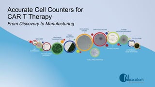
2. Accurate Cell Counting Methods, Fit for
Purpose, for CAR T Therapy
RESEARCH
DEVELOPMENT
MANUFACTURING
3. Accurate Cell Counting Methods, Fit for
Purpose, for CAR T Therapy
Fast Accurate Counting for Primary Samples
Overcoming morphology variability in cancer cell lines
Only one cell counter is needed throughout the (CAR)
Chimeric Antigen Receptor T-Cell Development
Improve consistency, accuracy and efficiency
Optimize Viral Vector Development with Image Cytometry
Improved transfection, transduction and viability
Accurate cell counting assay of apheresis materials -
suitable for stability program
Cell Counting Method Development Process
1
2
3
4
5
6
4. Accurate Cell Counters for CAR T Therapy
RESEARCH DEVELOPMENT MANUFACTURING
“Our Cellometer Auto 2000 has greatly improved the speed and accuracy of our
cell counting and has allowed us to analyze our cell samples in a variety of ways.
The AOPI feature has been integral in generating cell viable data for a few of our
new projects. The customer service team is also amazing! They are very
knowledgeable about their products and technology and are always available to
help us troubleshoot.” - NIH-NIDCD
“The Cellometer Auto 2000 and the AOPI Viastain gives us the ability to get an
accurate count of our live splenocytes while excluding the red blood cells.” - Eli Lilly
“…definitely the standard for cell counting. Results are consistent time and again,
the reliability is great when performing duplicate or triplicate counts…. I also enjoy
the ability to establish several different settings based on the type of cell, and
appreciate Nexcelom’s customer service!” - Cold Spring Harbor Laboratory
1 2 3 4 5 6
5. RESEARCH DEVELOPMENT MANUFACTURING
Fast Accurate Counting for Primary Samples
Mouse samples are variable in cell type and phenotypic characteristics.
• Cell counting accuracy can be affected by heterogeneous
cell populations
• Nuclear staining is critical for accurate cell counting
1
“The Cellometer Auto 2000 has greatly aided
the development of our tissue dissociation
assays. The unit allows fast quantification of
viable white blood cells from a heterogeneous
population following spleen, thymus, lymph
node, etc. dissolution which can then quickly
proceed to downstream flow-based analysis.
The time saved by this method has been
extremely valuable!” - Takeda Pharmaceuticals
Cellometer provides accurate
concentration and viability for cells that
vary in morphology and heterogeneity.
Ezeh PC, et al. PLoS One. 2014; 9(4):e93920 | Xu H, et al. Toxicol Lett. 2016; 262:55-61
6. RESEARCH DEVELOPMENT MANUFACTURING
Overcoming Morphology Variability in Cancer Cell Lines
Advanced imaging and analysis algorithms are necessary to address the cell morphology
diversity in the large tumor cell lines. It is important for the cell counter to measure cells
with these characteristics accurately.
2
“We use the Cellometer Auto 2000 daily to count
cells and assess viability of cells derived from a
variety of sources. [We] perform a lot of cell line
work, and need accurate counts and viability
assessments before using the cells in downstream
assays” – Regeneron Pharmaceuticals
Cellometer provides accurate
concentration and viability for cells that
vary in morphology and heterogeneity.
Rothenberg SM, et al. Cancer Res. 2010; 70(6): 2158-64
7. RESEARCH DEVELOPMENT MANUFACTURING
Only One Cell Counter is
Needed Throughout CAR T
Development
Cell counters and image cytometers are
critical throughout the CAR T process
• Accurate cell counting from Leukopaks
• Measuring transduction efficiencies
• Determining efficacy of engineered
CAR T-cells
3
Singh H, et al. PLoS One. 2013; 8(5): e64138
Wang X, et al. Cancer Gene Ther 2015; 22(2):85-94
8. RESEARCH DEVELOPMENT MANUFACTURING
Optimized Viral Vector Development with Image Cytometer
Accurate cell number and viability
measurement is necessary for viral
vector development and downstream
manufacturing processes.
• Assure correct plating density,
cell confluence
• Determine transduction
efficiencies (No trypsinization
required)
• Verify cell number and viability
for plating
4
“The Cellometer Auto 2000 has made functional testing of T cells a
much quicker and simpler process than before! When the cell
concentration of each population in a 96 well plate must be
determined, it is hard to imagine having to count the cells by hand
and doing this in a timely manner. Great product!”
- University of Texas at Austin
White KM, et al. ACS Infectious Diseases 2018; 4(2):146-157
Zhang Z, et al. BMC Biotechnology 2018; 18(1):4
9. RESEARCH DEVELOPMENT MANUFACTURING
Accurate Cell Counting Assay of
Apheresis Materials –
Suitable for Stability Program
Counting assay, of difficult to measure
apheresis material, for stability programs
including shipping, handling and storage
conditions.
• Measure viable and dead nucleated cells
• No RBC lysis necessary
• No staining-incubation period
5
“…We routinely process PBMCs from both fresh whole blood and from frozen stock. The Cellometer [Auto2000] has
made it much easier to get cell numbers and viability percentages for use in downstream applications …”
- Human Longevity, Inc
Manual / trypan blue method is not accurate for counting leukapheresis. RBCs are 3-12x the number of lymphocytes.
Proven Repeatability: 14 PMBC samples were tested using the Cellometer Auto2000. The resulting in a CV of < 6%.
Burchiel SW, et al. Inhal Toxicol. 2016; 28(2):61-70
10. RESEARCH DEVELOPMENT MANUFACTURING
Cell Counting Method Development Process
Counting is Crucial During All Stages of Cell-Based Product Development
6
Critical Process Parameters
• Cell growth and expansion
• Selection of target cells
• Pre-stimulation conditions
• Transduction conditions
Purity
• % CD3+ T cells
• %CAR T cells
• Residual tumor
burden
• Residual beads
Critical Quality Attributes
Safety
• Gram stain/sterility
• Mycoplasma
• Endotoxin level
• Copies of transgene insertion
• Replication Competent
Retrovirus/Lentivirus
Stability
• Shipping/Handling:
Cryopreserved CAR T
• Storage validation for
Apheresis materials:
Fresh/Frozen/Thawed
CAR T samples
• Vector
• Final product
Identity
• % CAR T cells
Potency
• In vitro Cytotoxic T
Lymphocyte
• Interferon-γ
secretion
Cellometer Cell Counting Celigo Imaging Cytometer
11. RESEARCH DEVELOPMENT MANUFACTURING
Cell Counting Method Development Process (continued)
Establishing Early and Robust Cell Counting Procedures During Cell-Based Product Development
is Crucial for Many of These Parameters
6
• Selection
• Development
• Optimization
PRE-CLINICAL PHASE I PHASE II PHASE III
• Methods
qualified
• Set tentative
release/stability
criteria
• Assay
optimization
• Consider
validation
acceptance
criteria
• Delineate/initiate
assay validation
parameters
• Full assay
validation by
licenser
(recommended
by Phase III)
• Biologics license
applications
≥70% cell viability is recommended for pre-clinical
safety studies and clinical dose.
Cellometer K2 can measure cell viability from 0%
viable to 100% viable cell population.
12. RESEARCH DEVELOPMENT MANUFACTURING
Cell Counting Method Development Process (continued)
Establishing Early and Robust Cell Counting Procedures During Cell-Based Product Development
is Crucial for Many of These Parameters
6
PHASE II
• Assay optimization
• Consider validation
acceptance criteria
• Delineate/initiate assay
validation parameters
“I am currently using the Cellometer K2 in our lab,
mostly to count T cells, PBMCs, and tumor cells. I use
them for cell culture, and later the cells are used for
further assays like ELISA and FACS. The instrument is
very accurate, especially with AO/PI.” - EMD Serono
• Linearity
• Accuracy
• Range
• Intermediate precision
• Precision: repeatability
and more …
Cellometer K2 Linearity Measurement
13. RESEARCH DEVELOPMENT MANUFACTURING
Cell Counting Method Development Process (continued)
Establishing Early and Robust Cell Counting Procedures During Cell-Based Product Development
is Crucial for Many of These Parameters
6
PHASE II
• Assay optimization
• Consider validation
acceptance criteria
• Delineate/initiate assay
validation parameters
Accuracy translates from cell lines to primary
samples.
• Linearity
• Accuracy
• Range
• Intermediate precision
• Precision: repeatability
and more …
Intermediate Precision: Instrument
Repeatability and Validation
14. RESEARCH DEVELOPMENT MANUFACTURING
Cell Counting Method Development Process (continued)
Establishing Early and Robust Cell Counting Procedures During Cell-Based Product Development
is Crucial for Many of These Parameters
6
PHASE II
• Assay optimization
• Consider validation
acceptance criteria
• Delineate/initiate assay
validation parameters
Cellometer cell counters have been facilitating
CAR T manufacturing with accurate cell counting
and analysis since 2010.
• Linearity
• Accuracy
• Range
• Intermediate precision
• Precision: repeatability
and more …
Precision: Repeatability of Cellometer K2
JinJ,etal.JofTranslMed2018;16(1):13|SinghH,etal.CancerGeneTher.(2015)22(2):95-100
Manuri,PVR,etal.HumanGeneTherapy.2010;21:427-437
15.
16. Innovation and Expertise in the Science of Cell Counting
Schedule a FREE on-line demonstration, on-site demonstration or technical seminar with a
Nexcelom Applications Specialist today.
Customer Support
Call 978-327-5340 or
E-mail info@nexcelom.com
For Europe
Call 44 (0) 161 232 4593 or
E-mail info@nexcelom.co.uk
For China
Call (+86) 21 5886 0038 or
E-mail support@nexcelom.com.cn