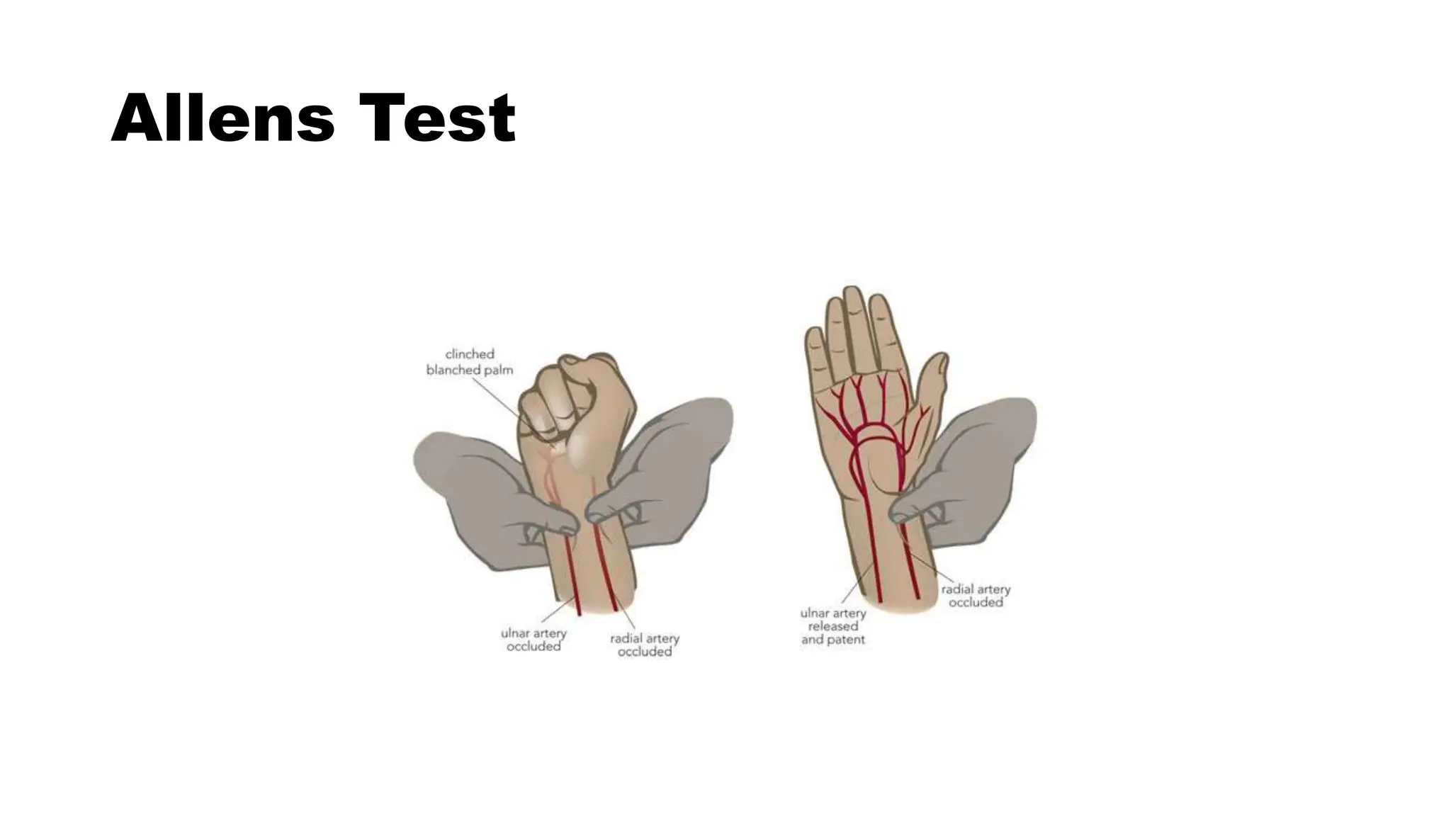
STEP BY STEP ABG
INTERPRETATION
INDICATION OF ABG
 Assess adequacy of ventilation and oxygenation
Assess changes in acid- base homeostasis
Helps in management of ICU patients.
SITE OF ABG
Radial Artery
Brachial Artery
Femoral Artery
Dorsalis Pedis Artery
Posterior Tibial artery
Allens Test
PROCEDURE
CONTRAINDICATION
Cellulitis or other infections over puncture site
Absence of palpable arterial pulse
Negative result of an Allen test/modified Allen test
Coagulopathies / anticoagulant therapy
History of arterial spasm following previous puncture
Severe Peripheral Vascular Disease
Arterial grafts
Dialysis shunt – choose another site
TECHNICAL ERRORS IN
SAMPLING EXCESSIVE HEPARIN
• Ideally pre-heparinised syringes should be used
• Syringes flushed with 1:1000 heparin and rinsed
• Do not leave excessive heparin in the syringe
• Heparin has dilutional effect:
• Low HCO3
 Low PCO2
 High Sr Na
TECHNICAL ERRORS IN
SAMPLING
• ALTERATION OF RESULTS WITH SIZE OF SYRINGE/NEEDLE AND VOL OF
SAMPLE
• Syringes must have > 50% blood
• Use only 3ml or smaller syringes
• 25% lower values if 1 ml sample taken in 10ml syringe (0.25ml
heparin in needle
TECHNICAL ERRORS IN SAMPLING
BODY TEMPERATURE
• Affects values of PCO2 and HCO3
• BG analysers controlled for normal body temperatures
TECHNICAL ERRORS IN SAMPLING
AIR BUBBLES IN BLOOD SAMPLE
• In air, PO2 is 150 mm of Hg & PCO2 is 0
• Contact with air bubble will lead to higher PO2 and lower PCO2
• Seal syringe immediately after sampling
TECHNICAL ERRORS IN SAMPLING
WBC COUNTS
• 0.01ml O2 consumed /dl/min
• Marked increase in high TLC/Platelet Count decreased PO2
• Chilling/immediate analysis
• ABG syringe must be transported earliest via cold chain
Blood Gas Norms
pH pCO2 pO2 HCO3 BE
Arterial 7.35-
7.45
35-45 80-100 22-26 -2 to +2
Venous 7.30-
7.40
43-50 ~45 22-26 -2 to +2
Normal Values
ANALYTE Normal Value Units
pH 7.35 - 7.45
PCO2 35 - 45 mm Hg
PO2 72 – 104 mm Hg`
[HCO3] 22 – 30 meq/L
SaO2 95-100 %
Anion Gap 12 + 4 meq/L
∆HCO3 +2 to -2 meq/L
Normal values for calculation
Normal pH=7.4
Normal PaCO2=40 mmHg
Normal HCO3=24mmol/L
Normal AG=12 meq/L
Blood Gas Norms
pH pCO2 pO2 HCO3 BE
Arterial 7.35-7.45 35-45 80-100 22-26 -2 to +2
Venous 7.30-7.40 43-50 ~45 22-26 -2 to +2
• Blood pH <6.8 or >7.8 not compatible with
life and indicates irreversible cell damage or
death
Step by step
interpretation
• STEP 1
• Validity of Gas
• STEP 2
• Assess for Oxygenation
• STEP 3
• Look at pH
• STEP 4
• Identify the Primary disorder
• STEP 5
• If respiratory disorder is present whether its Acute or Chronic
• STEP 6
• Metabolic compensation for respiratory disorder
• STEP 7
• Evaluate the type of Metabolic disorder
• STEP 8
• Respiratory compensation for Metabolic disorder
• STEP 9
• Gap- Gap approach
• Pre requisites prior to any arterial blood gas
interpretation, is detailed history of the patient and
clinical examination
FIRST STEP-CHECK FOR
VALIDITY
• At normal pH of 7.40 number of H+ ions are 40
• As H+ ions increase acidosis occurs; so if you multiply 40 by a factor of
1.25 you will get H+ ions equal to 50 which is pH of 7.3. As you
multiply further H+ ions by 1.25 you get H+ ions 62.5 which are equal
to pH of 7.2.
• Similarly as H+ ions fall alkalosis occurs; so if you multiply 40 by a
factor of 0.8 you get H+ ions equal to 32 which is pH of 7.5. As you
multiply further H + by 0.8 you get H+ ions 26 which correspond to pH
of 7.6.
• You calculate the H+ ions by putting the values of HCO3- and PaCO2 in
the Henderson-Hasselbach equation.
• H+=24 x [PaCO2/HCO3-]
• you calculate the H+ ions by this formulae and then compare this
with the above denoted normogram. The calculated value must
match with the normogram. This indicates the validity of ABG.
NORMAL NORMOGRAM
H+ ion (neq/l) pH
100 7.00
79 7.10
63 7.20
50 7.30
45 7.35
40 7.40
35 7.45
32 7.50
25 7.60
SECOND STEP- Assess for
Oxygenation
•At Room air- The normal alveolar arterial oxygenation gradient (PAO2-
PaO2) is 5-15mm Hg.
•Normal PaO2 for age = 109- (0.45 x Age in years)
DETERMINATION OF PaO2
PaO2 is dependent upon Age, FiO2,
Patm
As Age the expected PaO2
• PaO2 = 109 - 0.4 (Age)
As FiO2 the expected PaO2
• Alveolar Gas Equation:
• PAO2= (PB-P h2o) x FiO2- pCO2/R
O
X
Y
G
E
N
A
T
I
O
N
PAO2 = partial pressure of oxygen in alveolar gas, PB = barometric pressure
(760mmHg), Ph2o = water vapor pressure (47 mm Hg), FiO2 = fraction of
inspired oxygen, PCO2 = partial pressure of CO2 in the ABG, R = respiratory
quotient (0.8)
ON MECHANICAL VENTILATION
ARDS P/F RATIO
MILD ARDS 200-300
MODERATE 100-200
SEVERE <100
The normal PaO2/FiO2 ratio is 400-500
THIRD STEP-ACIDOSIS OR
ALKALOSIS
• Look at pH; normal pH is 7.35-7.45 for calculation purpose it is 7.4
• Acidemia - pH <7.40 is referred as acidemia
• The process in the tissue leading to this is referred as acidosis
• Alkalemia - pH >7.45 is alkalemia
• The process in the tissue leading to this is referred as alkalosis.
FOURTH STEP
• Identify the primary disorder - metabolic or respiratory.
• Where pH α HCO3
-/CO22(Kidney/ lungs) correspondingly looks at the HCO-
3
and PaCO2 and correspond it with pH accordingly.
• Respiratory
• Primary change is in PaCO2
• pH 1/PaCO2
• Metabolic
• Primary change is in HCO3
• pH  HCO3
• PaCO2>45 mm Hg identifies respiratory acidosis, HCO3<22mm Hg identifies
metabolic acidosis. Similarly PaCO2<35 mmHg identifies respiratory
alkalosis and HCO3
->26 mmHg identifies metabolic alkalosis.
IS PRIMARY DISTURBANCE RESPIRATORY OR
METABOLIC?
pH PCO2 or pH PCO2 METABOLIC
pH PCO2 or pH PCO2 RESPIRATORY
In primary respiratory disorders, the pH and PaCO2 change
in opposite directions; in metabolic disorders the pH and
PaCO2 change in the same direction.
STEP4-RESPIRATORY OR METABOLIC
• As already discussed pH α HCO3-/PaCO2
• Where HCO3- is directly proportional to function of kidney
• CO2 is directly proportional to lung function
FIFTH STEP
If there is a primary respiratory disturbance is it
acute or chronic
• Acute disorder pH falls/rises by 0.008 for every change in PaCO2
• Chronic disorder pH falls/rises by 0.003 for every change in
PaCO2
SIXTH STEP : COMPONSATION
FOR RESPIRATORY
Disorder CO2 change HCO3 change
Acute respiratory
acidosis
For every 10 mmHg
rise in CO2
HCO3 will rise by
1mmol/l
Chronic respiratory
acidosis
For every 10 mmHg
rise in CO2
HCO3 will rise by
4mmol/l
DISORDER CO2 CHANGE HCO3 CHANGE
Acute respiratory
alkalosis
For every 10
mmHg fall in CO2
HCO3 will fall by
2mmol/l
Chronic
respiratory
alkalosis
For every 10
mmHg fall in CO2
HCO3 will fall by
5mmol/l
SIXTH STEP : COMPONSATION
FOR RESPIRATORY
SEVENTH STEP
Evaluate for Metabolic disorder-whether Metabolic acidosis /
Alkalosis
• If Metabolic acidosis is there look for High AG / Normal AG
• Normal Anion Gap is 8-12 meq/l
• [AG= Na-(Cl-+HCO3-) ]
• The corrected AG=AG+2.5(4-albumin)
• WhereAlbumin levels are in gm/dL
HIGH ANION GAP (HAGMA)
M METHANOL
U UREMIA - ARF/CRF
D DIABETIC KETOACIDOSIS
P PARALDEHYDE, PROPYLENE
I ISONIAZIDE, IRON
L LACTIC ACIDOSIS
E ETHANOL, ETHYLENE GLYCOL
S SALICYLATE
OSMOLAR GAP
If the anion gap is elevated, consider calculating the osmolal gap in
compatible clinical situations
• Elevation in AG is not explained by an obvious case (DKA, lactic
acidosis, renal failure)
• Toxic ingestion is suspected
• OSM gap = Measured Osm – Cal. Plasma Osm
Cal. Plasma Osmolarity = 2[Na+] + [Gluc]/18 +
[BUN]/2.8
The OSM gap should be < 10 mOsm/kg
Osm gap > 10 mOsm/kg indicates presence of abnormal osmotically active
substance
 Ethanol
 Methanol
 Ethylene glycol
NORMAL ANION GAP (NAGMA)
Hypokalemic
a. GI losses of HCO3
i. Ureterosigmoidostomy
ii.Diarrhea
iii.Ileostomy
b. Renal losses of HCO3
i. Proximal RTA
ii.Carbonic Anhydrase Inhibitors
Normokalemic or hyperkalemic
a. Renal tubular disease
i. Acute tubular necrosis
ii. Chronic tubulointerstitial disease
iii. Distal RTA (type I and IV)
iv. Hypoaldesteronism
b. Pharmacological
i. Ammonium chloride,Cacl2 ingestion
ii. Hyperalimentation
iii. Dilutional acidosis
• Non AG metabolic acidosis
• If urine pH > 5.5 : Type 1 RTA
• If urine pH < 5.5 : Type 2 or Type 4 RTA
• Type 2 or Type 4 RTA can be later differentiated using serum K+ level
CONT…..
• Urinary NH4
+ levels can be estimated by calculating the urine anion
gap (UAG)
• UAG = [Na+ + K+]u – [Cl–]u
• [Cl–]u > [Na+ + K+], the urine gap is negative by definition
• Helps to distinguish GI from renal causes of loss of HCO3 by
estimating Urinary NH4+ (elevated in GI HCO3 loss but low in distal
RTA)
• Hence a -ve UAG (av -20 meq/L) seen in former while +ve value (av
+23 meq/L) seen in latter.
LOW ANION GAP
Reduction in unmeasured anions (hypoproteinemia)
Excess of unmeasued cations(lithium toxicity)
Excessively abnormal positively charged
Protein (Multiple Myeloma )
EIGHT STEP
• Look for respiratory compensation
• Metabolic acidosis : 1.5xHCO3+8±2
• Metabolic alkalosis : 0.7xHCO3+21±2
NINTH STEP-GAP- GAP OR DELTA
DELTA APPROACH
Delta AG= AG-12
Delta HCO3=24-HCO3
Gap-gap=delta AG- delta HCO3
Normally it should be ±4;
If gap is more than 4 then concomitant metabolic alkalosis is there and if
gap-gap is less than 4 then concomitant acidosis is there.
AG/ HCO3
- = 1  Pure High AG Met Acidosis
AG/ HCO3
- > 1  Assoc Metabolic Alkalosis
 AG/ HCO3
- < 1  Assoc N AG Met Acidosis
1. Loss of H+ ions (e.g. vomiting, diuretics)
2. Increased reabsorption of bicarbonate – Low intravascular volume
– Hypokalemia
– High pCO2
– Increased mineralocorticoids
3. Administration of alkali (in setting of renal impairment) e.g. Ringer’s lactate
where lactate gets metabolised to bicarbonates in liver adding to alkali pool.
METABOLIC ALKALOSIS
• Look for urinary chloride
• Urinary chloride is<20 meq/l it is chloride responsive i.e. in
hypovolemic patients
• If Urinary chloride>20 meq/l; it is chloride unresponsive.
RESPIRATORY ACIDOSIS
1. Airway/pulmonary parenchymal disease
Upper airway obstruction
Lower airway obstruction
 Pulmonary
Cardiogenic pulmonary edema
Pneumonia iii. ARDS
Pulmonary perfusion defect—PE—air/fat/tumor
2. Cns Depression -head injury ,medications such as narcotics,
sedatives or anesthesia
3. Neuromuscular Disease and Impairment
4. Ventilatory Restriction—due to pain, chest wall injury/
deformity, or abdominal distension
INCREASED CO2 PRODUCTION
 Large caloric loads
Malignant hyperthermia
Intensive shivering
Prolonged seizure activity
Thyroid storm
Extensive thermal injury (burns
1. CNS stimulation Fever, pain, Thyrotoxicosis, Cerebrovascular
accidents.
2. Hypoxemia Pneumonia, Pulmonary edema.
3. Drugs/hormones Medroxyprogesterone, Catecholamines,
Salicylates.
4. Miscellaneous Sepsis, Pregnancy
5. Psychological responses Anxiety or Fear
RESPIRATORY ALKALOSIS
Complications
Pain
Bruising and haematoma
Nerve damage
Aneurysm
Spasm
AV fistula
Infection
Air or thromboembolism
Anaphylaxis from local anaesthetic
KEEP DISTANCNE
BE SAFE
THANK YOU
• Anion Gap= measured cations- measured anions
ANION GAP(AG) = Na – (HCO3 + Cl)
Normal Value = 12 + 4 ( 8- 16 meq/l)
• ΔAG= MEASURED AG -12
IF ΔAG POSITIVE OR AG>16: METABOLIC ACIDOSIS
IF ΔAG NEGATIVE OR LOW AG:
Reduction in unmeasured anions (hypoproteinemia)
Excess of unmeasued cations(lithium toxicity)
Excessively abnormal positively charged
protien(multiple myeloma)
• Anion Gap= measured cations- measured anions
ANION GAP(AG) = Na – (HCO3 + Cl)
Normal Value = 12 + 4 ( 8- 16 meq/l)
• ΔAG= MEASURED AG -12
IF ΔAG POSITIVE OR AG>16: METABOLIC ACIDOSIS
IF Δ
• Albumin is the major unmeasured anion
• The anion gap should be corrected if there are gross changes in
serum albumin levels.
AG (CORRECTED) = AG + { (4 – [ALBUMIN]) × 2.5}
• Anion Gap = Measured AG – Normal AG
• Measured AG – 12
∆ HCO3 = Normal HCO3 – Measured HCO3
• 24 – Measured HCO3
• Ideally ∆Anion Gap = ∆HCO3
• For each 1 meq/L increase in AG, HCO3 will fall by 1 meq/L
• DELTA GAP = ΔAG-Δ HCO3
• DELTA GAP = (MEASURED AG-12) – (24-MEASURED HCO3)
• DELTA GAP = Na-Cl-36
• POSITIVE OR DELTA GAP > +6
METABOLIC ALKALOSIS
BICARBONATE RETENTION FOR RESPIRATORY ACIDOSIS
• NEGATIVE OR DELTA GAP < -6meq/l
NORMAL ANION GAP METABOLIC ACIDOSIS
 Look at pH
<7.35 - acidemia
>7.45 – alkalemia
STEP1- ACIDEMIA OR ALKALEMIA
ABG – Procedure and
Precautions
Pre-heparinised ABG syringes
Syringe should be FLUSHED with 0.05ml to 0.1ml of 1:1000 Heparin
solution and emptie
DO NOT LEAVE EXCESSIVE HEPARIN IN THE SYRINGE
HEPARIN DILUTIONAL HCO3
EFFECT PCO2
Use only 2ml or less syringe.
ABG – Procedure and
Precautions
Pre-heparinised ABG syringes
Syringe should be FLUSHED with 0.05ml to 0.1ml of 1:1000 Heparin solution and emptied.
DO NOT LEAVE EXCESSIVE HEPARIN IN THE SYRINGE
HEPARIN DILUTIONAL EFFECT H2CO3
PCO2
Use only 2ml or less syringe.
Ensure No Air Bubbles. Syringe must be sealed immediately after withdrawing
sample.
• Contact with AIR BUBBLES
Air bubble = PO2 150 mm Hg , PCO2 0 mm Hg
Air Bubble + Blood = PO2 PCO2
ABG Syringe must be transported at the earliest to the laboratory for EARLY
analysis via COLD CHAIN
ABG Analyser is controlled for Normal Body temperatures
Sample of hyperthermic patient >37°C, Measured values of PaO2
and PaCO2 are less than actual.
Hypothermic patient < 37°C measured values of PaO2 and PaCO2
are more than the actual values.
Every 1◦C ↓in temperature PCO2↓
pH ↑
Every1◦C ↑in temperature PCO2 ↑
pH ↓
Body temperature
ABG Sample should always be sent with relevant
information regarding O2, FiO2 status and Temp .
Before you withdraw a sample for ABG
After any change in FiO2 wait for 20min
And wait for 30 min after any change in ventilatory
parameters to ensure steady state.
Determination of PaO2
PaO2 is dependent upon Age, FiO2, Patm
As Age the expected PaO2
• PaO2 = 109 - 0.4 (Age)
As FiO2 the expected PaO2
• Alveolar Gas Equation:
• PAO2= (PB-P h2o) x FiO2- pCO2/R
O
X
Y
G
E
N
A
T
I
O
N
PAO2 = partial pressure of oxygen in alveolar gas, PB = barometric pressure
(760mmHg), Ph2o = water vapor pressure (47 mm Hg), FiO2 = fraction of
inspired oxygen, PCO2 = partial pressure of CO2 in the ABG, R = respiratory
quotient (0.8)
For calculation purposes
• Normal pH=7.4
• Normal PaCO2=40 mmHg
• Normal HCO3=24mmol/L
• Normal AG=12 meq/L
Is this ABG authentic ?
• pH = - log [H+]
Henderson-Hasselbalch equation
pH = 6.1 + log HCO3
-
0.03 x PCO2
pHexpected = pHmeasured = ABG is authentic
• STEP 0- IS THIS ABG AUTHENTIC?
• STEP1- ACIDEMIA OR ALKALEMIA
• STEP2-RESPIRATORY OR METABOLIC
• STEP3- IF RESPIRATORY-ACUTE OR CHRONIC?
• STEP4- IS COMPENSATION ADEQUATE?
• STEP5-IF METABOLIC- ANION GAP?
• STEP6- IF HIGH ANION GAP METABOLIC ACIDOSIS- Δ GAP?
• pH within normal range- No acid base disorder,
Fully compensated
or mixed disorder.
• pH out of the range- uncompensated
or partially compensated
Compensation
• PCO2 = (1.5 X [HCO3
-])+8 ±2
• For every 1mmol/l in HCO3 the PCO2 falls
by 1.25 mm Hg
METABOLIC
ACIDOSIS
• PCO2 = (0.7 X [HCO3
-])+ 21± 2
• For every 1mmol/l in HCO3 the PCO2 by
0.75 mm Hg
METABOLIC
ALKALOSIS
Metabolic Disorders – Compensation in these disorders leads to
a change in PCO2
Loss of H+ ions (e.g. vomiting, diuretics)
1. Increased reabsorption of bicarbonate – Low intravascular volume
– Hypokalemia
– High pCO2
– Increased mineralocorticoids
(aldosterone).
3. Administration of alkali (in setting of renal impairment) e.g. Ringer’s lactate where lactate gets
metabolised to bicarbonates in liver adding to alkali pool.
Airway/pulmonary parenchymal disease
a. Upper airway obstruction
b. Lower airway obstruction
c. Pulmonary -
i. Cardiogenic pulmonary edema
ii. Pneumonia iii. ARDS
iv. Pulmonary perfusion defect—PE—air/fat/tumor
2. CNS depression -head injury ,medications such as narcotics, sedatives, or
anesthesia
3. Neuromuscular disease and impairment
4. Ventilatory restriction—due to pain, chest wall injury/ deformity, or abdominal
distension.
• Increased CO 2 production
Large caloric loads
Malignant hyperthermia
Intensive shivering
Prolonged seizure activity
Thyroid storm
Extensive thermal injury (burns)
. CNS stimulation: Fever, pain, thyrotoxicosis, cerebrovascular accidents.
2. Hypoxemia: pneumonia, pulmonary edema.
3. Drugs/hormones: Medroxyprogesterone, catecholamines, salicylates.
4. Miscellaneous: Sepsis, pregnancy
5. Psychological responses, such as anxiety or fear.
COMPLICATIONS
• Pain
• Bruising and haematoma
• Nerve damage
• Aneurysm
• Spasm
• AV fistula
• Infection
• Air or thromboembolism
• Anaphylaxis from local anaesthetic
ABG intreptretation on clinical setup-1.pptx

ABG intreptretation on clinical setup-1.pptx

  • 1.
    STEP BY STEPABG INTERPRETATION
  • 2.
    INDICATION OF ABG Assess adequacy of ventilation and oxygenation Assess changes in acid- base homeostasis Helps in management of ICU patients.
  • 3.
    SITE OF ABG RadialArtery Brachial Artery Femoral Artery Dorsalis Pedis Artery Posterior Tibial artery
  • 4.
  • 6.
  • 7.
    CONTRAINDICATION Cellulitis or otherinfections over puncture site Absence of palpable arterial pulse Negative result of an Allen test/modified Allen test Coagulopathies / anticoagulant therapy History of arterial spasm following previous puncture Severe Peripheral Vascular Disease Arterial grafts Dialysis shunt – choose another site
  • 8.
    TECHNICAL ERRORS IN SAMPLINGEXCESSIVE HEPARIN • Ideally pre-heparinised syringes should be used • Syringes flushed with 1:1000 heparin and rinsed • Do not leave excessive heparin in the syringe • Heparin has dilutional effect: • Low HCO3  Low PCO2  High Sr Na
  • 9.
    TECHNICAL ERRORS IN SAMPLING •ALTERATION OF RESULTS WITH SIZE OF SYRINGE/NEEDLE AND VOL OF SAMPLE • Syringes must have > 50% blood • Use only 3ml or smaller syringes • 25% lower values if 1 ml sample taken in 10ml syringe (0.25ml heparin in needle
  • 10.
    TECHNICAL ERRORS INSAMPLING BODY TEMPERATURE • Affects values of PCO2 and HCO3 • BG analysers controlled for normal body temperatures
  • 11.
    TECHNICAL ERRORS INSAMPLING AIR BUBBLES IN BLOOD SAMPLE • In air, PO2 is 150 mm of Hg & PCO2 is 0 • Contact with air bubble will lead to higher PO2 and lower PCO2 • Seal syringe immediately after sampling
  • 12.
    TECHNICAL ERRORS INSAMPLING WBC COUNTS • 0.01ml O2 consumed /dl/min • Marked increase in high TLC/Platelet Count decreased PO2 • Chilling/immediate analysis • ABG syringe must be transported earliest via cold chain
  • 13.
    Blood Gas Norms pHpCO2 pO2 HCO3 BE Arterial 7.35- 7.45 35-45 80-100 22-26 -2 to +2 Venous 7.30- 7.40 43-50 ~45 22-26 -2 to +2
  • 14.
    Normal Values ANALYTE NormalValue Units pH 7.35 - 7.45 PCO2 35 - 45 mm Hg PO2 72 – 104 mm Hg` [HCO3] 22 – 30 meq/L SaO2 95-100 % Anion Gap 12 + 4 meq/L ∆HCO3 +2 to -2 meq/L
  • 15.
    Normal values forcalculation Normal pH=7.4 Normal PaCO2=40 mmHg Normal HCO3=24mmol/L Normal AG=12 meq/L
  • 16.
    Blood Gas Norms pHpCO2 pO2 HCO3 BE Arterial 7.35-7.45 35-45 80-100 22-26 -2 to +2 Venous 7.30-7.40 43-50 ~45 22-26 -2 to +2
  • 17.
    • Blood pH<6.8 or >7.8 not compatible with life and indicates irreversible cell damage or death
  • 18.
  • 19.
    • STEP 1 •Validity of Gas • STEP 2 • Assess for Oxygenation • STEP 3 • Look at pH • STEP 4 • Identify the Primary disorder • STEP 5 • If respiratory disorder is present whether its Acute or Chronic
  • 20.
    • STEP 6 •Metabolic compensation for respiratory disorder • STEP 7 • Evaluate the type of Metabolic disorder • STEP 8 • Respiratory compensation for Metabolic disorder • STEP 9 • Gap- Gap approach
  • 21.
    • Pre requisitesprior to any arterial blood gas interpretation, is detailed history of the patient and clinical examination
  • 22.
    FIRST STEP-CHECK FOR VALIDITY •At normal pH of 7.40 number of H+ ions are 40 • As H+ ions increase acidosis occurs; so if you multiply 40 by a factor of 1.25 you will get H+ ions equal to 50 which is pH of 7.3. As you multiply further H+ ions by 1.25 you get H+ ions 62.5 which are equal to pH of 7.2. • Similarly as H+ ions fall alkalosis occurs; so if you multiply 40 by a factor of 0.8 you get H+ ions equal to 32 which is pH of 7.5. As you multiply further H + by 0.8 you get H+ ions 26 which correspond to pH of 7.6.
  • 23.
    • You calculatethe H+ ions by putting the values of HCO3- and PaCO2 in the Henderson-Hasselbach equation. • H+=24 x [PaCO2/HCO3-] • you calculate the H+ ions by this formulae and then compare this with the above denoted normogram. The calculated value must match with the normogram. This indicates the validity of ABG.
  • 24.
    NORMAL NORMOGRAM H+ ion(neq/l) pH 100 7.00 79 7.10 63 7.20 50 7.30 45 7.35 40 7.40 35 7.45 32 7.50 25 7.60
  • 25.
    SECOND STEP- Assessfor Oxygenation •At Room air- The normal alveolar arterial oxygenation gradient (PAO2- PaO2) is 5-15mm Hg. •Normal PaO2 for age = 109- (0.45 x Age in years)
  • 26.
    DETERMINATION OF PaO2 PaO2is dependent upon Age, FiO2, Patm As Age the expected PaO2 • PaO2 = 109 - 0.4 (Age) As FiO2 the expected PaO2 • Alveolar Gas Equation: • PAO2= (PB-P h2o) x FiO2- pCO2/R O X Y G E N A T I O N PAO2 = partial pressure of oxygen in alveolar gas, PB = barometric pressure (760mmHg), Ph2o = water vapor pressure (47 mm Hg), FiO2 = fraction of inspired oxygen, PCO2 = partial pressure of CO2 in the ABG, R = respiratory quotient (0.8)
  • 27.
    ON MECHANICAL VENTILATION ARDSP/F RATIO MILD ARDS 200-300 MODERATE 100-200 SEVERE <100 The normal PaO2/FiO2 ratio is 400-500
  • 28.
    THIRD STEP-ACIDOSIS OR ALKALOSIS •Look at pH; normal pH is 7.35-7.45 for calculation purpose it is 7.4 • Acidemia - pH <7.40 is referred as acidemia • The process in the tissue leading to this is referred as acidosis • Alkalemia - pH >7.45 is alkalemia • The process in the tissue leading to this is referred as alkalosis.
  • 29.
    FOURTH STEP • Identifythe primary disorder - metabolic or respiratory. • Where pH α HCO3 -/CO22(Kidney/ lungs) correspondingly looks at the HCO- 3 and PaCO2 and correspond it with pH accordingly. • Respiratory • Primary change is in PaCO2 • pH 1/PaCO2 • Metabolic • Primary change is in HCO3 • pH  HCO3 • PaCO2>45 mm Hg identifies respiratory acidosis, HCO3<22mm Hg identifies metabolic acidosis. Similarly PaCO2<35 mmHg identifies respiratory alkalosis and HCO3 ->26 mmHg identifies metabolic alkalosis.
  • 30.
    IS PRIMARY DISTURBANCERESPIRATORY OR METABOLIC? pH PCO2 or pH PCO2 METABOLIC pH PCO2 or pH PCO2 RESPIRATORY In primary respiratory disorders, the pH and PaCO2 change in opposite directions; in metabolic disorders the pH and PaCO2 change in the same direction. STEP4-RESPIRATORY OR METABOLIC
  • 31.
    • As alreadydiscussed pH α HCO3-/PaCO2 • Where HCO3- is directly proportional to function of kidney • CO2 is directly proportional to lung function
  • 32.
    FIFTH STEP If thereis a primary respiratory disturbance is it acute or chronic • Acute disorder pH falls/rises by 0.008 for every change in PaCO2 • Chronic disorder pH falls/rises by 0.003 for every change in PaCO2
  • 33.
    SIXTH STEP :COMPONSATION FOR RESPIRATORY Disorder CO2 change HCO3 change Acute respiratory acidosis For every 10 mmHg rise in CO2 HCO3 will rise by 1mmol/l Chronic respiratory acidosis For every 10 mmHg rise in CO2 HCO3 will rise by 4mmol/l
  • 34.
    DISORDER CO2 CHANGEHCO3 CHANGE Acute respiratory alkalosis For every 10 mmHg fall in CO2 HCO3 will fall by 2mmol/l Chronic respiratory alkalosis For every 10 mmHg fall in CO2 HCO3 will fall by 5mmol/l SIXTH STEP : COMPONSATION FOR RESPIRATORY
  • 35.
    SEVENTH STEP Evaluate forMetabolic disorder-whether Metabolic acidosis / Alkalosis • If Metabolic acidosis is there look for High AG / Normal AG • Normal Anion Gap is 8-12 meq/l • [AG= Na-(Cl-+HCO3-) ] • The corrected AG=AG+2.5(4-albumin) • WhereAlbumin levels are in gm/dL
  • 36.
    HIGH ANION GAP(HAGMA) M METHANOL U UREMIA - ARF/CRF D DIABETIC KETOACIDOSIS P PARALDEHYDE, PROPYLENE I ISONIAZIDE, IRON L LACTIC ACIDOSIS E ETHANOL, ETHYLENE GLYCOL S SALICYLATE
  • 37.
    OSMOLAR GAP If theanion gap is elevated, consider calculating the osmolal gap in compatible clinical situations • Elevation in AG is not explained by an obvious case (DKA, lactic acidosis, renal failure) • Toxic ingestion is suspected • OSM gap = Measured Osm – Cal. Plasma Osm
  • 38.
    Cal. Plasma Osmolarity= 2[Na+] + [Gluc]/18 + [BUN]/2.8 The OSM gap should be < 10 mOsm/kg Osm gap > 10 mOsm/kg indicates presence of abnormal osmotically active substance  Ethanol  Methanol  Ethylene glycol
  • 39.
    NORMAL ANION GAP(NAGMA) Hypokalemic a. GI losses of HCO3 i. Ureterosigmoidostomy ii.Diarrhea iii.Ileostomy b. Renal losses of HCO3 i. Proximal RTA ii.Carbonic Anhydrase Inhibitors
  • 40.
    Normokalemic or hyperkalemic a.Renal tubular disease i. Acute tubular necrosis ii. Chronic tubulointerstitial disease iii. Distal RTA (type I and IV) iv. Hypoaldesteronism b. Pharmacological i. Ammonium chloride,Cacl2 ingestion ii. Hyperalimentation iii. Dilutional acidosis
  • 41.
    • Non AGmetabolic acidosis • If urine pH > 5.5 : Type 1 RTA • If urine pH < 5.5 : Type 2 or Type 4 RTA • Type 2 or Type 4 RTA can be later differentiated using serum K+ level
  • 42.
    CONT….. • Urinary NH4 +levels can be estimated by calculating the urine anion gap (UAG) • UAG = [Na+ + K+]u – [Cl–]u • [Cl–]u > [Na+ + K+], the urine gap is negative by definition • Helps to distinguish GI from renal causes of loss of HCO3 by estimating Urinary NH4+ (elevated in GI HCO3 loss but low in distal RTA) • Hence a -ve UAG (av -20 meq/L) seen in former while +ve value (av +23 meq/L) seen in latter.
  • 43.
    LOW ANION GAP Reductionin unmeasured anions (hypoproteinemia) Excess of unmeasued cations(lithium toxicity) Excessively abnormal positively charged Protein (Multiple Myeloma )
  • 44.
    EIGHT STEP • Lookfor respiratory compensation • Metabolic acidosis : 1.5xHCO3+8±2 • Metabolic alkalosis : 0.7xHCO3+21±2
  • 45.
    NINTH STEP-GAP- GAPOR DELTA DELTA APPROACH Delta AG= AG-12 Delta HCO3=24-HCO3 Gap-gap=delta AG- delta HCO3 Normally it should be ±4; If gap is more than 4 then concomitant metabolic alkalosis is there and if gap-gap is less than 4 then concomitant acidosis is there.
  • 46.
    AG/ HCO3 - =1  Pure High AG Met Acidosis AG/ HCO3 - > 1  Assoc Metabolic Alkalosis  AG/ HCO3 - < 1  Assoc N AG Met Acidosis
  • 47.
    1. Loss ofH+ ions (e.g. vomiting, diuretics) 2. Increased reabsorption of bicarbonate – Low intravascular volume – Hypokalemia – High pCO2 – Increased mineralocorticoids 3. Administration of alkali (in setting of renal impairment) e.g. Ringer’s lactate where lactate gets metabolised to bicarbonates in liver adding to alkali pool. METABOLIC ALKALOSIS
  • 48.
    • Look forurinary chloride • Urinary chloride is<20 meq/l it is chloride responsive i.e. in hypovolemic patients • If Urinary chloride>20 meq/l; it is chloride unresponsive.
  • 49.
    RESPIRATORY ACIDOSIS 1. Airway/pulmonaryparenchymal disease Upper airway obstruction Lower airway obstruction  Pulmonary Cardiogenic pulmonary edema Pneumonia iii. ARDS Pulmonary perfusion defect—PE—air/fat/tumor
  • 50.
    2. Cns Depression-head injury ,medications such as narcotics, sedatives or anesthesia 3. Neuromuscular Disease and Impairment 4. Ventilatory Restriction—due to pain, chest wall injury/ deformity, or abdominal distension
  • 51.
    INCREASED CO2 PRODUCTION Large caloric loads Malignant hyperthermia Intensive shivering Prolonged seizure activity Thyroid storm Extensive thermal injury (burns
  • 52.
    1. CNS stimulationFever, pain, Thyrotoxicosis, Cerebrovascular accidents. 2. Hypoxemia Pneumonia, Pulmonary edema. 3. Drugs/hormones Medroxyprogesterone, Catecholamines, Salicylates. 4. Miscellaneous Sepsis, Pregnancy 5. Psychological responses Anxiety or Fear RESPIRATORY ALKALOSIS
  • 53.
    Complications Pain Bruising and haematoma Nervedamage Aneurysm Spasm AV fistula Infection Air or thromboembolism Anaphylaxis from local anaesthetic
  • 54.
  • 55.
    • Anion Gap=measured cations- measured anions ANION GAP(AG) = Na – (HCO3 + Cl) Normal Value = 12 + 4 ( 8- 16 meq/l) • ΔAG= MEASURED AG -12 IF ΔAG POSITIVE OR AG>16: METABOLIC ACIDOSIS IF ΔAG NEGATIVE OR LOW AG: Reduction in unmeasured anions (hypoproteinemia) Excess of unmeasued cations(lithium toxicity) Excessively abnormal positively charged protien(multiple myeloma)
  • 56.
    • Anion Gap=measured cations- measured anions ANION GAP(AG) = Na – (HCO3 + Cl) Normal Value = 12 + 4 ( 8- 16 meq/l) • ΔAG= MEASURED AG -12 IF ΔAG POSITIVE OR AG>16: METABOLIC ACIDOSIS IF Δ
  • 57.
    • Albumin isthe major unmeasured anion • The anion gap should be corrected if there are gross changes in serum albumin levels. AG (CORRECTED) = AG + { (4 – [ALBUMIN]) × 2.5}
  • 58.
    • Anion Gap= Measured AG – Normal AG • Measured AG – 12 ∆ HCO3 = Normal HCO3 – Measured HCO3 • 24 – Measured HCO3 • Ideally ∆Anion Gap = ∆HCO3 • For each 1 meq/L increase in AG, HCO3 will fall by 1 meq/L
  • 59.
    • DELTA GAP= ΔAG-Δ HCO3 • DELTA GAP = (MEASURED AG-12) – (24-MEASURED HCO3) • DELTA GAP = Na-Cl-36 • POSITIVE OR DELTA GAP > +6 METABOLIC ALKALOSIS BICARBONATE RETENTION FOR RESPIRATORY ACIDOSIS • NEGATIVE OR DELTA GAP < -6meq/l NORMAL ANION GAP METABOLIC ACIDOSIS
  • 60.
     Look atpH <7.35 - acidemia >7.45 – alkalemia STEP1- ACIDEMIA OR ALKALEMIA
  • 61.
    ABG – Procedureand Precautions Pre-heparinised ABG syringes Syringe should be FLUSHED with 0.05ml to 0.1ml of 1:1000 Heparin solution and emptie DO NOT LEAVE EXCESSIVE HEPARIN IN THE SYRINGE HEPARIN DILUTIONAL HCO3 EFFECT PCO2 Use only 2ml or less syringe.
  • 62.
    ABG – Procedureand Precautions Pre-heparinised ABG syringes Syringe should be FLUSHED with 0.05ml to 0.1ml of 1:1000 Heparin solution and emptied. DO NOT LEAVE EXCESSIVE HEPARIN IN THE SYRINGE HEPARIN DILUTIONAL EFFECT H2CO3 PCO2 Use only 2ml or less syringe.
  • 63.
    Ensure No AirBubbles. Syringe must be sealed immediately after withdrawing sample. • Contact with AIR BUBBLES Air bubble = PO2 150 mm Hg , PCO2 0 mm Hg Air Bubble + Blood = PO2 PCO2 ABG Syringe must be transported at the earliest to the laboratory for EARLY analysis via COLD CHAIN
  • 64.
    ABG Analyser iscontrolled for Normal Body temperatures Sample of hyperthermic patient >37°C, Measured values of PaO2 and PaCO2 are less than actual. Hypothermic patient < 37°C measured values of PaO2 and PaCO2 are more than the actual values. Every 1◦C ↓in temperature PCO2↓ pH ↑ Every1◦C ↑in temperature PCO2 ↑ pH ↓ Body temperature
  • 65.
    ABG Sample shouldalways be sent with relevant information regarding O2, FiO2 status and Temp . Before you withdraw a sample for ABG After any change in FiO2 wait for 20min And wait for 30 min after any change in ventilatory parameters to ensure steady state.
  • 66.
    Determination of PaO2 PaO2is dependent upon Age, FiO2, Patm As Age the expected PaO2 • PaO2 = 109 - 0.4 (Age) As FiO2 the expected PaO2 • Alveolar Gas Equation: • PAO2= (PB-P h2o) x FiO2- pCO2/R O X Y G E N A T I O N PAO2 = partial pressure of oxygen in alveolar gas, PB = barometric pressure (760mmHg), Ph2o = water vapor pressure (47 mm Hg), FiO2 = fraction of inspired oxygen, PCO2 = partial pressure of CO2 in the ABG, R = respiratory quotient (0.8)
  • 67.
    For calculation purposes •Normal pH=7.4 • Normal PaCO2=40 mmHg • Normal HCO3=24mmol/L • Normal AG=12 meq/L
  • 68.
    Is this ABGauthentic ? • pH = - log [H+] Henderson-Hasselbalch equation pH = 6.1 + log HCO3 - 0.03 x PCO2 pHexpected = pHmeasured = ABG is authentic
  • 69.
    • STEP 0-IS THIS ABG AUTHENTIC? • STEP1- ACIDEMIA OR ALKALEMIA • STEP2-RESPIRATORY OR METABOLIC • STEP3- IF RESPIRATORY-ACUTE OR CHRONIC? • STEP4- IS COMPENSATION ADEQUATE? • STEP5-IF METABOLIC- ANION GAP? • STEP6- IF HIGH ANION GAP METABOLIC ACIDOSIS- Δ GAP?
  • 70.
    • pH withinnormal range- No acid base disorder, Fully compensated or mixed disorder. • pH out of the range- uncompensated or partially compensated
  • 71.
    Compensation • PCO2 =(1.5 X [HCO3 -])+8 ±2 • For every 1mmol/l in HCO3 the PCO2 falls by 1.25 mm Hg METABOLIC ACIDOSIS • PCO2 = (0.7 X [HCO3 -])+ 21± 2 • For every 1mmol/l in HCO3 the PCO2 by 0.75 mm Hg METABOLIC ALKALOSIS Metabolic Disorders – Compensation in these disorders leads to a change in PCO2
  • 72.
    Loss of H+ions (e.g. vomiting, diuretics) 1. Increased reabsorption of bicarbonate – Low intravascular volume – Hypokalemia – High pCO2 – Increased mineralocorticoids (aldosterone). 3. Administration of alkali (in setting of renal impairment) e.g. Ringer’s lactate where lactate gets metabolised to bicarbonates in liver adding to alkali pool.
  • 73.
    Airway/pulmonary parenchymal disease a.Upper airway obstruction b. Lower airway obstruction c. Pulmonary - i. Cardiogenic pulmonary edema ii. Pneumonia iii. ARDS iv. Pulmonary perfusion defect—PE—air/fat/tumor 2. CNS depression -head injury ,medications such as narcotics, sedatives, or anesthesia 3. Neuromuscular disease and impairment 4. Ventilatory restriction—due to pain, chest wall injury/ deformity, or abdominal distension.
  • 74.
    • Increased CO2 production Large caloric loads Malignant hyperthermia Intensive shivering Prolonged seizure activity Thyroid storm Extensive thermal injury (burns)
  • 75.
    . CNS stimulation:Fever, pain, thyrotoxicosis, cerebrovascular accidents. 2. Hypoxemia: pneumonia, pulmonary edema. 3. Drugs/hormones: Medroxyprogesterone, catecholamines, salicylates. 4. Miscellaneous: Sepsis, pregnancy 5. Psychological responses, such as anxiety or fear.
  • 76.
    COMPLICATIONS • Pain • Bruisingand haematoma • Nerve damage • Aneurysm • Spasm • AV fistula • Infection • Air or thromboembolism • Anaphylaxis from local anaesthetic

Editor's Notes

  • #6 \Modified Allen’s test This is done before puncturing dorsalis pedis artery. Elevate patient’s feet. Occlude dorsalis pedis artery; then blanch the great toe by compressing the toenail for several seconds. Release pressure on the nail and observe for flushing (rapid return of color indicates adequate collateral flow). Compress the posterior tibial artery if it is being used as the puncture site (pediatric population).