

Recommended
More Related Content
What's hot
What's hot (20)
Viewers also liked
Viewers also liked (20)
Similar to A wayang kulit
Similar to A wayang kulit (20)
Recently uploaded
Recently uploaded (20)
A wayang kulit
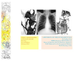
- 1. CHEST RADIOGRAPHS, A REVISION OF THE FUNDAMENTALS A WAYANG KULIT Dr Ng Kian Seng Part One MBBS (Singapore) MCGP (Malaysia) Second Edition Master Of Medicine (Internal Medicine, Singapore) February 2012 FAFP (Malaysia) Cert In Occupational Medicine Ph D (Theology, USA)
- 2. Hippocrates of Cos, Father of Medicine ANATOMY IN THE CHEST RADIOGRAPH
- 3. A Normal Chest Radiograph Some examiners like you to call x ray films ‘Radiographs’; strictly speaking you can’t actually see the x rays themselves.
- 4. Anatomy in the Chest Radiograph The right main bronchus is slightly larger than the left & comes off at a less acute angle than the left (hence septic material & foreign substances are more likely to be inhaled into the right lung than into the left).
- 5. Chest Radiograph, PA View, No 1 Apex Of Carina Trachea Lung Right para-tracheal stripe Aortic arch Main Pulmonary Artery Left Atrial appendage Descending thoracic aorta Left ventricle Gastric Air Bubble
- 6. Chest Radiograph, PA View, No 2 Right upper lobe pulmonary vein Horizontal fissure Right hilum Right lower lobe pulmonary artery Right atrium Right Right Costophrenic Cardiophrenic Angle Angle
- 7. Chest Radiograph, PA View, No 3 Spinous Process Scapula Anterior Rib Clavicle Posterior Rib Right Main Bronchus Left Main Bronchus Breast Diaphragm Lung Tissue Soft Tissue Retrocardiac Superimposed Vertebra On diaphragm
- 8. Anatomy Of the Heart In The Chest Radiograph
- 9. MEDIASTINAL SILHOUETTE IN MIDDLE AGE & THE ELDERLY
- 10. Aorto-pulmonary Window Aorto-pulmonary window. The aorto-pulmonary window lies between the arch of the aorta and the pulmonary arteries. It contains the ligamentum arteriosum, the recurrent laryngeal nerve, lymph nodes, and fatty tissue.
- 11. RIGHT PARA-TRACHEAL STRIPE From the level of the clavicles to the azygous vein the right edge of the trachea is seen as a thin white stripe. This appearance is created by air of low density (blacker) lying either side of the comparatively dense (whiter) tracheal wall. If this stripe is thickened (normally less than 5 mm) this may represent pathology such as a paratracheal mass or enlarged lymph node. The left side of the trachea is not so well defined because of the position of the aortic arch and great vessels.
- 12. Anatomy in the Lateral Chest X-ray 1. Ascending thoracic Aorta 2. Sternum 3. Right ventricle 4. Left ventricle 5. Left atrium 6. Gastric air bubble 7. Right Hemidiaphragm 8. Left Hemidiaphragm 9. Right upper lobe bronchus 10. Left upper lobe bronchus 11. Trachea.
- 13. Anatomy in the Lateral Chest X-ray In the lateral CXR, you will see the Right Hemidiaphragm in its entirety But where the Left Hemidiaphragm is concerned, you can only see a part of it because anteriorly it “merges” with the inferior border of the heart.
- 14. NAME THE STRUCTURES IN THE LATERAL CHEST X-RAY 1.Trachea 2. Aortopulmonary window 3. Sternum 4. Right ventricle 5. Right Hemidiaphragm 6. Left Hemidiaphragm 7. Left atrium 8. Scapula 9. Right Upper 9 Lobe Bronchus 10. Left upper 10 Lobe Bronchus
- 15. THE MEDIASTINUM The mediastinum is divided by a plane passing from the sternal angle to T4-T5 into: Superior mediastinum and The inferior mediastinum The inferior mediastinum is further subdivided into three regions namely: Anterior mediastinum Middle mediastinum Posterior mediastinum These divisions are for descriptive purposes, they merge into each other imperceptibly. There are no distinct boundaries between them.
- 16. ZONES OF THE CHEST RADIOGRAPH Apex to a line drawn through UPPER the lower borders of the ZONE anterior ends of the 2nd costal cartilage. From the 1st line to one drawn MIDDLE through the lower borders of the ZONE 4th costal cartilage & includes the Hila of the lungs From the 2nd line to the LOWER bases of the lungs. ZONE
- 17. THE FISSURES OF THE LUNGS Oblique Fissure From 4 th Dorsal spine sweeping down obliquely to the 6th rib in mid mammary line or the 6th costochondral junction, anteriorly. Horizontal Fissure. From the 4th costo chondral junction to meet Oblique Fissure at the mid axillary line.
- 18. THE LOBES & FISSURES OF THE LUNGS Base of the Lung: 6th costochondral junction, obliquely to the 10th rib in Anterior Axillary Line, then horizontally to 12th thoracic vertebra
- 19. OBLIQUE FISSURE , HORIZONTAL FISSURE Oblique Fissure : From 4th Dorsal spine sweeping down obliquely to the 6th rib in mid mammary line or the 6th Costochondral junction, anteriorly. Horizontal Fissure. Runs from the 4th costochondral junction to meet Oblique Fissure at the mid axillary line.
- 20. THE RIGHT & LEFT OBLIQUE FISSURES From 4th dorsal spine sweeping down obliquely to the 6th rib in mid mammary line or the 6th costochondral junction, anteriorly.
- 21. THE HORIZONTAL FISSURE Horizontal Fissure. Runs from the 4th costochondral junction to meet Oblique Fissure at the Mid Axillary Line.
- 22. WHAT IS THE ABNORMALITY HERE?
- 23. ACESSORY FISSURE, THE AZYGOS FISSURE The azygos lobe appears starting in a teardrop shape at around the level of T5 to the right of the midline as a pale line curving outward . and upward and then back in to meet the root of the neck, the line is the infolding of the pleura. Also described as a “curvilinear opacity, Inverted comma, tadpole.”
- 24. Hippocrates of Cos, Father of Medicine THE BLACK & WHITE RADIOLOGICAL TERMS
- 25. RADIODENSITY SCALE Radiodensity : Physical quality of an object that determines how much radiation it absorbs from the X-Ray beam. Radiodensity is determined by composition ( atomic weight) and thickness radioLucent = bLack radiopaquE = whitE
- 26. RADIODENSITY SCALE “WHITE IMAGES” “BLACK IMAGES” The greater the density, the lesser The lesser the density, the greater the penetration of the X-Rays the penetration of the X-Rays through the object. through the object The film remains less exposed The film is more exposed White Image Black Image Term used : Radiodense Term Used : Radiolucent Or Radiopaque Term Density Appearance Example Radiolucent Low Black Air, Fat Radiodense High White Bone, Barium (Opaque)
- 27. RADIODENSE VERSUS RADIOLUCENT RADIOLUCENT RADIODENSE RADIOPAQUE
- 28. Collage, Shanghai Girls Series By Ng Kian Seng Copyright : Please Do Not Post This PowerPoint On The Net
Editor's Notes
- Note that the lower zones reach below the diaphragm. This is because the lungs pass behind the dome of the diaphragm into the posterior sulcus of each hemithorax. Normal lung markings can be seen below the well defined edges of the diaphragm.
- costochondral junction