
Recommended
Recommended
More Related Content
What's hot
What's hot (20)
Similar to Chest Radiology.ppt
Similar to Chest Radiology.ppt (20)
More from Maheen Fatima
More from Maheen Fatima (17)
Recently uploaded
Recently uploaded (20)
Chest Radiology.ppt
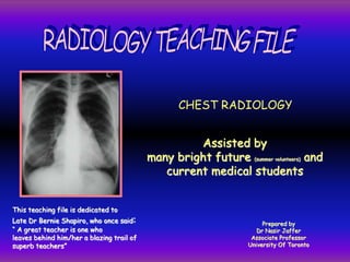
- 1. CHEST RADIOLOGY Assisted by many bright future (summer volunteers) and current medical students Prepared by Dr Nasir Jaffer Associate Professor University Of Toronto This teaching file is dedicated to Late Dr Bernie Shapiro, who once said: “ A great teacher is one who leaves behind him/her a blazing trail of superb teachers”
- 2. NOMENCLATURE IN PULMONARY RADIOLOGY “You see what you look for and you recognise what you know” In the line drawing on your right, what figure do you see? In radiology many terms are used to identify abnormalities on a radiograph. The following slides illustrate a few of these used in pulmonary radiology The figure is either a beautiful young lady or an old woman
- 3. A medical student’s famous words!
- 4. Reading Chest radiograph The classic question that every medical student asks, “Can you please teach me how to read a chest film” Before you look at a chest film, you should have provisional clinical diagnosis after taking a history and physical examination The chest X-ray findings should confirm you physical findings rather than be an aid to make your physical findings later !. (Before you bother a radiologist please read this!) The most important question you should first ask is “ Why did I order the chest X-ray on this patient?”
- 5. Pearls: Reading A Chest Radiograph Chest radiographs are done in a Postero-anterior (PA) and Left Lateral positions The PA view is done to minimize cardiac magnification Lateral view is done to allow three dimensional interpretation of the chest film findings (see later) In sick patients, an antero-posterior (AP) chest film is done, either supine or upright depending how immobile the patient is. On AP view, it is sometimes hard to assess cardiac enlargement, pleural effusion, and lung parenchyma, especially if the patient did not take good inspiratory effort
- 6. PA Chest radiograph X-ray tube The patient is facing the x-ray film cassette, and x-ray beam enters the patient’s back
- 7. 3: Is there any rotation of the chest? Assessed by checking the upper thoracic spinous process (oval) in relation to the medial ends of the clavicles (lines ‘a’) - this CXR is rotated to left 1 4 7 10 a a 1: Adequate penetration of the mediastinum-is the thoracic spine seen? 2: Has the patient taken a good inspiratory effort? About 8-10 posterior thoracic ribs should be seen through the lungs
- 8. Left Lateral Chest Radiograph X-ray tube The patient’s left side is against the x-ray film cassette, with x-ray beam entering the patient’s right side Lt Rt
- 9. Lateral view The patient’s left side is against the film and the X-ray beam travels from the right to left side This view is used to localise A lung lesion (nodule or consolidation etc) to a specific segment of lung, Other pathology if you know your anatomy of lateral CXR (See later) Assess cardiac enlargement (left/right ventricle, left atrium) Hilar enlargement, thoracic spine etc
- 10. On this PA CXR, there is a lesion near the aortic arch (arrow) The question is = Is it anterior or posterior or part of aorta, or is it in lung? Click left mouse key to proceed for answer On the lateral CXR, the lesion is localised to the posterior mediastinum, probably a neurofibroma The three dimensionality of PA & Lat CXR
- 12. ANATOMY OF THE LUNGS 1: LUNG UNIT: Primary pulmonary lobule arises from last respiratory bronchiole, consisting of alveolar ducts, atria, alveolar sacs and alveoli & vessels. Secondary pulmonary lobule is the smallest portion of lung surrounded by connective tissue septa, as an irregularly polyhedral shape measuring 1 to 2.5 cm (visible as Kerley B lines when abnormally filled with fluid). RIGHT LUNG LEFT LUNG Right Upper Lobe Anterior. Posterior & Apical segments Left Upper Lobe Apico-posterior, & Anterior Right Middle Lobe Medial and Lateral segments Lingular Lobe Superior & Inferior segments Right Lower Lobe Superior, Anterior, Medial, Lateral and Posterior basal segments Left Lower Lobe Superior, Antero-medial, Lateral and Posterior basal segments 2. LOBAR AND BRONCHIAL SEGMENTAL ANATOMY: The diagrams following this slide, show the various bronchial and lobar segments. A summary is listed below.
- 13. Anatomy of Bronchial Segments LUL Right Lung Left. Lung
- 14. Rt Lt Anterior Lateral Medial Left Lung Lateral Medial Right Lung Posterior Anatomy of Pulmonary Segments Lt Rt
- 15. NORMAL CHEST RADIOGRAPH P.A view Lateral view P.A view: Patient’s anterior chest is placed against the X-film, and the X-ray beam travels from posterior to anterior Left Lateral View: The patient’s left side is against the film and the X-ray beam travels from the right to left side
- 16. LATERAL CXR - NORMAL ANATOMY LIV Left innominate vein TR Trachea SVC Superior venacava AA Ascending Aorta RVO Right ventricular outflow tract MiF Minor fissure RV Right ventricle LLH Left lung against heart border (bare area of heart & mediastinum on left side) L Ma F Left major fissure R Ma F Right major fissure IVC Inferior venacava St Stomach RD Right hemidiaphragm LD Left hemidiaphragm LV left ventricle Co PV Confluence of pulmonary veins RPA Right pulmonary artery LPA Left pulmonary artery l u b Left upper lobe bronchus r u b Right upper lobe bronchus Ao A Aortic arch T-E St Tracheo-esophageal stripe LEGEND FOR LATERAL CXR rub lub T-E St
- 17. In the earlier days of chest radiology, before the introduction of CT & MRI, abnormalities on CXR were classified under lung, mediastinal, pleural and extra thoracic regions. A differential diagnosis was then given depending in which area and which organ in the area was affected. An example a middle mediastinal mass near the diaphragm at esophageal hiatus was most likely esophageal in origin e.g hiatus hernia. There are 3 classifications of the mediastinum 1:Anatomic 2: Fraser’s (radiological) 3: Felson’s (radiological)
- 18. ANATOMIC CLASSIFICATION The mediastinum is divided into 4 parts Superior mediastinum Apex of thorax to a plane passing through the manubrio- sternal junction and fourth dorsal vertebral body Anterior mediastinum Is anterior to heart & great vessels Middle mediastinum Contains heart & great vessels, mediastinal lymph nodes Posterior mediastinum Contains descending thoracic aorta, azygous/hemiazygous veins,esophagus, thoracic duct, splanchnic nerves & posterior mediastinal lymph nodes
- 19. FELSON’S CLASSIFICATION CLASSIFICATION OF MEDIASTINUM FELSON’S RADIOLOGICAL CLASSIFICATION Posterior Middle Anterior Felson’ classification divided the thorax into 3 compartments, Anterior, Middle and Posterior. Anterior: The boundaries were the sternum anteriorly and a line drawn from the anterior tracheal wall and behind the heart Middle: The compartment bounded by the trachea/posterior heart border and a line drawn 1cm posterior to the anterior portion of thoracic spine. Posterior: The space behind the posterior limit of middle mediastinum 1 cm
- 20. FRASER’S CLASSIFICATION The Fraser’s classification used 4 compartments. Anterior : Anterior to the heart Middle : Contained heart great vessels & trachea Posterior: Area in front of anterior aspect of spine and behind trachea Paravertebral: Containing spine, nerves & descending thoracic Aorta CLASSIFICATION OF MEDIASTINUM FRASER’S RADIOLOGICAL CLASSIFICATION
- 21. CLASSIFICATION OF MEDIASTINUM IN THE ERA OF COMPUTED TOMOGRAPHY With the introduction of Computed Tomography and Magnetic Resonance Imaging of the chest, it has become imperative that one become familiar once again with anatomy of the lung, and mediastinal structures. Many of the small mediastinal structures such as azygous vein are visualized on these two imaging modalities. These cross-sectional images require cross-sectional anatomical knowledge gained from dissection studies. The following slides illustrate some of the anatomy of many important structures seen on the CT and MRI images of your patients. Esopahgus Azygous vein Ascending Aorta Pulmonary outflow tract CT SCAN OF THE MEDIASTINUM AT TRACHEAL BIFURCATION
- 22. Heart Descending thoracic aorta Main pulmonary artery Internal thoracic vessels Left superior intercostal vein Aortic arch Sympathetic chain Left recurrent laryngeal nerve B V V MEDIASTINAL ANATOMY FROM THE LEFT SIDE Left pulmonary veins Left main stem bronchus
- 23. MEDIASTINAL ANATOMY FROM THE RIGHT SIDE ANTERIOR POSTERIOR Diaphragm Azygous vein Sympathetic chain Right superior intercostal vein Ascending aorta Superior Venacava RIGHT HILUM containing Pulmonary artery(A)) Vein (V) Bronchus (B) Right Phrenic nerve A V B
- 25. ACINAR PATTERN (CXR) Radiology: Round or elliptical ill-defined 4-8mm opacities in lung Microscopic: Portion of lung distal to terminal bronchial (respiratory bronchial, alveolar duct, alveolar sac and alveoli) is the acinus CXR close up of acinar pattern
- 26. ACINAR PATTERN (CT SCAN) Round or elliptical ill-defined 4-8mm opacities in lung CT scan of right upper lobe showing typical acinar pattern (arrow)
- 27. AIR BRONCHOGRAM Air containing bronchus peripheral to the hilum surrounded by airless lung CXR CT Scan Air Bronchogram
- 28. HONEYCOMB PATTERN: Ring shadows of air spaces 5 to 10mm in diameter and 2-3mm wall thickness, resembling honeycomb due to ‘end stage” lung HRCT CXR Elderly patient with chronic shortness of breath Path Diagnosis : Hamman Rich Pulmonary firbosis
- 29. RETICULAR PATTERN A collection of innumerable small linear opacities that together produce an appearance resembling a net. The pattern can be fine, medium or coarse. LUL RUL Fine Medium
- 30. RETICULONODULAR Collection of innumerable small, linear and nodular opacities together producing a net with small superimposed nodules. Close up of reticulonodular pattern CXR CT
- 31. BULLA: Gas containing avascularity of lung measuring 1cm or more in diameter, 1mm thickness BLEB: Gas containing space within or contiguous to visceral pleura usually at lung apex EMPHYSEMA: Abnormally expanded air spaces distal to terminal bronchiole with destruction of walls of involved air spaces. There are 2 types - Panlobular & Centrilobular. Bulla CT of bulla
- 32. MILIARY PATTERN: Tiny discrete 2mm or less, uniform opacities, e.g miliary TB
- 33. ATELECTASIS Three types of atelectasis are Passive: With process such as pneumothorax or effusion, lung retracts with decrease in volume. Cicatrization: When lung compliance decreases as in pulmonary fibrosis, there is a decrease in lung volume Adhesive: When action of alveolar surfactant is disturbed, e.g respiratory distress(ARDS), there is wide spread collapse of alveoli Definition: Less than normal inflation of all or a portion of lung with decrease in lung volume. Plate or discoid atelectasis is a linear opacity which represents diminished volume in a portion of lung usually lower lobes (Green arrow)
- 34. HILUM OVERLAY SIGN (Case A) The hilum (pulmonary artery-yellow) is well visualized as normal if a pulmonary lesion (Pink) is anterior or posterior to it = HILUM OVERLAY SIGN (Case B) A hilar lesion (lymphadenopathy-blue outline) or enlarged pulmonary artery) will enlarge the hilum Case A Case B
- 35. SILHOUETTE SIGN: The effacement of an anatomic soft tissue border within the thorax by a process of equal or similar density in contiguous lung or pleural; e.g pneumonia in medial segment of right middle lobe will silhouette the right heart border, lingula the left heart border, and the lower lobes silhouette the hemidiaphragm Pneumonia of the left lower lobe has obscured the posterior portion of the left hemidiaphragm Rt Lt
- 37. History: Young patient with cough and night sweats 1:What is your diagnosis 2: Give differential diagnosis for upper lobe fibrosis CASE 1
- 38. DIAGNOSIS: Acute tuberculous pneumonia (Bronchogenic spread) Radiographic findings: Bilateral upper lobe fibrosis with air space disease in some areas Differential Diagnosis of Upper Lobe Fibrosis * Tuberculosis * Radiation (e.g breast carcinoma) * Sarcoidosis * Chronic extrinsic allergic alveolitis * Histoplasmosis * Progressive Massive Fibrosis (pneumoconiosis) * Ankylosing spondylitis ( may have spine changes) Note: Activity of disease can only be determined by history, and pulmonary changes from previous CXR CASE 1
- 40. This slide is hyperlinked to CXR images of the different forms of lung TB. Place cursor on text in boxes
- 41. Radiologic Patterns of Primary TB include 1: Parenchymal disease: Homogeneous, ill-defined airspace consolidation. (Primary TB rarely cavitates) 2: Nodal disease: Hilar and paratracheal adenopathy are common 3: Airway disease: Atelectasis may be present from intrinsic (ie exudate) or extrinsic (ie adenopathy) bronchial obstruction 4: Pleural disease: Effusions are often a presenting symptom of primary TB 5: Calcification: Parenchymal granulomas often calcify 6: The Ranke complex: Which consists of a parenchymal scar (the Ghon focus) and calcified hilar or paratracheal lymph nodes.
- 42. Radiologic Patterns of Post-Primary TB include: 1: Local exudative or fibrocaseous TB: Apical and posterior segments of upper lobes and superior segments of lower lobes 2: Cavitation: Usually thick walled, may disappear with treatment 3: Acute Bronchopneumonia: Dissemination of caseaus material from one segment to other lobes (bronchogenic spread) 4: Miliary TB : Numerous tiny discrete foci uniformly distributed throughout lungs 5: Bronchiectasis: Due to either compression by large lymph nodes or infection of bronchial wall infection leading to bronchiectasis 6: Tuberculoma: A round/oval opacity ½-4 cm in diameter with smooth borders 7: Pleural Effusion: Empyema or secondary to bronchopleural fistula formation
- 43. PULMONARY TUBERCULOSIS This young patient has primary tuberculosis; Left upper lobe consolidation and left hilar (white arrow) and right paratracheal (green arrow) lymphadenopathy Primary Form
- 44. PULMONARY TUBERCULOSIS The CXR shows 1: Loss of volume of the right upper lobe (open arrow showing displaced hilum) 2: Apical pleural thickening (green arrow head) 3: Calcified right hilar lymph nodes (red arrow) Fibrocalcific Pattern
- 45. PULMONARY TUBERCULOSIS Post primary tuberculosis 1: Ranke complex a)calcified hilar lymph node b) Gohn’s focus (arrow) 2: Autonephrectomy of left kidney (green arrow) Post Primary Pattern Gohn’s focus
- 46. PULMONARY TUBERCULOSIS CXR showing fine miliary pattern scattered throughout both lungs. Close up shows the fine miliary pattern. (Note: sometimes also seen in patients with BCG treatment of superficial bladder tumors Close up Left upper lobe Miliary Pattern
- 47. History: Young patient with sudden onset of shortness of breath 1: What is your diagnosis? 2: What is the management of this patient? Case 2
- 48. DIAGNOSIS: TENSION PNEUMOTHORAX Radiographic Findings: Right sided pneumothorax with marked contralateral shift of the mediastinum. Management: The patient should have a chest tube inserted immediately to prevent vascular collapse. Tension pneumothorax is a clinical diagnosis. Etiology of pneumothorax * Spontaneous (rupture of bleb, bullae) * Iatrogenic (artificial ventilaton,lung biopsy, complication of central line insertions) * Traumatic (rib fractures) * Lung disease (metastasis -osteogenic sarcoma; COPD,asthma,honeycomb lung etc) * Mediastinal emphysema (asthma, esophageal rupture etc) * Pneumoperitoneum (through defects in diaphragm- peuroperitoneal foramen) Case 2
- 49. Case 3 History: Middle aged patient with high fevers and productive cough for 2 weeks What is your diagnosis? Management:If these findings persisted after antibiotic treatment, what is the next management
- 50. Radiographic Findings: There is consolidation of the right middle lobe with no air bronchogram; there is also some expansion of the lobe. Management: Other diagnosis should be considered when treatment fails * Inappropriate antiobiotic treatment * Obstructive pneumonitis (endobronchial lesion - tumor or foreign body) * Unusual lobar ‘expanding’ pneumonias (e.g Klebsiella pneumonia, Legionella pneumonia) * Neoplasm (bronchogenic carcinoma) * Chronic aspiration = (e.g elderly patients who take mineral oil as laxatives = lipoid pneumonia) Case 3 DIAGNOSIS: Right middle lobe pneumonia, with possible lung abscess
- 51. History: 26 year old male with several day history of fever, SOB Case 5
- 52. Diagnosis : Left Lower Lobe Pneumonia Left lower lobe airspace infiltrates, with the “spine sign” and “silhoutte sign” (See next slide) The Radiology of Pneumonia The radiographic presentation of pneumonia is quite variable, depending, among other factors, on etiologic agent. Most bacterial pneumonias show lobar, segmental, or patchy airspace infiltrates. Interstitial pneumonias are most commonly due to atypical organisms, including Mycoplasma, viruses, and PCP. Radiographic findings Case 5
- 53. 1: Air-bronchograms: See slide 25 2: “Silhoutte sign” Loss of an interface between lung and diaphgram or lung and cardiac border e.,g RML pneumonia silhoutte right heart border 3: “Spine Sign” On lateral CXR, the vertebral bodies appear ‘denser’ towards the lower thoracic spine due to overlying lower lobe lung disease.
- 54. March 8 Sep 21 History: A pigeon breeder, with dry cough. She presented several times with same findings until last CXR done on Sept 21. What is your diagnosis? Case 6
- 55. Case 6 Radiographic findings: Initial CXR shows basal alveolar pattern. The last CXR after patient removed all the birds from her home is normal. BIRD-FANCIER’S LUNG: * Occurs in people handling birds including budgerigars, pigeons, parakeets, parrots, hens,ducks and geese. * Antigen consists of bird sera, droppings or feathers. * Major source is probably bird serum excreted in lumen of bird’s gut, and inhaled in form of droppings. Radiology: Diffuse reticulonodular or nodular; commonly associated with an air space component in the acute stage. Rarely, long term exposure may result in ‘honeycomb lung’. DIAGNOSIS: Extrinsic allergic alveolitis (bird fanciers disease)
- 56. Case 7 History: Young patient with occasional cough. What is your diagnosis? Where is this lesion located (in the hilum or lung)
- 57. Case 7 DIAGNOSIS: Hamartoma in the right middle lobe Radiographic findings: Soft tissue density nodule overlying but not obscuring the right hilum (hilum overlay sign), therefore in the right middle lobe. Differential diagnosis: Granuloma (Tuberculoma) Malignant tumor: Bronchogenic carcinoma, metastasis, alveolar cell carcinoma, Benign: Adenoma, hamartoma, carcinoid Round pneumonia Infections: Echinococcal nodule Congenital: Bronchogenic cyst, Lobar sequestration. (See next slide for discussion)
- 58. HAMARTOMA About 96% occur in patients over 40 years of age Develops from an embryologic rest, containing cartilage, epithelium,, fibrous connective tissue, smooth muscle, adipose tissue and sometimes bone. They are well defined, lobulated, usually less than 4 cm, and are intrapulmonary in location, within 2 cm of pleura. Calcification (‘popcorn’, or punctate) occur in 30% of cases.
- 59. Case 8 History: Young patient with weight loss, cough and night sweats. What is your differential diagnosis
- 60. Case 8 DIAGNOSIS: HODGKIN’S LYMPHOMA Radiographic findings: Mediastinal lymphadenopathy, involving both hila, paratracheal, and anterior mediastinum Differential diagnoses: * Lymphoma * Sarcoidosis * Infectious mononucleosis * Metastasis (seminomas etc) HODGKIN’S LYMPHOMA Pathology: 4 cell types- lymphocytic predominence, nodular sclerosis, mixed cellularity and lymphocytic depletion. Radiology: Mediastinal lymphadenpathy,common; parenchymal involvement with consolidation and air bronchogram and sometimes masses are seen. Obstructive pneumonitis due to endobronchial lesion uncommon. Pleural effusion occurs in 30 % of cases. 15% may have bone involvement (ivory vertebra)
- 61. Case 9 History: Elderly patient with weight loss and cough. What are the findings. What is the next best imaging modality for the patient?
- 62. Case 9 Radiographic findings: Consolidation of the collapsed right lower lobe. There is displacement of right major fissure and hilum inferiorly. A right hilar mass is seen both on the P.A and lateral views. DIAGNOSIS: RIGHT LOWER LOBE COLLAPSE DUE TO HILAR MASS - BROCNCHOGENIC CARCINOMA. Next Investigation: A CT scan was performed showing collapsed right lower lobe with narrowing of the bronchus by enlarged hilar mass Right lower lobe bronchus
- 63. RADIOLOGY OF LOSS OF VOLUME OF DIFFERENT LOBES The roentgenologic signs of atelectasis are A: Direct signs Displacement of fissures B: Indirect signs Local increase in density Elevation of hemidiaphragm Compensatory overinflation Displacement of hila Approximation of ribs Absence of air bronchogram The following cases are typical examples of lobar collapse
- 64. RADIOGRAPHIC FINDINGS LOSS OF VOLUME OF DIFFERENT LOBES
- 65. RIGHT UPPER LOBE COLLAPSE Right minor fissure (arrow) and hilum displaced up with a triangular opacity seen along the upper lobe area.
- 66. RIGHT MIDDLE LOBE COLLAPSE A triangular ‘pancake’ shaped density obscuring right heart border. The minor fissure (single arrow) and lower half of major fissure (green arrow) approximate with progressive collapse. P.A view Lateral view
- 67. RIGHT LOWER LOBE COLLAPSE Findings seen in both right and left lower lobes are similar The major fissure is displaced posteriorly and medially. The X-ray findings are On P.A film there is a triangular opacity at posterior costo-vertebral angle (arrows) Lateral CXR, there is obliteration of the posterior third of the respective diaphgram P.A Lateral
- 68. RLL collapse
- 69. LEFT UPPR LOBE COLLAPSE The Lingula and Left upper lobe collapse together (single LUL bronchus) Lateral CXR: Distinctive ‘pancake’(outline) opacity in retrosternal air space. CXR Findings: PA view large opacity (outline) silhouetting left heart border (arrows). P.A Lateral
- 70. History: Young patient with back pain Questions: Where is this lesion located? What is your differential diagnosis? Case 10
- 71. Diagnosis: Posterior mediastinal neurofibroma Radiographic Findings: A smooth soft tissue mass seen in the upper posterior mediastinum
- 72. History: A 43 year old IV drug user presents with worsening shortness of breath on exertion. 1. Describe the chest X-ray findings (June 6, 1979) and give a diagnosis. 2. What are some of the complications? (see chest x-ray from May 28, 1985) June 6, 1979 May 28, 1985 Case 11
- 73. • Pneumoconiosis represents the reaction of the lung to inhaled dust particles resulting in alteration of lung parenchymal structure. •Talc is a fibrous silicate containing hydrate magnesium silicate. •In drug users who inject talc-containing solutions intravenously, they develop multiple small pulmonary nodules, conglomerate masses, and premature bullous emphysema. •Pathologically: • It is a granulomatous pulmonary arteritis • Perihilar ground glass alveolar involvement predominately of the upper lobes of the lung a charasteristic finding Natural History: Progression to a chronic stage, in which extensive deposition or alteration of collagen results in fibrosis. DIAGNOSIS: TALC PULMONARY FIBROSIS DUE TO DRUG ABUSE
- 74. History: Middle aged man with cough and weight loss with occasional hemoptysis. 1. Describe Findings 2. Which systemic syndromes are associated with 1o lung tumors (cancers)? Systemic Syndromes Associated with Primary Lung Tumors Hypercalcimia (PTH) Epidermoid Cushing’s (ACTH) Small Cell Carcinoid (Serotonin) Carcinoid 2. 1: Radiographic Findings: There is complete collapse of the Right upper lobe, with cephalad displacement of minor fissure (white arrow)and elevation of right hilum, and a triangular opacity representing collapsed lobe. Case 12
- 75. History: 55 year old woman with a 2 month history of dyspnea, hemoptysis, and weight loss. Case 13
- 76. Diagnosis Bronchioloalveolar carcinoma of the lung Bronchioloalveolar Carcinoma of the Lung A distinct form of adenocarcinoma of the lung Characterized by growth along the walls of preexisting normal lung air spaces without disturbance or destruction of lung parenchyma Radiographically, can appear as a solitary pulmonary nodule, diffuse nodules, or as airspace consolidation One of a few lung tumors that demonstrates the air bronchogram sign The Air Bronchogram Sign Characteristic of alveolar disease Caused by air-filled bronchi surrounded by non-aerated lung tissue Can be seen in pneumonia, pulmonary edema, nonobstructive atelectasis, bronchiectasis, and more rarely in fibrosing alveolitis, sarcoidosis, lymphoma, and bronchioloalveolar carcinoma. The mechanism of the establishment of the air bronchogram sign in bronchioloalveolar carcinoma is unknown. Clasically, it was thought to be caused by mucus secretion into the alveoli by the tumor, but also may be due to neoplastic cell desquamation or papillary tumor projection into alveoli. Source: Wong, Weisbrod, Chamberlain, and Herman. Bronchioloalveolar Carcinoma and the Air Bronchogram Sign: A New Pathologic Explanation. Journal of Thoracic Imaging, 9(3) 141-144, 1994.
- 77. Different pattern of bronchioloalveolar carcinoma There is a small soft tissue density nodule (arrow)
- 78. History: 66 year old with dry cough and fever. Case 14
- 79. Radiologic Findings Left upper lobe consolidation as well as diffuse right lung airspace consolidation with multiple areas of cavitation, including a large right lower lobe cavitating lesion. Further Diagnostic Workup The patient underwent lung biopsy, where caseating granulomas positive for acid-fast bacilli (Mycobacterium Tuberculosis) were found. Note that this patient complained of lower back pain. No spinal films were included in this teaching case. Tuberculous involvement of the spinal tissues is known as Pott’s disease.
- 80. Case 15 History: Young patient with a cough had a CXR done shown above. What is your diagnosis? Look at the CT and now what is your diagnosis? CT
- 81. Pathogenesis: Majority of thymic tumors are solid lymphoepithelial neoplasms (some are cystic) Occasionally they are fat-filled and referred to as thymolipomas (2-9% of thymomas). Thymomas are the most common anterior mediastinal neoplasms (1/3 are malignant) They can occur at any age but are rare in children. Arise from both epithelial and thymocytic elements of the thymic parenchyma. Virtually impossible to determine if they are benign or malignant from histology. More pertinent to prognosis from gross characteristics of local invasion or complete encapsulation. Clinical Symptoms: Close relationship between thymic tumors and myasthenia gravis. In the absence of myasthenia gravis most patients are asymptomatic. Distant metastases are rare. Recurrence rate following resection is high. Following resection, 50% of myasthenia gravis patients show some improvement. Radiologic Findings: Most are seen near the junction of the heart and great vessels. They are round or oval and the margins are smooth or lobulated. May protrude to one or both sides of the mediastinum and often displace the heart and great vessels posteriorly. It may be solid or cystic. Calcification may be visualized at the periphery of the lesion or throughout its substance. Because thymolipomas tend to grow large in size and are soft and pliable, they tend to slump towards the diaphragm which may leave the superior mediastinum clear. THYMIC TUMORS
- 82. History: Young asthmatic patient presents with shortness of breath and cough You are provided with CXR What are your findings and you diagnosis? Case 16
- 83. Radiographic findings: 1: Left upper lobe collapse (see section on lobar collapse) (Arrow heads) 2: Localised bronchiectasis in Right upper lobe (Flash arrow)
- 84. Pathogenesis: Asperigillus fumigantus is a fungus causing infections of the ear, nose lungs and other organs. The fungus increases the consistency of mucus, leading to mucoid impaction of bronchi distal to the lobar bronchus resulting in dilatation. Plugs are 2.5 to 6 cm in length and up to 3 cm in width. There is upper lobe predominance. Pulmonary infiltrates consisting of areas of acute eosinophilic infiltration of the alveoli can be seen. Clinical Symptoms: Most are asymptomatic . A productive cough resulting expectoration of the mucous casts, and fever or chest pain. Radiologic Findings: Mucus-filled bronchi appearing as bandlike opacities with rounded distal margins extending from the hilum. Usually in the upper lobes (seldom in the main-stem or first order bronchi) with second order bronchi involvement, the findings resembles a cluster of grapes. There is proximal bronchiectasis which is difficult to visualize until the mucus plugs being expectorated. (Our case has bronchiectasis in RUL) Peripheral bronchi are generally normal. Atelectasis or obstructive pneumonitis may be seen. Cavitation occurs in roughly 15% of cases. Pulmonary infiltrates, usually patchy and lobar or segmental in mid lung zones are commonly seen in asthmatic patients. Areas of consolidation measure 1 to 5 cm and are commonly migratory. Diagnosis: Diagnosis can be made on the basis of the roentgenographic findings (especially if there is proximal bronchiectasis associated with mucous impaction). Spasmodic asthma and peripheral eosinophilia are also very helpful. BRONCHOPULMONARY ASPERGILLOSES
- 85. History: Elderly patient with right apical and shoulder pain and right Horner’s syndrome Case 17
- 86. Diagnosis: Superior sulcus tumor (Bronchogenic carcioma) Anatomical Landmarks: The anatomy of superior sulcus: laterally by the first rib, anteriorly by the first costal cartilage and manubrium, and posteriorly by the head of the first rib and the body of the first thoracic vertebra, containing the following: the subclavian & jugular veins, the phrenic & vagus nerve, subclavian & common carotid arteries, the recurrent laryngeal nerves, the eighth cervical and first thoracic nerves, the sympathetic chain and stellate ganglion, and the first four ribs and upper vertebrae. Clinical features: First described by Pancoast in 1932 A triad of 1) Pain (shoulder, upper and lower arm), 2) Horner's syndrome, 3) Tumor in apices of lung 4) Secondary findings: atrophy of hand muscles, hemoptysis, weight loss, fever, chest pain, thrombophlebitis. Pathology: Commonly it is non-small cell bronchogenic carcinoma (squamous cell carcinoma). Others include small cell carcinoma, metastasis and benign processes.
- 87. Radiological features: A plain CXR, CT scan, MRI of chest and upper abdomen (to check adrenal & liver mets) are useful in staging the tumor The characteristic radiographic features include: 1: An apical cap > 5 mm in thickness (or a 5 mm asymmetry of the apical caps on the two sides) or an apical mass. 2: Bone destruction. (1,2 & 3rd ribs and vertebral bodies) 3: Secondary features: Pleural effusion, pneumothorax, bilateral parynchymal lesions resulting from contralateral hematogenous metastases, unilateral diaphragmatic (phrenic nerve) paralysis and a diffuse or local reticulonodular pattern caused by lymphangitic carcinomatosis. 4: Distant metastasis (adrenal, liver etc) Management: Depends on several factors. * Cell type of the tumor: Squamous cell carcinoma usually localised and hence slightly better prognosis * Stage of the tumor Therapy both surgical and to a lesser extent nonsurgical (radiation, chemotherapy, and immunotherapy) influences survival. A) Surgery: Resection of the tumor can be curative but sometimes for palliation.. Surgical resection can vary from simple wedge resection of the tumor to total pneumonectomy. B) Irradiation or chemotherapy (or both)
- 88. History: Elderly patient with severe cough and high fever What is your differential diagnosis? Case 18
- 89. Differential Diagnosis: This is a lobar expanding pneumonia a) Klebsiella Pneumonia b) Lipoid Pneumonia (chronic aspiration of mineral oil used as a laxative especially by elderly)) c) Bronchioloalveolar carcinoma d) Obstructive pneumonitis (endobronchial lesion causing pneumonitis) e) Legionella pneumonia f) Lymphoma
- 90. Patient was admitted to Intensive Care, due to respiratory failure He also developed a pneumothorax requiring a right chest tube You are provided a CXR done two weeks post admission. What is your diagnosis now? Pneumonia has undergone cavitation
- 91. DIAGNOSIS: Klebsiella Pneumonia Causative organism: An encapsulated, non-motile gram negative rod, aerobic micro-organism, K pneumoniae is found in normal human gastrointestinal tract; also acquired through the ingestion of contaminated food or water. Of the 82 serotypes, most frequent ones involved in acute fulminating are K. pneumoniae, K. rhinoscleromatis, K. ozaenae and K. oxytoca. K. rhinoscleromatis. It causes a chronic destructive granulomatous infection confined to the nose and may extend to the upper airways with the result of airway obstruction. K. ozaenae was associated with long term colonization in patients with cystic fibrosis. Route of infection: It is commonly through aspiration of oral secretions which usually go down to the most gravity dependent sites in the lungs. Site: It is more often unilateral, on the right side. It affects the most gravity dependent segments of the lungs (The superior portion of the lower lobes and the posterior segment of the upper lobes). Clinical features: - Usually men, 6th decade chronic alcoholics, diabetics or in patients with chronic lung disease. - Sudden in onset (mortality rate up to 50% in first 48 hrs) - Moderate fever, malaise, chills - Productive cough of purulent material sometimes bloody - Pleurisy - Dyspnea, tachypnea, cyanosis, even hypotension (septic shock)
- 92. Radiological Findings: They share the same general radiological features of acute lobar pneumonia .So in uncomplicated cases it appears as homogeneous parenchymal consolidation containing an air bronchogram but with no precise segmental distribution unless the whole lobe is involved. Three features to aid in differentiation from other types of pneumonia: 1) Tendency to form voluminous inflammatory exudate which increases the lung volume markedly with the result of bulging of interlobar fissures 2) Tendency to cavitate and form abscess. 3) High frequency of pleural effusion and empyema Treatment: Immediate and intensive antibiotic therapy. It is susceptible to Tetracyclines, Cloramphenicol, cephalosporins, Septra, Aminoglycosides. Complications: Abscess and cavity formation, massive lung gangrene, empyema, bronchpulmonary fistula, pneumothorax, pneumatocele formation, pericardial involvement, haematogenous spread i.e. meninges, chronic persistent infection with +ve culture (may resemble radiologically fibroproductive tuberculosis) DIAGNOSIS: Klebsiella Pneumonia
- 93. History: Young patient with shortness of breath on exertion. What are your radiographic findings? P.A Lateral Close up close up of RLL Case 20
- 94. Diagnosis: Alpha 1 Anti-trypsin deficiency Radiographic Findings: 1: Bilateral emphysema 2: Young patient with no exogenous risk factors
- 95. - Alpha 1-antitrypsin : A protease inhibitor, rises in inflammatory reaction deficient in some patients with the early onset of emphysema - Gene Type : most common gene type is MM >2.5g/ml MZ & MS range : 0.5g/l - 2.5g/l, ZZ & SS 0.0g/l - 0.5g/l - Clinical manifestation: 1: Progressive dyspnea, minimal cough, chronic bronchitis in smokers, panacinar process predominates at the lung base 2: 10% of children with ZZ type children develop significant liver disease; neonatal hepatitis, progressive cirrhosis. 3: Adults patients are usually asymptomatic 4: Cirrhosis, which may progress from a micro to macronodular state 5: Complications include hepatocellular carcinoma ALPHA 1-ANTITRYPSIN DEFICIENCY
- 96. History: Elderly patient with a chronic cough Case 21
- 97. Pneumoconiosis Definition: Accumulation of inorganic dust particles that are less than 5 m in diameter in the lungs, resulting in tissue reactions due to their presence Diagnosis: Requires (1) exposure history, (2) abnormal CXR findings, (3) Pulmonary Function Test findings (decreased diffusion, restrictive pattern) Symptoms: May be asymptomatic or present with dyspnea, productive sputum, cough
- 98. Pneumoconiosis CXR: Used for diagnosis and to monitor progression Findings include: (1) small opacities (rounded, irregular) (2) large opacities (3) atelactasis (4) pleural thickening (5) pleural calcification (6) hilar node involvement
- 99. Pneumoconiosis Varieties include: Silicosis: Usually in miners, foundry workers, sandblasters Generally need > 20 years exposure CXR with (1) reticulonodular pattern throughout the lung, predominantly in upper lobes and parahilar regions, with characterisitc onion-skin arrangement, and (2) possible hilar node involvement Asbestosis Usually in work dealing with insulation, construction Generally need 10-20 years exposure, but may present with less time CXR shows (1) fibrosis with linear streaking, predominantly at the bases, (2) possible pleural thickening ± calcification, and (3) possible pleural effusion Coal Miner’s Pneumoconiosis (CWP) CXR shows multiple nodular opacities, predominantly in upper lobes
- 100. CXR Close up RLL Right lower lobe mass Right lower lobe mass History: Young Greek patient with a cough Case 22
- 101. Same patient three weeks later Right lower lobe pulmonary nodule cavitates
- 102. Same patient a month later: The pulmonary nodule has ruptured into the pleural space CT showing consolidation in RLL and larger right pleural effusion
- 103. Pathogenesis: Hydatid disease is caused by the parasite Echinococcus granulosus Majority occur in immigrants from the Mediterranean regions, Yugoslavia, Russia, Africa, Argentinia, Chile, Uruguay and Australia.. It is endemic to northern Canada and Alaska. The ova are ingested and develop into larvae in the duodenum, pass into the bloodstream and then, most often, to the lungs and liver. It may be as long as 20 years before symptoms develop. Thoracic hydatid cysts occur mainly in the lung parenchyma with 2-5% developing in the mediastinum, pleura or diaphragm. In its early stages it is a spherical or oval mass surrounded by a layer of endogenous fibrous material (pericyst). The hydatid cyst itself is composed of two layers (the exocyst and germinal layer). Pulmonary hydatid cysts may rupture into the surrounding bronchi replacing the contents of the cyst with air. Sometimes only the pericyst ruptures allowing communication between the bronchial tree and the potential space between the exocyst and pericyst. Cysts rarely rupture into the pleural space producing a pyopneumothorax. Clinical Symptoms: The majority are asymptomatic. With cyst rupture, patients develop cough, expectoration, chest pain and fever. An acute hypersensitivity reaction may also develop with urticaria, pruritis and possibly hypotension. Sputum may become purulent and contain fragments of hydatid membrane. Diagnosis: Diagnosis can be made from the radiologic findings. Laboratory aids include the occasional identification of hooklets in the sputum which is part of the attachment organ of the tapeworm. Serologic tests and a skin test (the Casoni test) are also useful. HYDATID DISEASE OF THE LUNG
- 104. HYDATID DISEASE OF THE LUNG Radiologic Findings: * Characteristically presents as a solitary sharply circumscribed spherical or oval mass surrounded by normal lung. * 20-30% of patients have multiple cysts. * They range from 1 to greater than 10 cm in diameter. * The doubling time is 16 to 20 weeks. * The majority are found in the lower lobe, more often posterior, and slightly more common in the right. * The fluid content of the cysts is evident by the change in shape on radiographs at maximal inspiration and expiration or in erect and recumbant positions. With the rupture in the pericyst only, a rim of air around the periphery of the cyst can be visualized. * When all layers rupture, an air fluid level is often seen within the cyst. * Cyst fluid in the surrounding parenchyma may simulate pneumonic consolidation.
- 105. History: Young patient with shortness of breath. A CXR is provided. Case 23
- 106. See slide 61 for detailed explanation of lobar collapse
- 107. History: Elderly patient with hemoptysis and cough Case 25
- 108. A CT scan of the chest is shown RUL bronchus
- 109. Diagnosis: Right Upper lobe collapse due to an endobronchial carcinioma
- 110. History: Patient with shortness of breath. A CXR is shown Case 26
- 111. RADIOGRAPHIC FINDINGS: 1: Total opacification of left hemithorax 2: Shift of mediastinum to the left DIFFERENTIAL DIAGNOSIS 1: Collapse of lung due to endobronchial obstructing lesion 2: Previous total pneumonectomy
- 112. A CT scan was done shown below
- 113. Diagnosis: Complete collapse of Left lung. Etiology: Large bronchogenic carcinoma
- 114. History: Young patient with neck swelling and dysphagia Case 27
- 115. Diagnosis: Enlarged Thyroid gland obstructing esophagus Radiographic Findings: Large superior mediastinal mass predominantly on the left side and extending above the clavicles Differential diagnosis: Lymphoma Goitre Aneurysm of Aortic arch and left subclavian artery Esophageal lesion (Tumor, Zenker’s filled with fluid) Bronchogenic carcinoma (Left upper lobe; less likely)
- 116. History: Young patient with a heart murmur now presents with fever and cough Case 28
- 117. RADIOGRAPHIC FINDINGS: 1: Multiple ill defined soft tissue nodules 2: Normal heart DIFFERENTIAL DIAGNOSIS: 1: Septic pulmonary nodules 2: Pulmonary metastasis 3: Infectious nodules (TB, Fungus) 4. Autoimmune diseases (Wegener’s) 5: Sarcoidosis
- 118. Septic Pulmonary Emboli Pathogenesis: – cardiac origin - bacterial endocarditis of tricuspid valve, congenitally malformed tricuspid valve/congenital heart disease, rheumatic endocarditis, ventricular septal defects – septic thrombophlebitis: arm veins - IV drug users, arterio-venous shunts (e.g. hemodialysis) tonsillar and internal jugular veins - acute pharyngeal infection/abscesses pelvic veins - pelvic infections and veins near indwelling catheters – bacteremia - commonly staphylococcus aureus and streptococci species – soft tissue infection - e.g. staphylococcus osteomyelitis Risk Factors: – drug addiction, alcoholism, dermal infection, immunologic deficiency (e.g. lymphoma) Clinical Presentation: Common symptoms of pulmonary embolism - sudden onset of cough, dyspnea, chest pain (retrosternal angina pectoris and/or pleuritc chest pain), hemoptysis, apprehension, diaphoresis, syncope Common signs of pulmonary embolism - tachypnea, tachycardia, febrile, crackles, pleural rub, increased breath sound, accentuated P2 with new atypical murmur, phlebitis, edema, cyanosis
- 119. Roentgenographic findings in Septic Pulmonary Emboli Findings of septic pulmonary embolism: – Multiple peripheral, ill-defined, round or wedge shaped opacities of variable size – Consolidation (subsegmental ill defined infiltrates) – Cavitations are common, often thin-walled with no fluid level. – Pleural effusions are present in some cases – Cavities with central loose body simulating fungal ball are present (rarely). – Hilar or mediastinal lymph node enlargement (rarely)
- 120. Case 29 History: Elderly woman with a chronic cough Radiographic Findings: Ill defined soft tissue mass obscuring the right heart border ( P.A view) Green arrow head A triangular shaped density with inferior displacement of minor fissure and anterior displacement of lower half of the right major fissure ( Flash arrow) a b
- 121. Right Middle Lobe collapse Diagnosis: To see a more detailed understanding of lobar collapse see slide 60 (use the action button on left)
- 122. Case 30 History: Patient with shortness of breath
- 123. . For detailed description of lobar collapse see slide 61.
- 124. History: 31 year old male with cough and SOB Case 31
- 125. Diagnosis : Stage 2 sarcoidosis Radiographic findings: 1: Bilateral hilar lymphadenopathy 2: Bilateral upper lobe interstitial pattern Close up RUL
- 126. Sarcoidosis Definition: An idiopathic, multi-systemic disease characterized by the presence of noncaseating granuloma often in multiple organs, including the lung, lymph nodes, liver, spleen, skin and bone. Epidemiology: most common among age 20-40 North American Blacks (female predominance) and North European Whites Pathogenesis: Unknown, probably involves an infectious etiology and immunologic reactions to unidentified antigens. Morphologically, it consists of noncaseating multi-nucleated giant cell granuloma often preceded by active alveolitis which matures into Langerhans cells with subsequent complete resolution or residual parenchymal fibrosis. Clinical Presentation: – 50% are asymptomatic with an abnormal CXR – Constitutional symptoms: insidious onset of fever, weight loss, night sweat, weakness & malaise – Pulmonary symptoms: dry cough, dyspnea, hemoptysis (rare), – Peripheral lymphadenopathy, hepatosplenomegaly – Cutaneous manifestation: lupus pernio, psoriasis like plaques, erythema nodosum – Bone & Joint involvement: single/recurrent/migratory mono/polyarticular arthritis – Occular manifestation: uveitis, possibly with conjunctival, scleral, retinal and lens involvement – CNS involvement: cranial nerve palsy, hypothalamic hormonal disturbances – Miscellaneous (rare): exocrine glands, kidney, GI tract and skeletal muscle involvement
- 127. There is a radiographic staging of Sarcoidosis for descriptive purposes Stage 0: No demonstrable abnormality Stage 1: Hilar and mediastinal lymph node enlargement with no pulmonary abnormality Stage 2: Hilar and mediastinal lymph node enlargement plus diffuse pulmonary disease Stage 3: Diffuse pulmonary disease only with no node enlargement Stage 4: Pulmonary Fibrosis/End stage lung
- 128. Lymphadenopathy: often symmetrical and bilateral, tracheobronchial, paratracheal and bronchopulmonary. Lobulated and calcified (rare) Pulmonary Disease: 3 Distinctive patterns (1) Reticulonodular pattern: ranging from purely nodular to purely reticular pattern with fine/coarse reticular network and miliary pattern (rare) (2) Acinar pattern: discrete or coalescent ill-defined 6-7mm opacities, may be associated with air bronchogram and frequently a reticulonodular pattern giving “acinar shadow”. Upper zone fibrosis and bullae may be seen. Pulmonary fibrosis may be seen as disease progresses. (3) Large Nodules: Sharply marginated nodular opacities with average diameter > 1cm Other roentgenographic abnormalities: – cavitation (expulsion of necrotic tissue, rare) – atelectasis (obstructive pneumonitis) – pleural effusion – spontaneous pneumothorax Radiologic manifestations of Sarcoid ( Contd)
- 129. Rt Paratracheal (yellow) Bilateral hilar (blue) & ductal lymphadenopathy (green) Definition: Hilar and mediastinal lymph node enlargement with no pulmonary abnormality
- 130. Definition: Hilar lymphadenopathy bilaterally & bilateral upper lobe interstitial lung disease
- 131. Close up RUL Definition: Diffuse pulmonary disease only with no node enlargement
- 132. Close up of RLL •bilateral diffuse coarse interstitial disease (arrow) • loss of volume (lower lobes) •Reticulonodular pattern in R middle lobe and fibrosis in RLL Definition:Pulmonary Fibrosis/End stage lung
- 133. Numerous rounded coalescing non-caseating granulomas (arrow) A granuloma consists of epitheliod histiocytes, mulitnucleated giant cells, lypmhocytes plasma cells, fibroblasts and collagen Sarcoid histology of lung
- 134. Lymphadenopathy seen only in * Rt Paratracheal (widening of Rt paratracheal stripe ; pink arrow) * Rt hilar lymphadenopathy (lumpy bumpy Rt hilum ; yellow arrow) ASYMMETRICAL LYMPHADENOPATHY
- 135. Close up of right upper lobe FINE INTERSTITIAL PARENCHYMAL PATTERN There is a diffuse fine interstitial pattern in both lungs
- 136. NODULAR SARCOID There are multiple soft tissue pulmonary nodules along with bilateral hilar, and bilateral paratracheal lymphadenopathy Close up LUL
- 137. SARCOID CHANGES OF BONE Rt Lt Rt Lt Close up of the bone changes •Cystic leisons osteolytic and/or osteoclastic (arrow) • Lacelike trabecular pattern (arrowhead)