COLLEGE OF NURSINGSIR C.J
INSTITUTE OF PSCHIATRY HYDERBAD
ANATOMY &
PHYSIOLOGY
A s s i g n e d b y : M A ’ A M T A S L E E M L A G H A R I
R E P R O D U C T I O
N
SachaN
Raichand
Lal Chand
Amrat Kumar
Khalid
Presented by:
2.
Reproductive System
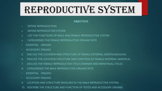
OBJECTIVES
1. DEFINEREPRODUCTION
2. DEFINE REPRODUCTIVE SYSTEM
3. LIST THE FUNCTIONS OF MALE AND FEMALE REPRODUCTIVE SYSTEM
4. CATEGORISED THE FEMALE REPRODUCTIVE ORGANS INTO
• ESSENTIAL ORGANS
• ACCESSORY ORGANS
5. DISCUSS THE LOCATION AND STRUCTURE OF FEMALE EXTERNAL GENITALIA(VULVA)
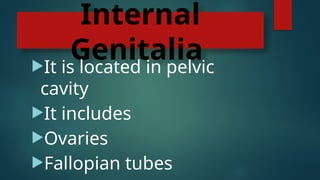
6. DISCUSS THE LOCATION STRUCTURE AND FUNCTION OF FEMALE INTERNAL GENITALIA
7. DISCUSS THE FEMALE REPRODUCTIVE CYCLE (OVARIAN AND MENSTRUAL CYCLE)
8. CATEGORISED THE MALE REPRODUCTIVE ORGANS INTO
• ESSENTIAL ORGANS
• ACCESSORY ORGANS
9. LOCATION AND STRUCTURE INVOLVED IN THE MALE REPRODUCTIVE SYSTEM
10. DESCRIBE THE STRUCTURE AND FUNCTION OF TESTIS AND ACCESSORY ORGANS
3.
Reproduction
The process ofproducing of a springs
that are biological are genetically
similar to the parent organism.
Act of producing a new individual from
at least one parent.
Production of new one species are
organism.
It has two types sexual reproduction
4.
Reproductive
System
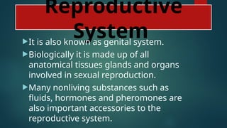
It is alsoknown as genital system.
Biologically it is made up of all
anatomical tissues glands and organs
involved in sexual reproduction.
Many nonliving substances such as
fluids, hormones and pheromones are
also important accessories to the
reproductive system.
5.
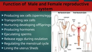
Function of Maleand Female reproductive
system
Producing sex cells (sperms/egg)
Transporting sex cells
Nurturing developing offSprings
Producing hormones
Ejaculating sperms
Release eggs during ovulation
Regulating the menstrual cycle
Lining the uterus Sheds
6.
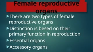
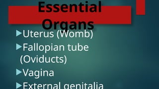
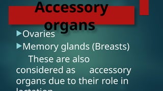
Female reproductive
organs
There aretwo types of female
reproductive organs
Distinction is besed on their
primary function in reproduction
Essential organs
Accessory organs
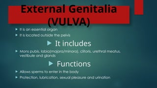
External Genitalia
(VULVA)
Itis an essential organ
It is located outside the pelvis
It includes
Mons pubis, labia(majora/minora), clitoris, urethral meatus,
vestibule and glands
Functions
Allows sperms to enter in the body
Protection, lubrication, sexual pleasure and urination
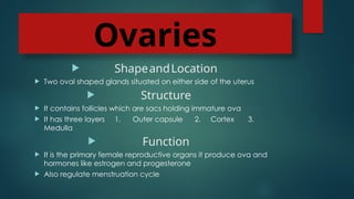
Ovaries
ShapeandLocation
Twooval shaped glands situated on either side of the uterus
Structure
It contains follicles which are sacs holding immature ova
It has three layers 1. Outer capsule 2. Cortex 3.
Medulla
Function
It is the primary female reproductive organs it produce ova and
hormones like estrogen and progesterone
Also regulate menstruation cycle
13.
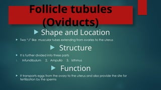
Follicle tubules
(Oviducts)
Shapeand Location
Two “J” like muscular tubes extending from ovaries to the uterus
Structure
It is further divided into three parts
1. Infundibulum 2. Ampulla 3. Isthmus
Function
It transports eggs from the ovary to the uterus and also provide the site for
fertilization by the sperms
14.
Uterus
Shape andLocation
Pear shaped hollow muscular organ located in the pelvic cavity situated between the bllader and
rectum
Structure
It is further divided into three parts
1. Fundus 2. Body 3. Cervix
Function
Provide a suitable environment to a fertilized egg to implant and develop into uterus during pregnancy
Shades its lining(endometrium) during the menstruation if fertilization does not occur
It is considered as a house of fetus during pregnancy
15.
Female
Reproductive
cycle
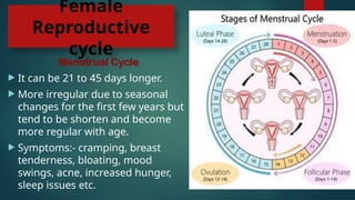
Menstrual Cycle
Itcan be 21 to 45 days longer.
More irregular due to seasonal
changes for the first few years but
tend to be shorten and become
more regular with age.
Symptoms:- cramping, breast
tenderness, bloating, mood
swings, acne, increased hunger,
sleep issues etc.
16.
Male reproductive
organs
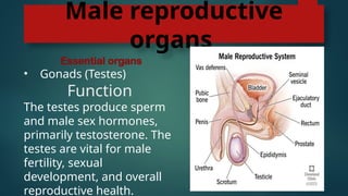
Essential organs
•Gonads (Testes)
Function
The testes produce sperm
and male sex hormones,
primarily testosterone. The
testes are vital for male
fertility, sexual
development, and overall
reproductive health.
17.
Essential Organs
Location
Apair aap testisis placed in a structure called as scrotum which is located outside
the abdominal cavity
Structure
Tunica albuginea: A tough, white fibrous capsule that surrounds the testis
Lobules: The testis is divided into lobules that contain seminiferous tubules
Seminiferous tubules: Tiny U-shaped tubes that contain germ cells and Sertoli cells
Rete testis: A network of uncoiled, interconnected channels that receive sperm
from the seminiferous tubules
Lyding cells: Produce testosterone and other androgens
18.
Accessory organs
Epididymis
Locatedat the back of the testicle and covered by the
visceral tunica vaginalis
It is divided into three parts: the head, body, and cauda
The head is round and located at the upper end of the
testis
The body is thinner than the head and located at the back
of the testis
The epididymis stores and carries sperm
It also ensures that spermatozoa are properly matured
19.
Vas deferens
Thevas deferens, also known as the ductus
deferens.
Long muscular tube runs from the epididymis into
the pelvic cavity behind your bladder and connects
to your urethra through a structure called the
ejaculatory duct.
The vas deferens transports sperm from the
epididymis to the urethra
During ejaculation, the vas deferens contracts to
propel sperm forward
20.
Seminal Vesicle
Locatedin the pelvis, behind the bladder and above the prostate
gland
Each seminal vesicle is a coiled, blind-ending tube that gives off
several irregular pouches
They are normally around 3 to 5 cm in length and 1 cm in diameter
They are composed of three layers: an inner mucosal layer, a
muscular layer, and an outer adventitial layer
During ejaculation, the muscular tissue inside of the seminal vesicles
pushes fluid out of the glands and into the forming semen
The seminal vesicles join with the vas deferens to become the
ejaculatory duct
21.
Prostate gland
Locatedin the male body, below the bladder and in front of the
rectum
The prostate is usually about the size of a walnut, but can enlarge
with age, covered by a stretchy connective tissue called prostatic
fascia
Made up of muscular and glandular tissue, it has 5 lobes anterior,
posterior, two lateral, and one median
The prostate produces fluid that mixes with sperm and other fluids to
form semen
The prostate’s muscles contract during ejaculation to help expel
semen
22.
Bulbourethral Gland
Alsoknown as Cowper’s glands, are located in the deep perineal
pouch, in the urogenital diaphragm, and near the membranous
urethra
Made up of small mucinous glands and surrounded by the
bulbocavernosus muscle
Each gland has multiple lobules that are made up of acini
The acini open into a central canal
The glands drain into the urethra through ducts
The bulbourethral glands produce a mucus-like fluid that lubricates
the urethra during sexual intercourse. This fluid is also known as pre-
ejaculate


![Reproductive%20 System[1]](https://cdn.slidesharecdn.com/ss_thumbnails/reproductive20system1-1220708198883512-8-thumbnail.jpg?width=640&height=640&fit=bounds)


