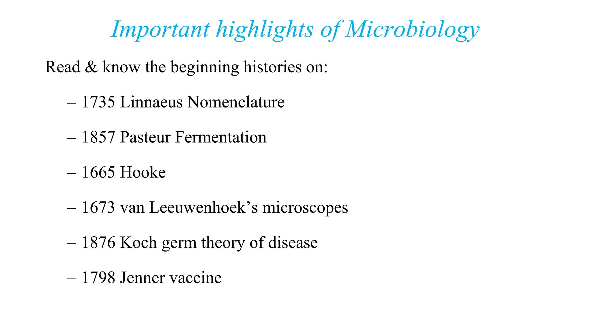
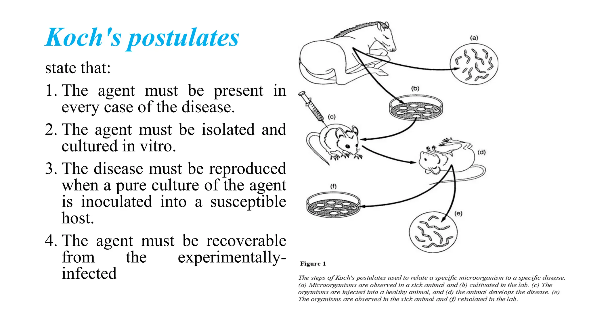
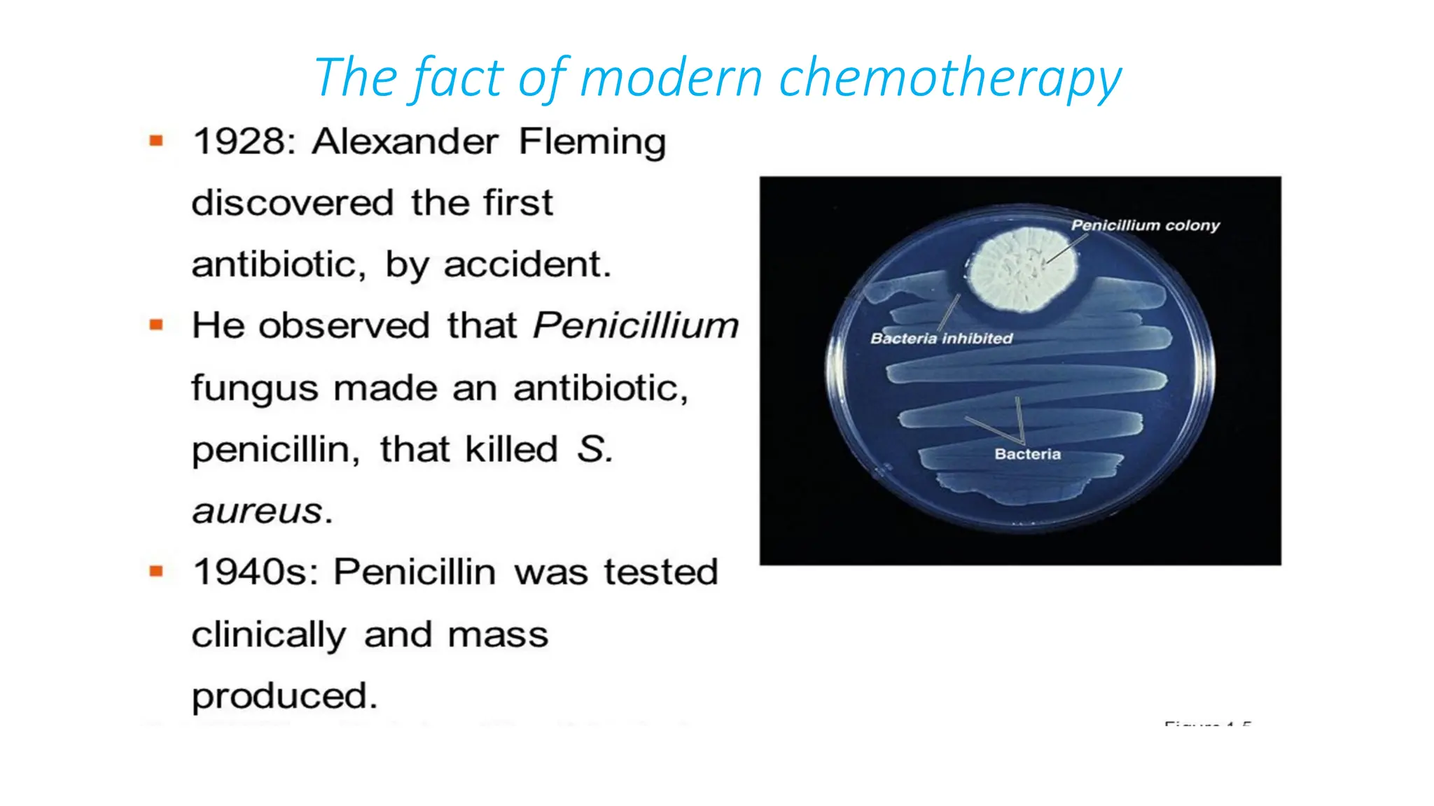
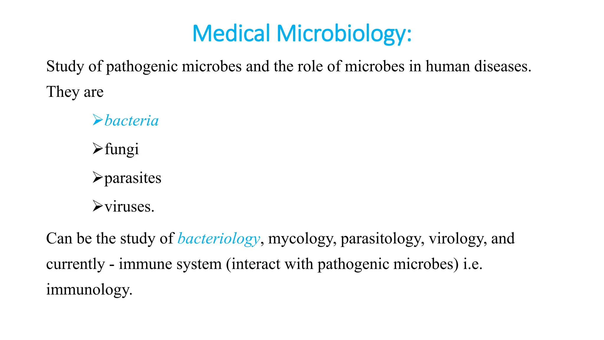
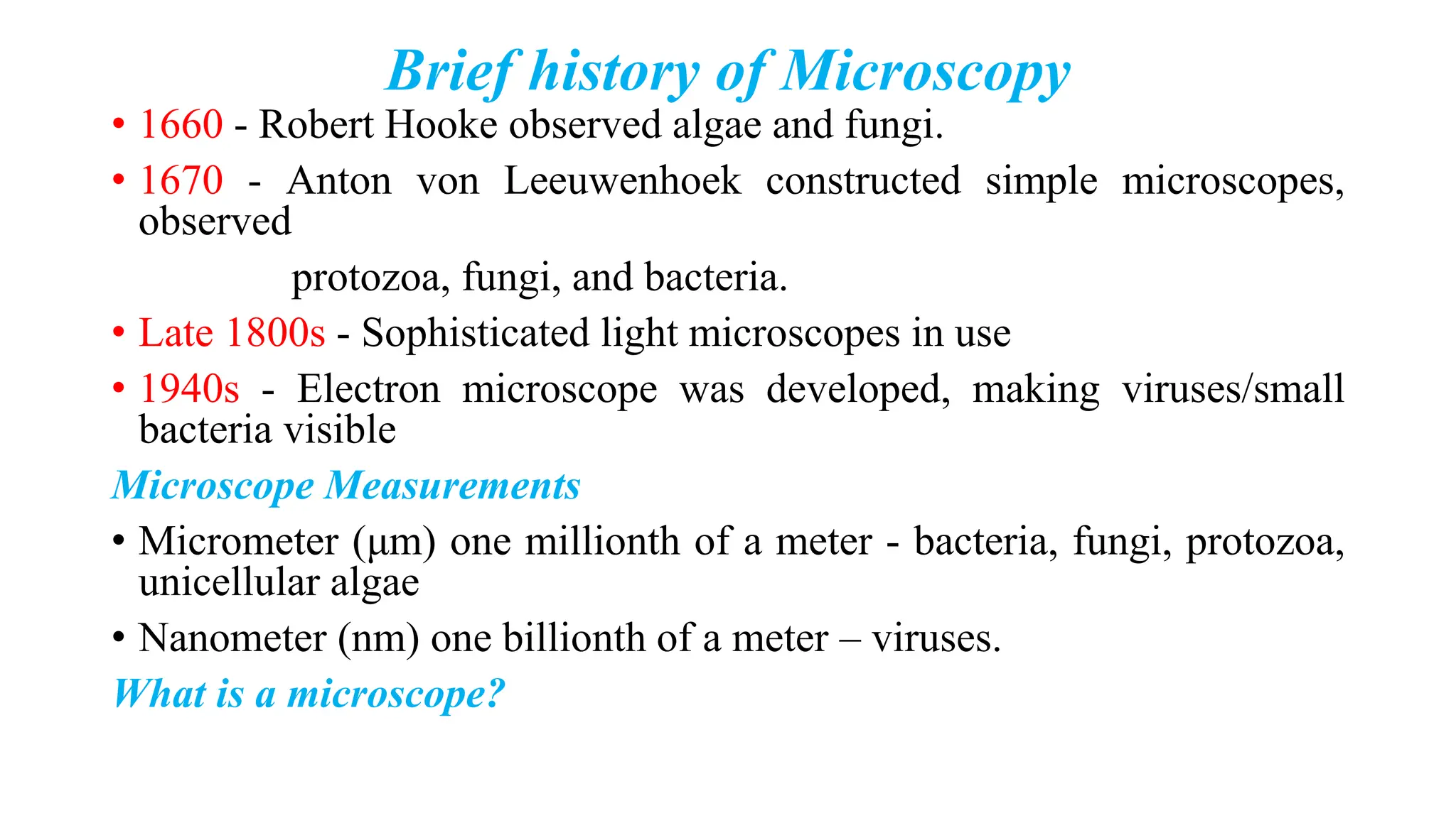
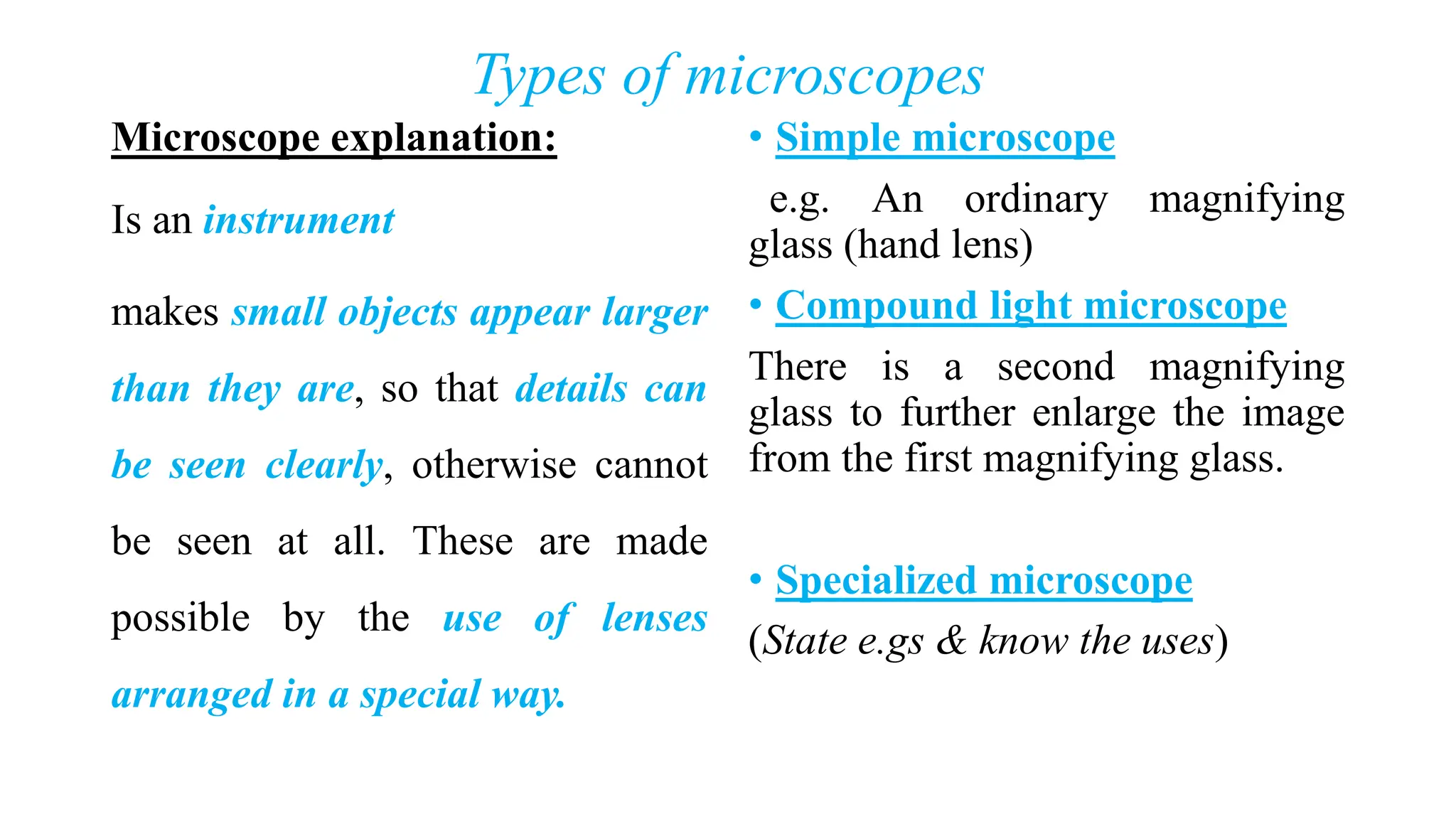
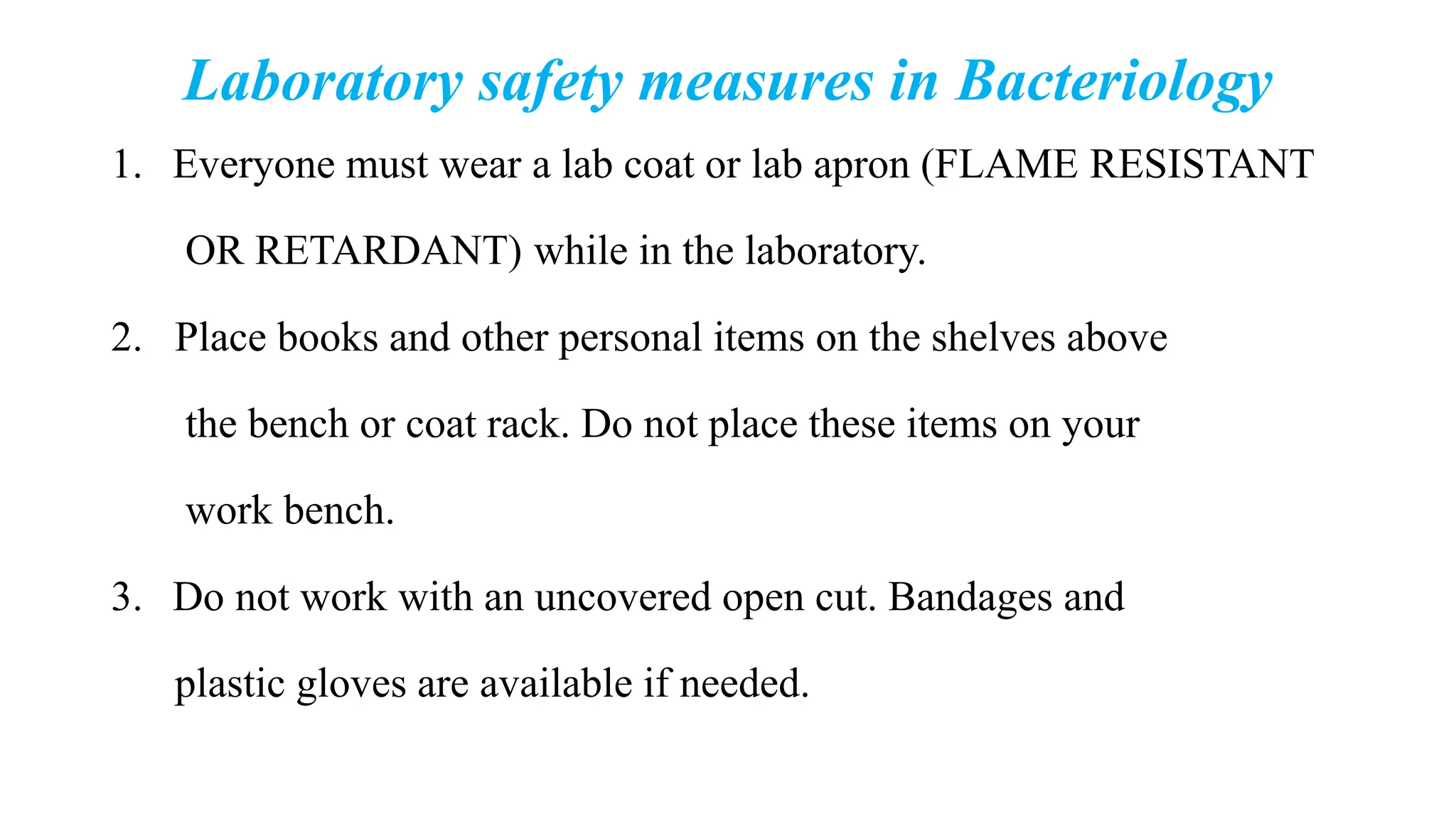
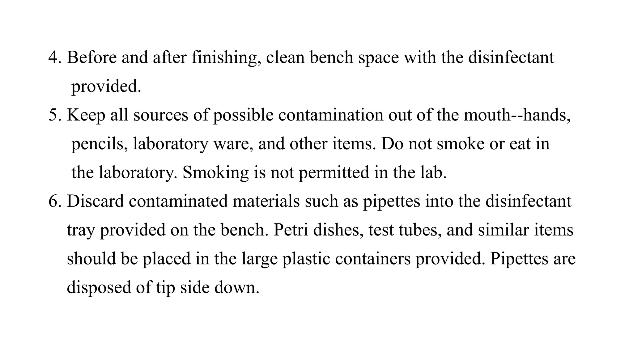
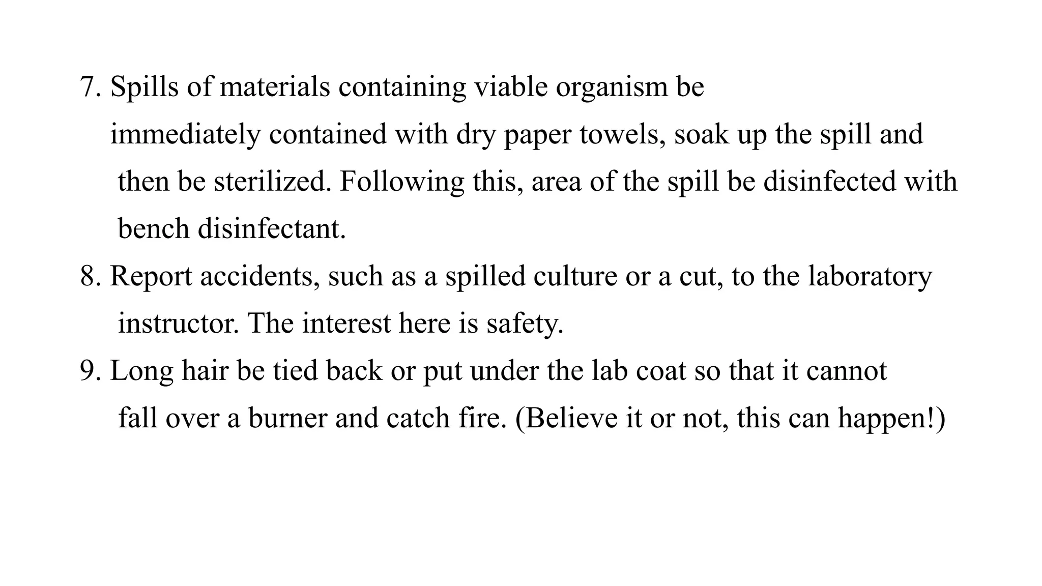
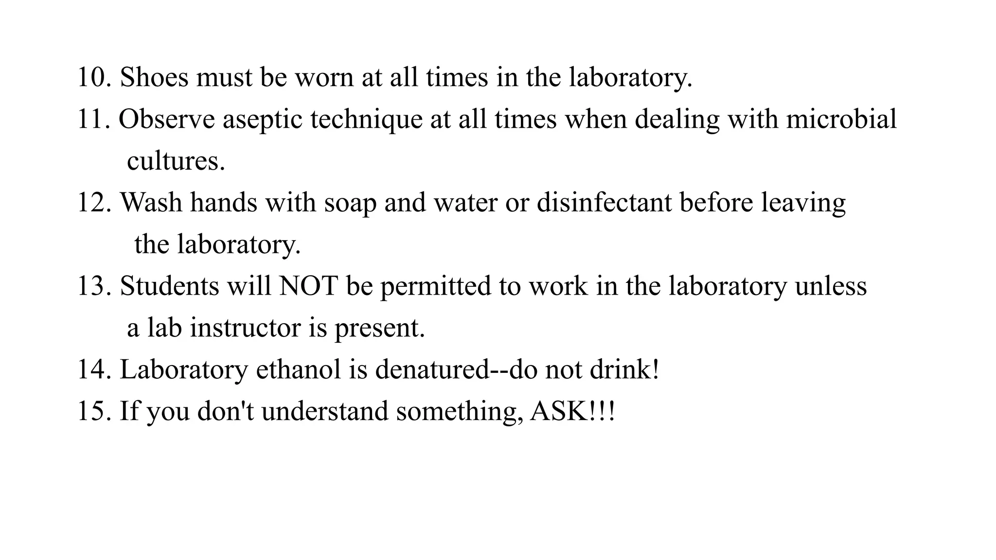
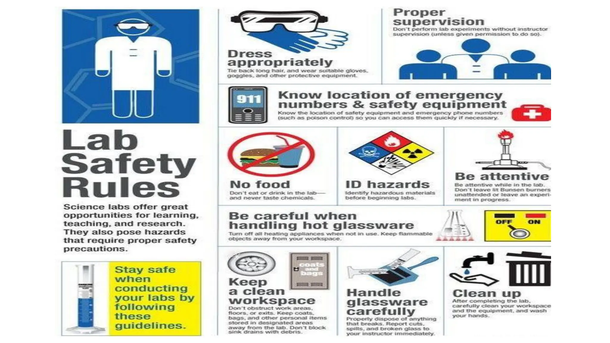
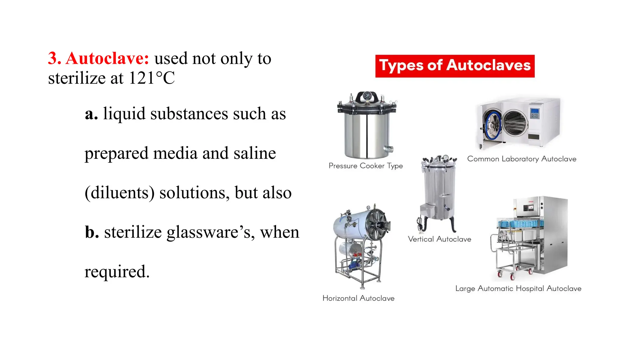
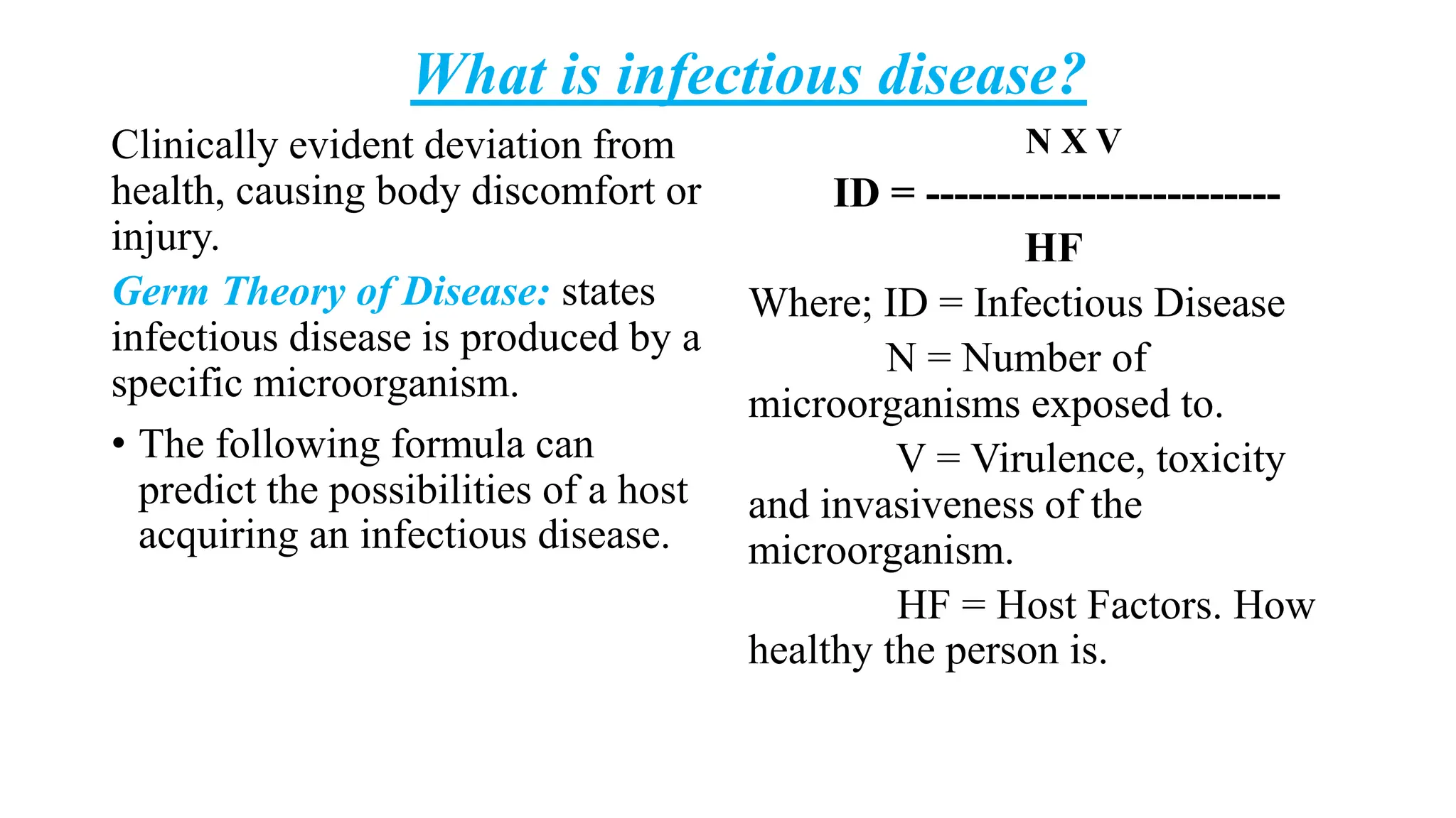
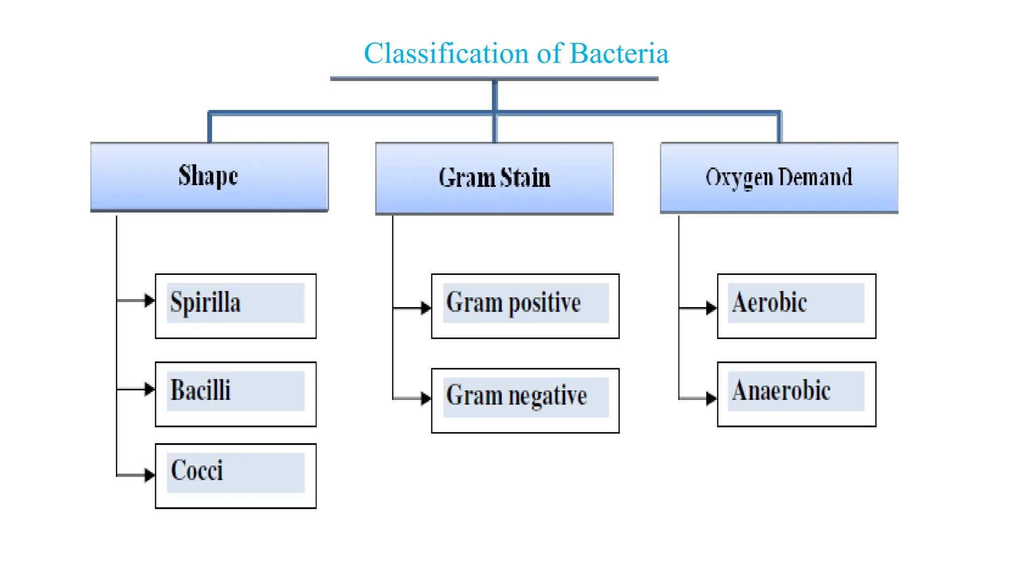
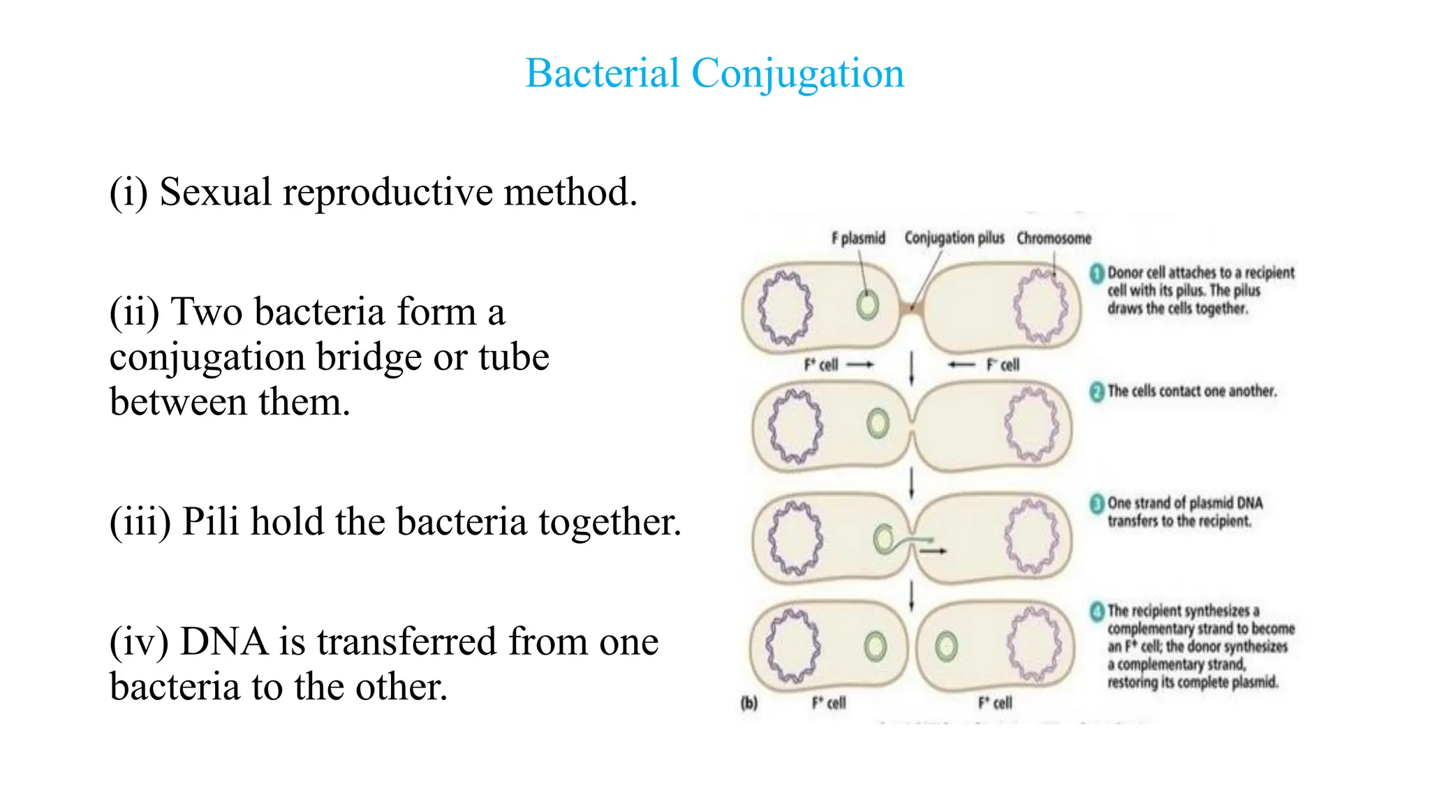
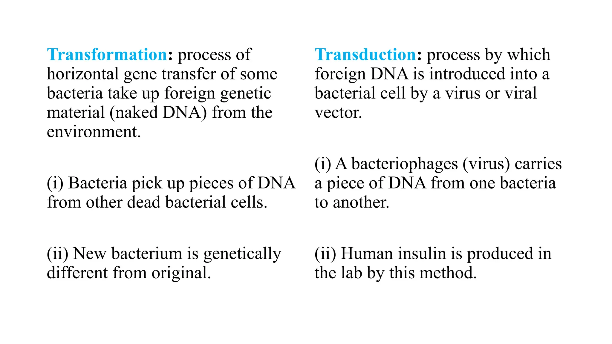
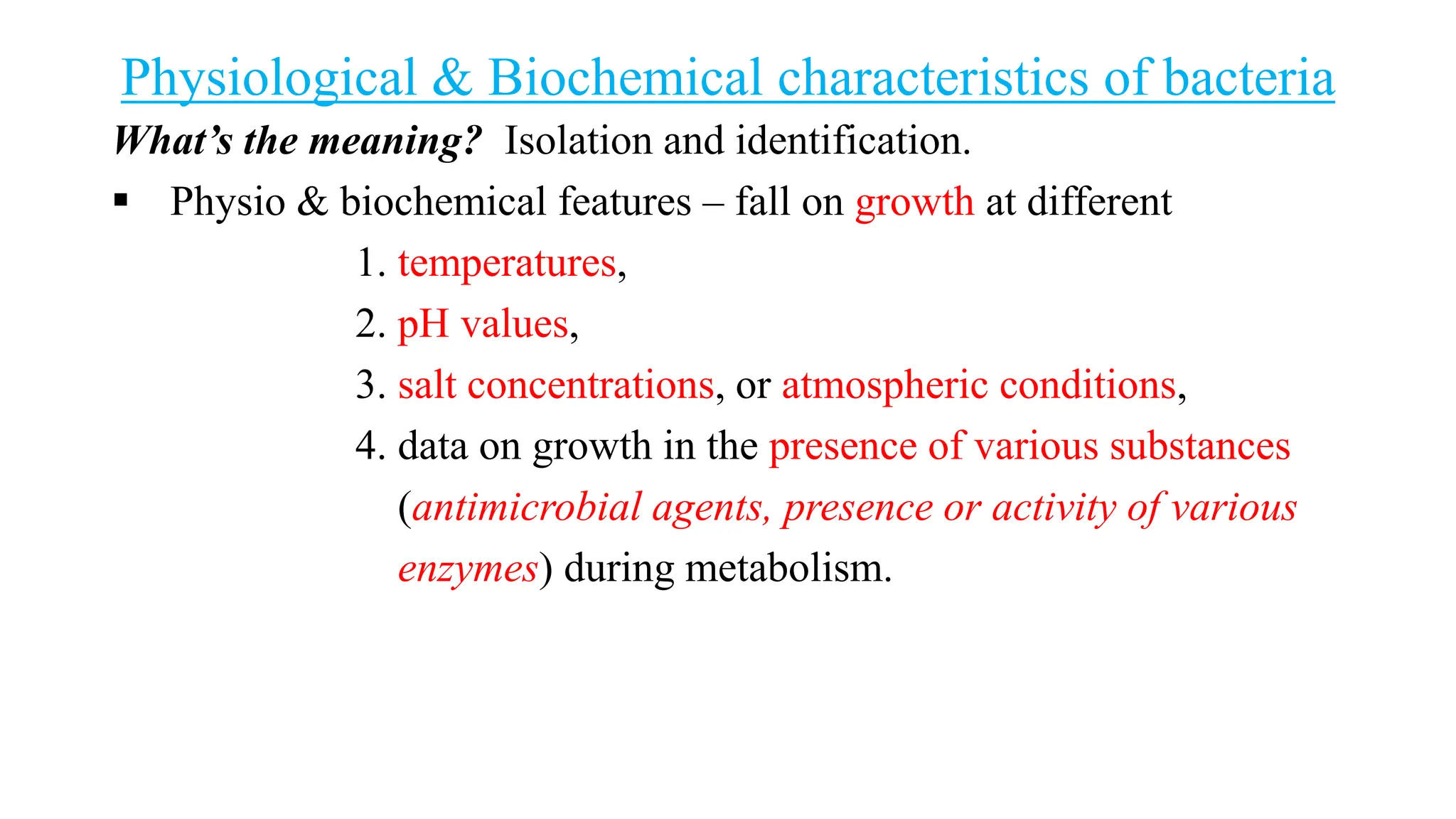
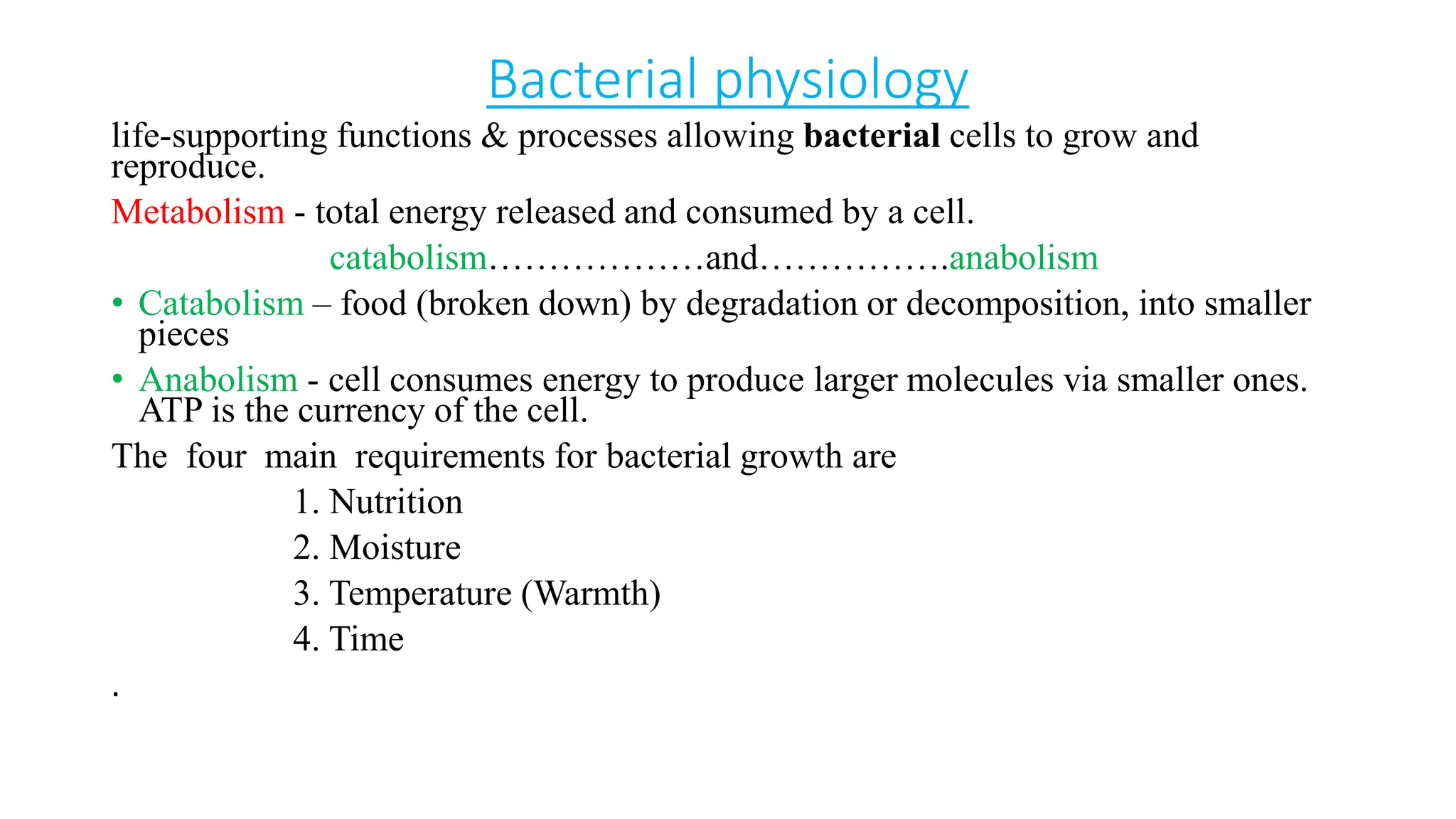
General Microbiology
(Bacteriology)
Lecture code: MLAB 212
By
Dr Francis S. Codjoe
Dept. of Medical Lab Sciences (Microbiology Unit)
Learning outcomes
Completion these lectures successfully:
• Understand and explain important historical Microbiological facts (Bact)
• Identify hazards in the Microbiology laboratory (Bact)
• Know safety measures needed in the laboratory (Bact)
• Know the methods of collection and transport of clinical samples for pathogens
identification.
• Know basic structure of bacteria and common examples
• Understand classification, colonial morphology, normal flora, transmission, aerobic &
anaerobic bacteria, microaerophilic rods & cocci and nutrition and biochemical
characteristics of bacteria & others.
Reading List
1. Gladwin, M. & Trattler, W. (2007). Clinical Microbiology Made
Ridiculously Simple. 4th Ed, MedMaster Inc.
2. Murray, P.R. (2017). Basic Medical Microbiology, 1st Edition, Elsevier
3. Brooks, G., Carroll, K.C., Butel, J., Morse, S. (2010). Jawetz, Melnick,
& Adelberg's Medical Microbiology, 25th Ed (LANGE Basic Science).
McGraw-Hill Medical, US.
4. Ford, M. (2010). Medical microbiology. Oxford: Oxford University Press.
5. Hawkey, P., Deirdre, L. (2003). Medical Bacteriology, A Practical
Approach, 2nd Edition, Oxford Univ. Press.
6. Mahon, C. R., Lehman, D. C., & Manuselis, G. (2011). Textbook of
diagnostic microbiology (4th ed.). Maryland Heights, Mo.:
Saunders/Elsevier.
Important highlights of Microbiology
Read & know the beginning histories on:
– 1735 Linnaeus Nomenclature
– 1857 Pasteur Fermentation
– 1665 Hooke
– 1673 van Leeuwenhoek’s microscopes
– 1876 Koch germ theory of disease
– 1798 Jenner vaccine
The “Spontaneous Generation” Debate
Understand the ff. debates:
1. microorganisms arise from lifeless matter such as beef broth.
2. the germ theory - the living organisms arise from
preexisting life, is called biogenesis.
Spontaneous generation controversy:
1688: Francesco Redi (1626-1678)
Italian physician refuted the idea of spontaneous generation
• showed that rotting meat carefully kept from flies will not
spontaneously produce maggots.
• 1861: Louis Pasteur's (1822-1895) famous experiments with swan-
necked flasks finally proved microorganisms do not arise by
spontaneous generation.
This eventually led to:
• Development of sterilization
• Development of aseptic technique
Koch's postulates
state that:
1. The agent must be present in
every case of the disease.
2. The agent must be isolated and
cultured in vitro.
3. The disease must be reproduced
when a pure culture of the agent
is inoculated into a susceptible
host.
4. The agent must be recoverable
from the experimentally-
infected
Koch's postulates
This eventually led to:
 Development of pure culture techniques
 Stains, agar, culture media, Petri dishes
Why Study Microbiology?
Are you interested in:
• Studying, preventing and controlling
infectious disease?
• Tracking down agents of infectious disease?
• Prevention and treatment of potential
bioterrorist attacks?
• Production of new products through
biotechnology?
• Production of fermented foods such
as cheese and yogurt?
• Understanding how elements are
cycled in our environment?
• Cleaning up toxic wastes in our
environment?
The Golden age of Microbiology
• 1857-1914
• Beginning with Pasteur’s work, discoveries included the relationship
between microbes and disease, immunity, and antimicrobial drugs.
• Leading to the ability to halt epidemics by interrupting the spread of
microorganisms.
Proofs: Fermentation and Pasteurization
• Pasteur showed - microbes responsible for fermentation
• Fermentation - conversion of sugar to alcohol to make beer and wine.
• Microbial growth - responsible for spoilage of food.
• Bacteria - use alcohol and produce acetic acid spoil wine by turning it
to vinegar (acetic acid).
• This application of a high heat
for a short time is called
pasteurization.
• Pasteur demonstrated that
bacterial spoilage be killed by
heat that was not hot enough to
evaporate the alcohol in wine.
Discoveries of the main pathogens
1877 Robert Koch Bacillus anthracis (anthrax)
1879 Albert Neisser Neisseria gonorrhea
1881 Alexander Ogston Staphylococcus aureus (pyogenic infections)
1882 Carl Gessard Pseudomonas aeruginosa (various)
1882 Robert Koch Mycobacterium tuberculosis
1882 Frederick Fehleisen Streptococcus pyogenes
1883 Theodor Klebs Corynebacterium diphtheriae
1884 Friedrich Loeffler Corynebacterium diphtheriae
1884 Arthur Nicolaier Clostridium tetani (anaerobe)
1884 Robert Koch Vibrio cholerae
1884 George Gaffky Salmonella typhi
1885 Gustav Hauser Proteus vulgaris (various)
1885 Theodor Escherich Escherichia coli (normal flora)
Truth of modern chemotherapy
• Treatment with chemicals - chemotherapy.
• Chemotherapeutic agents - treat infectious disease can be synthetic
drugs or antibiotics.
• Antibiotics - chemicals produced by bacteria and fungi that inhibit or
kill other microbes.
• Quinine - from tree bark was long used to treat malaria.
• 1910: Paul Ehrlich developed a synthetic arsenic drug, salvarsan, to
treat syphilis.
• 1930s: Sulfonamides were synthesized.
The fact of modern chemotherapy
Medical Microbiology:
Study of pathogenic microbes and the role of microbes in human diseases.
They are
bacteria
fungi
parasites
viruses.
Can be the study of bacteriology, mycology, parasitology, virology, and
currently - immune system (interact with pathogenic microbes) i.e.
immunology.
Brief history of Microscopy
• 1660 - Robert Hooke observed algae and fungi.
• 1670 - Anton von Leeuwenhoek constructed simple microscopes,
observed
protozoa, fungi, and bacteria.
• Late 1800s - Sophisticated light microscopes in use
• 1940s - Electron microscope was developed, making viruses/small
bacteria visible
Microscope Measurements
• Micrometer (μm) one millionth of a meter - bacteria, fungi, protozoa,
unicellular algae
• Nanometer (nm) one billionth of a meter – viruses.
What is a microscope?
Types of microscopes
Microscope explanation:
Is an instrument
makes small objects appear larger
than they are, so that details can
be seen clearly, otherwise cannot
be seen at all. These are made
possible by the use of lenses
arranged in a special way.
• Simple microscope
e.g. An ordinary magnifying
glass (hand lens)
• Compound light microscope
There is a second magnifying
glass to further enlarge the image
from the first magnifying glass.
• Specialized microscope
(State e.gs & know the uses)
A typical compound light microscope
Dark-field microscope
Understand & know the ff:
1. The principle of dark field
microscopy
2. When would you use a dark
field microscope
3. State microbes which dark
field microscope could be use
to examine.
Treponema pallidum (syphilis)
Microscope magnifications
• Calculation:
– Objective power x ocular power = total power
• Par centred - one in which the object in the centre of view
will remain in the centre when the objective is rotated.
• Par focal - one which, in focus with one objective, when the
objective is rotated, will remain in focus.
• Microscopic measurement
– Micrometer? Why must we calibrate it?
Developments in Medical Microbiology
• Diagnostics - lab investigations
• Prevention - measures to
control spread
• Use as a tool – biotechnologies
and genetics
• Vigilance and Surveys – checks
on infections and outbreaks
Gram stain of N. gonorrhoeae
Laboratory safety measures in Bacteriology
1. Everyone must wear a lab coat or lab apron (FLAME RESISTANT
OR RETARDANT) while in the laboratory.
2. Place books and other personal items on the shelves above
the bench or coat rack. Do not place these items on your
work bench.
3. Do not work with an uncovered open cut. Bandages and
plastic gloves are available if needed.
4. Before and after finishing, clean bench space with the disinfectant
provided.
5. Keep all sources of possible contamination out of the mouth--hands,
pencils, laboratory ware, and other items. Do not smoke or eat in
the laboratory. Smoking is not permitted in the lab.
6. Discard contaminated materials such as pipettes into the disinfectant
tray provided on the bench. Petri dishes, test tubes, and similar items
should be placed in the large plastic containers provided. Pipettes are
disposed of tip side down.
7. Spills of materials containing viable organism be
immediately contained with dry paper towels, soak up the spill and
then be sterilized. Following this, area of the spill be disinfected with
bench disinfectant.
8. Report accidents, such as a spilled culture or a cut, to the laboratory
instructor. The interest here is safety.
9. Long hair be tied back or put under the lab coat so that it cannot
fall over a burner and catch fire. (Believe it or not, this can happen!)
10. Shoes must be worn at all times in the laboratory.
11. Observe aseptic technique at all times when dealing with microbial
cultures.
12. Wash hands with soap and water or disinfectant before leaving
the laboratory.
13. Students will NOT be permitted to work in the laboratory unless
a lab instructor is present.
14. Laboratory ethanol is denatured--do not drink!
15. If you don't understand something, ASK!!!
The Most Important Lab Safety Rule
Know the Location of Safety Equipment
Dress for the Lab
Don't Eat or Drink in the Laboratory
 Having drinks in the lab risks your experiment, too. You could spill a
drink on your research or lab notebook.
 Eating and drinking in the lab is a form of distraction. If you are
eating, you aren't concentrating on your work.
Don't Taste or Sniff Chemicals
Don't Play Mad Scientist in the Laboratory
Dispose of Lab Waste Properly
Know What to Do With Lab Accidents
Leave Experiments at the Lab
Don't Experiment on Yourself
Science - using the scientific method. One needs data on multiple
subjects to draw conclusions, but using yourself as a subject and self
experimenting is dangerous, not to mention bad science.
Microbiology (Bacteriology) Lab. Equipment
1. Hot-air oven for Dry-heat
Sterilization
Used for sterilization of
glassware’s, such as test tubes,
pipettes and petri dishes at 160ºC.
2. Drying oven
At 100°C till the glassware’s dry
up completely.
3. Autoclave: used not only to
sterilize at 121°C
a. liquid substances such as
prepared media and saline
(diluents) solutions, but also
b. sterilize glassware’s, when
required.
4. Bacteriological Incubator:
usual temp. of incubation is 37°C.
Maintains optimal temp., humidity
and other conditions such as the
CO2 and oxygen content of the
atmosphere inside.
5. Fridge (Refrigerator): 6. Deep-fridge: Used to store
chemicals and preserve samples at
very low sub-zero temperatures.
7. Electronic Top-pan Balance:
• Used for weighing large quantities of media
and other chemicals, where precise weighing
is not of much importance.
8. Electronic Analytical Balance:
• It is used to weigh small quantities of
chemicals and samples precisely and quickly.
9. Double-pan Analytical Balance:
• Used to weigh chemicals and samples
precisely. Weighing takes more time, used in
emergency only.
10. Distilled Water Plant:
• Used in the preparation of media and reagents.
11. pH Meter: for determining the
pH of liquid media, liquid samples
and buffers.
12. Hot Plate:
• Used to heat chemicals and
reagents.
13. Shaking Water Bath:
• For heating at very precise
temperatures when required
14 Vortex Mixer:
• Used for thorough mixing of
liquids in test tubes.
15. Laminar Flow Chamber:
• Used for aseptic transfer of
sterilized materials, as well as for
inoculation of microbes.
16. Membrane Filtration
Apparatus: used to sterilize e. g.
urea disintegrate and lose their
original properties
17. Microscope: used for visual
observation of morphology,
motility, staining and fluorescent
reactions of bacteria
18. Centrifuge: used to spin down
to obtain deposits
19. Computers:
• Generally used for analysis of
results.
20. Spectrophotometer:
• For measuring the differences in
colour intensities of solutions.
21. Automatic Bacteria Identification System:
• Used for automatic computer-assisted identification of bacteria.
22. Electrical Devices:
• Used for the fluctuation of electric voltage in the lab i.e. to prolong the
equipment otherwise be damaged them.
Lab. Accessories
Tools supporting laboratory activities.
1. function to provide optimal efficiency and utility to lab workers
and scientists,
2. complimenting all major laboratory equipment.
E.gs stoppers, stands, clamps (double jaw clamp, extension
clamp, utility clamp, water bath clamp and nester
extension clamp), test chambers, lab frame rods and
slide holders.
Cleaning of Pipettes
1. Place pipettes delivery end down, in a glass cylinder (graduate) in
cleaning solution and allow them to stand overnight. (Steam may break
the glass cylinder).
2. Used pipettes be washed immediately. Grease cannot be removed
with water be treated with 10% NaOH and then with cleaning solution.
3. Rinse with tap water, followed by distilled water.
4. Rinse with alcohol. (Alcohol may be used repeatedly.)
5. Drain.
6. Autoclave if possible
Cleaning Other Glassware
1. Glassware containing liquefiable solid media - best cleaned by
heating and pouring out the material while in liquid condition, then
treating as above. (Solid media when liquefied by heat should never
be thrown in the sink, as it will solidify when cold and clog up the
traps and drains.).
2. Flasks, test tubes, Petri dishes, etc., containing cultures, must be
heated 1hr in flowing steam before cleaning. Cultures containing
spores should be autoclaved previous to cleaning.
3. If cultures or media have become dry, add water before heating.
Special care must be used in cleaning glassware in which mercuric
chloride or any other disinfectant has been used.
General rules for collection and transportation
of specimens
• Apply strict aseptic techniques throughout the procedure.
• Wash hands before and after the collection.
• Collect the specimen at the appropriate phase of disease.
• Make certain specimen is representative of the infectious process (e.g.
sputum is the specimen for pneumonia and not saliva), adequate in
quantity for the desired tests to be performed.
Receipt, handling and Transportation of specimen
Laboratory diagnosis of an infectious disease begins:
• With the collection of a clinical specimen for examination or
processing in the laboratory (the right one, collected at the right time,
transported in the right way to the right laboratory).
• Proper collection of an appropriate clinical specimen is the first step
in obtaining an accurate laboratory diagnosis of an infectious disease.
Guidelines for the collection and transportation of specimens should be
made available to clinicians in a lucidly written format. The guidelines
must emphasize two important aspects:
1. Collection of the specimen before the administration of
antimicrobial agents.
2. Prevention of contamination of the specimen with
externally present organisms or normal flora of the body.
Receipt and specimen Information
1. Check the patient demographic information on lab. request form
whether tallies with the specimen container.
2. Minimum number of information elements must be checked;
a. Patient's name
b. Date of birth
c. Hospital number or
d. Room number in the hospital
Receipt and specimen Information
A. This helps match the report with the patient, assuring that
the proper treatment be given to the right person.
B. This works better when the hospital computer system be
networked for in-patients.
3. Collect or place the specimen aseptically in a sterile and/or
appropriate container.
4. Ensure that the outside of the specimen container is clean and
uncontaminated.
5. Close the container tightly so that its contents do not leak during
transportation.
6. Label and date the container appropriately and complete the
requisition form.
7. Arrange for immediate transportation of the specimen to the
laboratory.
Criteria for rejection of specimens
The following are some examples:
1. Missing or inadequate identification.
2. Insufficient quantity.
3. Specimen collected in an inappropriate container.
4. Contamination suspected.
5. Inappropriate transport or storage.
6. Unknown time delay.
7. Haemolysed blood sample.
Storage and Postage of specimens
Bacteria Colonial Morphology
A. Bacteria are small (1-8µ)
B. Primary shapes (important for
identification and making
diagnosis)
1. Rods
2. Cocci
3. Spirochetes
4. Others (vibrio's, filamentous,
coccobacilli)
Normal Flora
Why is it called the Normal flora?
1. Majority of the organisms concerned are bacteria.
2. Normal flora may be categorized into two types:
1. Resident flora - always present
2. Transient flora - only present for short period of time
3. Humans have approximately
A. 1013 cells in the body
B. 1014 bacteria associated with them,
C. majority found in the large bowel.
4. Body parts exposed to, or communicate with external
environment:
skin,
nose and mouth,
intestinal
urogenital tracts.
NB: Internal organs and tissues are normally sterile.
Predominant bacteria at various anatomical locations in adults
Anatomical Location Predominant bacteria
Skin staphylococci and corynebacteria
Conjunctiva sparse, Gram-positive cocci and Gram-negative rods
Oral cavity
teeth streptococci, lactobacilli
mucous membranes streptococci and lactic acid bacteria
Upper respiratory tract
nasal membranes staphylococci and corynebacteria
pharynx (throat) streptococci, neisseria, Gram-negative rods and cocci
Lower respiratory tract none
Gastrointestinal tract
stomach Helicobacter pylori (up to 50%)
small intestine lactics, enterics, enterococci, bifidobacteria
colon bacteroides, lactics, enterics, enterococci, clostridia, methanogens
Urogenital tract
anterior urethra sparse, staphylococci, corynebacteria, enterics
vagina lactic acid bacteria during child-bearing years; otherwise mixed
Examples of bacterial specific adherence to host cells or tissue
Bacterium Bacterial adhesin Attachment site
Streptococcus pyogenes Cell-bound protein (M-protein) Pharyngeal epithelium
Streptococcus mutans Cell- bound protein (Glycosyl transferase) Pellicle of tooth
Streptococcus salivarius Lipoteichoic acid Buccal epithelium of tongue
Streptococcus pneumoniae Cell-bound protein (choline-binding protein) Mucosal epithelium
Staphylococcus aureus Cell-bound protein Mucosal epithelium
Neisseria gonorrhoeae N-methylphenylalanine pili Urethral/cervical epithelium
Enterotoxigenic E. coli Type-1 fimbriae Intestinal epithelium
Uropathogenic E. coli P-pili (pap) Upper urinary tract
Bordetella pertussis Fimbriae ("filamentous hemagglutinin") Respiratory epithelium
Vibrio cholerae N-methylphenylalanine pili Intestinal epithelium
Treponema pallidum Peptide in outer membrane Mucosal epithelium
Mycoplasma Membrane protein Respiratory epithelium
Chlamydia Unknown Conjunctival or urethral epithelium
Advantages of Normal flora
1. The normal flora synthesizes and excretes vitamins - especially
vitamin K and vitamin B12.
2. Prevent colonization by pathogens - competing for attachment sites
or for essential nutrients.
3. Antagonize other bacteria through the production of substances -
inhibit or kill non-indigenous species.
4. Stimulate the development of certain tissues, i.e., caecum and
certain lymphatic tissues (Peyer's patches) in the GI tract.
5. Stimulate the production of natural antibodies - induce an
immunological response, e.g. an antibody-mediated immune (AMI)
response.
Disadvantages of normal flora
1. Bacterial synergism - a member of the normal flora and a potential
pathogen helps in establishment of infection.
2. Competition for nutrients - gastrointestinal tract flora get utilizable
nutrients before absorption to the body.
3. Induction of a low grade toxaemia - Minute amounts of bacterial
toxins (e.g. endotoxin) be found in the circulation.
4. The normal flora may be agents of disease – many of the normal
flora are potential pathogens. E.gs. Neisseria meningitidis,
Streptococcus pneumoniae, E. coli, Salmonella etc., etc.
5. Transfer to susceptible hosts - at a point of weak immune status of
the host.
6. Important causes of hospital acquired infection - patients exposed
to invasive treatment. E. g. Patient with burns.
7. Overgrowth by potential normal flora change the local
environment (e.g. increases in stomach or vaginal pH).
Q. (What happens when the normal flora is absent?)
Student should answer.
Associations between Humans and the Normal Flora
Three types relationships between host and normal flora
1. Commensalism (Commensals) - no harm, no benefit to host
2. Mutualism (Mutualistic): beneficial relationship, both microbe
and host benefit
3. Opportunistic (Opportunists): potential pathogens producing
infection when host defenses depressed or when normal flora
disturbed
What is infectious disease?
Clinically evident deviation from
health, causing body discomfort or
injury.
Germ Theory of Disease: states
infectious disease is produced by a
specific microorganism.
• The following formula can
predict the possibilities of a host
acquiring an infectious disease.
N X V
ID = -------------------------
HF
Where; ID = Infectious Disease
N = Number of
microorganisms exposed to.
V = Virulence, toxicity
and invasiveness of the
microorganism.
HF = Host Factors. How
healthy the person is.
Ultrastructure of a Bacterial Cell
Classification of Bacteria
Bacterial Metabolism
Adenosine Triphosphate ( ATP) - complex organic chemical that
provides energy to drive many processes in living cells, e.g. muscle
contraction
Metabolism:
The sum of all chemical reactions within a living organism
Metabolism = Catabolism + anabolism
Catabolism:
Breakdown more complex organic molecules to simpler substances.
Release energy (ATP; stored and used to power anabolic chemical
reactions)
Anabolism:
Chemical reaction of simpler substances combined to form more
complex molecules. Require energy (ATP)
Bacterial Respiration
Two main types -
Respiration: Bacterial Examples
Obligate aerobe - requires free oxygen to survive e.g. Pseudomonas
aeruginosa.
Facultative anaerobe – lives with or without free oxygen e.g.
Streptococcus pyogenes.
Obligate anaerobe - requires a substance other than oxygen (NO3-,
SO4-2, CO3-2) as H-acceptor - sensitive to oxygen inhibition e.g.
Clostridium tetani.
Microaerophilic organism – Grows best in the presence of only a trace
of free oxygen e.g. Campylobacter jejuni.
Carboxyphilic organism – lives and grows best in an atmosphere which
contains a small amount of CO2 e.g. Neisseria meningitides. Traces of
CO2 - help the growth of most bacteria.
Rickettsia & Chlamydia
Rickettsiae and Chlamydiae - obligate intracellular organisms.
(classified as bacteria).
Rickettsia General features: -parasites of gut cells of athropods.
-transmission - athropod to animal.
Differences
Similarities
Structure and characteristics
Rickettsial infections of the CNS
Rickettsia and diseases
Chlamydia: Types, Species & Natural Hosts
Bacterial Genetics
Bacterial Reproduction and Genetic Recombination
(i) Most bacteria reproduce asexually by binary fission (chromosome
replicates and then the cell divides)
(ii) Bacteria replicate (double in number) every 20 minutes under ideal
conditions.
(iii) Bacteria contain much less DNA than eukaryotes.
(iv) Bacterial plasmids are used in genetic engineering to carry new genes
into other organisms.
(v) Bacteria recombine genetic material in 3 ways – transformation,
conjugation, and transduction.
Bacterial Conjugation
(i) Sexual reproductive method.
(ii) Two bacteria form a
conjugation bridge or tube
between them.
(iii) Pili hold the bacteria together.
(iv) DNA is transferred from one
bacteria to the other.
Transformation: process of
horizontal gene transfer of some
bacteria take up foreign genetic
material (naked DNA) from the
environment.
(i) Bacteria pick up pieces of DNA
from other dead bacterial cells.
(ii) New bacterium is genetically
different from original.
Transduction: process by which
foreign DNA is introduced into a
bacterial cell by a virus or viral
vector.
(i) A bacteriophages (virus) carries
a piece of DNA from one bacteria
to another.
(ii) Human insulin is produced in
the lab by this method.
Bacteria: Pathogenic Factors
Physiological & Biochemical characteristics of bacteria
What’s the meaning? Isolation and identification.
 Physio & biochemical features – fall on growth at different
1. temperatures,
2. pH values,
3. salt concentrations, or atmospheric conditions,
4. data on growth in the presence of various substances
(antimicrobial agents, presence or activity of various
enzymes) during metabolism.
Bacterial physiology
life-supporting functions & processes allowing bacterial cells to grow and
reproduce.
Metabolism - total energy released and consumed by a cell.
catabolism………………and…………….anabolism
• Catabolism – food (broken down) by degradation or decomposition, into smaller
pieces
• Anabolism - cell consumes energy to produce larger molecules via smaller ones.
ATP is the currency of the cell.
The four main requirements for bacterial growth are
1. Nutrition
2. Moisture
3. Temperature (Warmth)
4. Time
.
Nutrition
A. Carbon - building blocks of cell
components
B. Nitrogen - production of proteins,
nucleic acids
C. Hydrogen - occur in organic
compounds
D. Oxygen - involved in the production
of energy
E. Minerals - trace Elements required in
small amount.
Some e.gs & functions
Carbon - make carbohydrate, proteins,
nucleic acids and lipids, again serving as
a source of energy. Source in the culture
media: 1) Sugars and amino acids. 2)
Sugars.
Nitrogen - make proteins and nucleic
acids.
Source in the culture media: 1) Amino
acids and proteins. 2) Ammonium ions.
3) Nitrate ions.
Phosphorus - make nucleic acid and
certain types of lipids. Source in the
culture media: Phosphate ions.
Sulphur • To make proteins. Source in
the culture media: As Sulphate ions
2. Special metabolites ( growth
factors )
A - Substances required for growth
that the cell cannot produce using
the basic requirements already
listed
(Ex. : vitamins, amino acids,
carbohydrates, blood factors )
B - Organisms may be described
as being fastidious
2-types bacteria based on source
of nutrients.
1. Autotrophs - utilize inorganic
compounds (C – CO2,
carbonates; N – NH2, NO2):
Not pathogenic medically
2. Heterotrophs – utilize organic
compounds (C – CHO, lipids,
N – proteins): Usually
pathogenic
Moisture and Temperature
Sufficient moisture to provide bacteria
• Approx. 80% of water/dry weight
needed in order to grow.
• 2-5% phosphorous and the remainder
made up of various minerals and
combinations of oxygen and hydrogen
in organic compounds.
Bacteria require special growth
temperatures for isolation and culture
(e.g. Campylobacter - 42°C, Listeria -
4°C). Psychrophilic - grow best at low
temp. (15- 20°C). Mesophilic - grow best
at (30-37°C). Thermophilic - grow at (50-
60°C). Most bacteria are mesophilic;
30°C optimal for many free – living
forms and the body temp. of warm –
blood animal – 37°C. Some grow well at
100°C - 120°C are called Extremophiles.
Other Factors Affecting Growth
pH Level
Acidity or alkalinity of foods affect bacterial growth. Most
Bacteria like neutral conditions (pH of 7). Conc of H+, OH- ions
1--------------------------------------------7---------------------------------------14
Acid Neutral Basic
Divided into 4 here:
1. Acidophiles - 0 to 5
2. Neutrophiles – 5 to 8
3. Alkalinophiles – 8 to 12
4. Optimum pH 7.0 to 7.2
Aeration (Oxygen)
• obligate aerobes - free oxygen to survive, e.g. obligatory aerobe
Pseudomonas aeruginosa.
• Obligate anaerobes, required a substance other than oxygen as
hydrogen acceptor and sensitive to oxygen inhibition e.g. Clostridium
tetani.
• A microaerophilic organism – Grows best in the presence of only a
trace of free oxygen, e.g. Campylobacter jejuni.
• A carboxyphilic organism – lives and grows best in an atmosphere
with a small amount of CO2, e.g. Neisseria meningitides.
• A facultative anaerobe – can live with or without free oxygen, e.g.
Streptococcus pyogenes, Salmonella sp. and Staphylococcus aureus.
Osmotic pressure (op)
A - Exerted by solutes in water
B - Increase o. p. outside cell – water leaves cell
( very high o. p. - dehydrates cell )
C. Decreased o. p. outside cell – water enters cells
( very low o. p. – lysis of cells)
D. Halophiles - require the presence of 3% NaCl
( extreme halophiles - 20 to 30% NaCl )
Light (radiation)-A and Competition-B
A
1- Very small group
photosynthetic bacteria
(cyanobacteria)
- require UV light
2- Non-photosynthetic bacteria
(eubacteria)
- UV light is lethal (causes
mutations).
B
A number of different bacteria
present in food, may
compete for the same nutrients.
1. Pathogens - often not in
competition.
2. Spoilage bacteria - die in high
numbers.
Biochemical characteristics
Help provide certain reactions - useful for classification and
identification.
E.g. Catalase test - enzyme found in most bacteria. Catalyses the
breakdown of hydrogen peroxide to release free oxygen.
2 H2O2 ---------> 2 H2O + O2
Procedure: drop of H2O2 unto glass slide with a loopful of growth from
each culture (test bacterium and +ve control bacterium) to be tested.
Development of immediate froth of bubbles indicative of a positive
catalase test. Test performed on a blood-free medium.
(+) results - Staphylococcus aureus.
(-) results - Streptococcus faecalis, Clostridium, Lactobacillus,
Staphylococci & Streptococci tests
Catalase test Coagulase test
Staphylothrombin - catalyzes the
breakdown of fibrinogen to
insoluble fibrin.
Few other biochemical tests to know!
Read more using the reading list
Thank you!!

a lecturer slides on bacteriology show their characteristics

  • 1.
    General Microbiology (Bacteriology) Lecture code:MLAB 212 By Dr Francis S. Codjoe Dept. of Medical Lab Sciences (Microbiology Unit)
  • 2.
    Learning outcomes Completion theselectures successfully: • Understand and explain important historical Microbiological facts (Bact) • Identify hazards in the Microbiology laboratory (Bact) • Know safety measures needed in the laboratory (Bact) • Know the methods of collection and transport of clinical samples for pathogens identification. • Know basic structure of bacteria and common examples • Understand classification, colonial morphology, normal flora, transmission, aerobic & anaerobic bacteria, microaerophilic rods & cocci and nutrition and biochemical characteristics of bacteria & others.
  • 3.
    Reading List 1. Gladwin,M. & Trattler, W. (2007). Clinical Microbiology Made Ridiculously Simple. 4th Ed, MedMaster Inc. 2. Murray, P.R. (2017). Basic Medical Microbiology, 1st Edition, Elsevier 3. Brooks, G., Carroll, K.C., Butel, J., Morse, S. (2010). Jawetz, Melnick, & Adelberg's Medical Microbiology, 25th Ed (LANGE Basic Science). McGraw-Hill Medical, US. 4. Ford, M. (2010). Medical microbiology. Oxford: Oxford University Press. 5. Hawkey, P., Deirdre, L. (2003). Medical Bacteriology, A Practical Approach, 2nd Edition, Oxford Univ. Press. 6. Mahon, C. R., Lehman, D. C., & Manuselis, G. (2011). Textbook of diagnostic microbiology (4th ed.). Maryland Heights, Mo.: Saunders/Elsevier.
  • 4.
    Important highlights ofMicrobiology Read & know the beginning histories on: – 1735 Linnaeus Nomenclature – 1857 Pasteur Fermentation – 1665 Hooke – 1673 van Leeuwenhoek’s microscopes – 1876 Koch germ theory of disease – 1798 Jenner vaccine
  • 5.
    The “Spontaneous Generation”Debate Understand the ff. debates: 1. microorganisms arise from lifeless matter such as beef broth. 2. the germ theory - the living organisms arise from preexisting life, is called biogenesis.
  • 6.
    Spontaneous generation controversy: 1688:Francesco Redi (1626-1678) Italian physician refuted the idea of spontaneous generation • showed that rotting meat carefully kept from flies will not spontaneously produce maggots. • 1861: Louis Pasteur's (1822-1895) famous experiments with swan- necked flasks finally proved microorganisms do not arise by spontaneous generation.
  • 7.
    This eventually ledto: • Development of sterilization • Development of aseptic technique
  • 8.
    Koch's postulates state that: 1.The agent must be present in every case of the disease. 2. The agent must be isolated and cultured in vitro. 3. The disease must be reproduced when a pure culture of the agent is inoculated into a susceptible host. 4. The agent must be recoverable from the experimentally- infected
  • 9.
    Koch's postulates This eventuallyled to:  Development of pure culture techniques  Stains, agar, culture media, Petri dishes Why Study Microbiology? Are you interested in: • Studying, preventing and controlling infectious disease? • Tracking down agents of infectious disease? • Prevention and treatment of potential bioterrorist attacks? • Production of new products through biotechnology? • Production of fermented foods such as cheese and yogurt? • Understanding how elements are cycled in our environment? • Cleaning up toxic wastes in our environment?
  • 10.
    The Golden ageof Microbiology • 1857-1914 • Beginning with Pasteur’s work, discoveries included the relationship between microbes and disease, immunity, and antimicrobial drugs. • Leading to the ability to halt epidemics by interrupting the spread of microorganisms.
  • 11.
    Proofs: Fermentation andPasteurization • Pasteur showed - microbes responsible for fermentation • Fermentation - conversion of sugar to alcohol to make beer and wine. • Microbial growth - responsible for spoilage of food. • Bacteria - use alcohol and produce acetic acid spoil wine by turning it to vinegar (acetic acid).
  • 12.
    • This applicationof a high heat for a short time is called pasteurization. • Pasteur demonstrated that bacterial spoilage be killed by heat that was not hot enough to evaporate the alcohol in wine.
  • 13.
    Discoveries of themain pathogens 1877 Robert Koch Bacillus anthracis (anthrax) 1879 Albert Neisser Neisseria gonorrhea 1881 Alexander Ogston Staphylococcus aureus (pyogenic infections) 1882 Carl Gessard Pseudomonas aeruginosa (various) 1882 Robert Koch Mycobacterium tuberculosis 1882 Frederick Fehleisen Streptococcus pyogenes 1883 Theodor Klebs Corynebacterium diphtheriae 1884 Friedrich Loeffler Corynebacterium diphtheriae 1884 Arthur Nicolaier Clostridium tetani (anaerobe) 1884 Robert Koch Vibrio cholerae 1884 George Gaffky Salmonella typhi 1885 Gustav Hauser Proteus vulgaris (various) 1885 Theodor Escherich Escherichia coli (normal flora)
  • 14.
    Truth of modernchemotherapy • Treatment with chemicals - chemotherapy. • Chemotherapeutic agents - treat infectious disease can be synthetic drugs or antibiotics. • Antibiotics - chemicals produced by bacteria and fungi that inhibit or kill other microbes. • Quinine - from tree bark was long used to treat malaria. • 1910: Paul Ehrlich developed a synthetic arsenic drug, salvarsan, to treat syphilis. • 1930s: Sulfonamides were synthesized.
  • 15.
    The fact ofmodern chemotherapy
  • 16.
    Medical Microbiology: Study ofpathogenic microbes and the role of microbes in human diseases. They are bacteria fungi parasites viruses. Can be the study of bacteriology, mycology, parasitology, virology, and currently - immune system (interact with pathogenic microbes) i.e. immunology.
  • 17.
    Brief history ofMicroscopy • 1660 - Robert Hooke observed algae and fungi. • 1670 - Anton von Leeuwenhoek constructed simple microscopes, observed protozoa, fungi, and bacteria. • Late 1800s - Sophisticated light microscopes in use • 1940s - Electron microscope was developed, making viruses/small bacteria visible Microscope Measurements • Micrometer (μm) one millionth of a meter - bacteria, fungi, protozoa, unicellular algae • Nanometer (nm) one billionth of a meter – viruses. What is a microscope?
  • 18.
    Types of microscopes Microscopeexplanation: Is an instrument makes small objects appear larger than they are, so that details can be seen clearly, otherwise cannot be seen at all. These are made possible by the use of lenses arranged in a special way. • Simple microscope e.g. An ordinary magnifying glass (hand lens) • Compound light microscope There is a second magnifying glass to further enlarge the image from the first magnifying glass. • Specialized microscope (State e.gs & know the uses)
  • 19.
    A typical compoundlight microscope
  • 20.
    Dark-field microscope Understand &know the ff: 1. The principle of dark field microscopy 2. When would you use a dark field microscope 3. State microbes which dark field microscope could be use to examine.
  • 21.
  • 22.
    Microscope magnifications • Calculation: –Objective power x ocular power = total power • Par centred - one in which the object in the centre of view will remain in the centre when the objective is rotated. • Par focal - one which, in focus with one objective, when the objective is rotated, will remain in focus. • Microscopic measurement – Micrometer? Why must we calibrate it?
  • 23.
    Developments in MedicalMicrobiology • Diagnostics - lab investigations • Prevention - measures to control spread • Use as a tool – biotechnologies and genetics • Vigilance and Surveys – checks on infections and outbreaks Gram stain of N. gonorrhoeae
  • 24.
    Laboratory safety measuresin Bacteriology 1. Everyone must wear a lab coat or lab apron (FLAME RESISTANT OR RETARDANT) while in the laboratory. 2. Place books and other personal items on the shelves above the bench or coat rack. Do not place these items on your work bench. 3. Do not work with an uncovered open cut. Bandages and plastic gloves are available if needed.
  • 25.
    4. Before andafter finishing, clean bench space with the disinfectant provided. 5. Keep all sources of possible contamination out of the mouth--hands, pencils, laboratory ware, and other items. Do not smoke or eat in the laboratory. Smoking is not permitted in the lab. 6. Discard contaminated materials such as pipettes into the disinfectant tray provided on the bench. Petri dishes, test tubes, and similar items should be placed in the large plastic containers provided. Pipettes are disposed of tip side down.
  • 26.
    7. Spills ofmaterials containing viable organism be immediately contained with dry paper towels, soak up the spill and then be sterilized. Following this, area of the spill be disinfected with bench disinfectant. 8. Report accidents, such as a spilled culture or a cut, to the laboratory instructor. The interest here is safety. 9. Long hair be tied back or put under the lab coat so that it cannot fall over a burner and catch fire. (Believe it or not, this can happen!)
  • 27.
    10. Shoes mustbe worn at all times in the laboratory. 11. Observe aseptic technique at all times when dealing with microbial cultures. 12. Wash hands with soap and water or disinfectant before leaving the laboratory. 13. Students will NOT be permitted to work in the laboratory unless a lab instructor is present. 14. Laboratory ethanol is denatured--do not drink! 15. If you don't understand something, ASK!!!
  • 28.
    The Most ImportantLab Safety Rule
  • 29.
    Know the Locationof Safety Equipment
  • 30.
  • 31.
    Don't Eat orDrink in the Laboratory  Having drinks in the lab risks your experiment, too. You could spill a drink on your research or lab notebook.  Eating and drinking in the lab is a form of distraction. If you are eating, you aren't concentrating on your work.
  • 32.
    Don't Taste orSniff Chemicals
  • 33.
    Don't Play MadScientist in the Laboratory
  • 34.
    Dispose of LabWaste Properly
  • 35.
    Know What toDo With Lab Accidents
  • 36.
  • 37.
    Don't Experiment onYourself Science - using the scientific method. One needs data on multiple subjects to draw conclusions, but using yourself as a subject and self experimenting is dangerous, not to mention bad science.
  • 39.
    Microbiology (Bacteriology) Lab.Equipment 1. Hot-air oven for Dry-heat Sterilization Used for sterilization of glassware’s, such as test tubes, pipettes and petri dishes at 160ºC. 2. Drying oven At 100°C till the glassware’s dry up completely.
  • 40.
    3. Autoclave: usednot only to sterilize at 121°C a. liquid substances such as prepared media and saline (diluents) solutions, but also b. sterilize glassware’s, when required.
  • 41.
    4. Bacteriological Incubator: usualtemp. of incubation is 37°C. Maintains optimal temp., humidity and other conditions such as the CO2 and oxygen content of the atmosphere inside.
  • 42.
    5. Fridge (Refrigerator):6. Deep-fridge: Used to store chemicals and preserve samples at very low sub-zero temperatures.
  • 43.
    7. Electronic Top-panBalance: • Used for weighing large quantities of media and other chemicals, where precise weighing is not of much importance. 8. Electronic Analytical Balance: • It is used to weigh small quantities of chemicals and samples precisely and quickly. 9. Double-pan Analytical Balance: • Used to weigh chemicals and samples precisely. Weighing takes more time, used in emergency only. 10. Distilled Water Plant: • Used in the preparation of media and reagents.
  • 44.
    11. pH Meter:for determining the pH of liquid media, liquid samples and buffers. 12. Hot Plate: • Used to heat chemicals and reagents.
  • 45.
    13. Shaking WaterBath: • For heating at very precise temperatures when required 14 Vortex Mixer: • Used for thorough mixing of liquids in test tubes.
  • 46.
    15. Laminar FlowChamber: • Used for aseptic transfer of sterilized materials, as well as for inoculation of microbes. 16. Membrane Filtration Apparatus: used to sterilize e. g. urea disintegrate and lose their original properties
  • 47.
    17. Microscope: usedfor visual observation of morphology, motility, staining and fluorescent reactions of bacteria 18. Centrifuge: used to spin down to obtain deposits
  • 48.
    19. Computers: • Generallyused for analysis of results. 20. Spectrophotometer: • For measuring the differences in colour intensities of solutions.
  • 49.
    21. Automatic BacteriaIdentification System: • Used for automatic computer-assisted identification of bacteria.
  • 50.
    22. Electrical Devices: •Used for the fluctuation of electric voltage in the lab i.e. to prolong the equipment otherwise be damaged them.
  • 51.
    Lab. Accessories Tools supportinglaboratory activities. 1. function to provide optimal efficiency and utility to lab workers and scientists, 2. complimenting all major laboratory equipment. E.gs stoppers, stands, clamps (double jaw clamp, extension clamp, utility clamp, water bath clamp and nester extension clamp), test chambers, lab frame rods and slide holders.
  • 52.
    Cleaning of Pipettes 1.Place pipettes delivery end down, in a glass cylinder (graduate) in cleaning solution and allow them to stand overnight. (Steam may break the glass cylinder). 2. Used pipettes be washed immediately. Grease cannot be removed with water be treated with 10% NaOH and then with cleaning solution. 3. Rinse with tap water, followed by distilled water. 4. Rinse with alcohol. (Alcohol may be used repeatedly.) 5. Drain. 6. Autoclave if possible
  • 53.
    Cleaning Other Glassware 1.Glassware containing liquefiable solid media - best cleaned by heating and pouring out the material while in liquid condition, then treating as above. (Solid media when liquefied by heat should never be thrown in the sink, as it will solidify when cold and clog up the traps and drains.). 2. Flasks, test tubes, Petri dishes, etc., containing cultures, must be heated 1hr in flowing steam before cleaning. Cultures containing spores should be autoclaved previous to cleaning. 3. If cultures or media have become dry, add water before heating. Special care must be used in cleaning glassware in which mercuric chloride or any other disinfectant has been used.
  • 54.
    General rules forcollection and transportation of specimens • Apply strict aseptic techniques throughout the procedure. • Wash hands before and after the collection. • Collect the specimen at the appropriate phase of disease. • Make certain specimen is representative of the infectious process (e.g. sputum is the specimen for pneumonia and not saliva), adequate in quantity for the desired tests to be performed.
  • 55.
    Receipt, handling andTransportation of specimen Laboratory diagnosis of an infectious disease begins: • With the collection of a clinical specimen for examination or processing in the laboratory (the right one, collected at the right time, transported in the right way to the right laboratory). • Proper collection of an appropriate clinical specimen is the first step in obtaining an accurate laboratory diagnosis of an infectious disease.
  • 56.
    Guidelines for thecollection and transportation of specimens should be made available to clinicians in a lucidly written format. The guidelines must emphasize two important aspects: 1. Collection of the specimen before the administration of antimicrobial agents. 2. Prevention of contamination of the specimen with externally present organisms or normal flora of the body.
  • 57.
    Receipt and specimenInformation 1. Check the patient demographic information on lab. request form whether tallies with the specimen container. 2. Minimum number of information elements must be checked; a. Patient's name b. Date of birth c. Hospital number or d. Room number in the hospital
  • 58.
    Receipt and specimenInformation A. This helps match the report with the patient, assuring that the proper treatment be given to the right person. B. This works better when the hospital computer system be networked for in-patients.
  • 59.
    3. Collect orplace the specimen aseptically in a sterile and/or appropriate container. 4. Ensure that the outside of the specimen container is clean and uncontaminated. 5. Close the container tightly so that its contents do not leak during transportation. 6. Label and date the container appropriately and complete the requisition form. 7. Arrange for immediate transportation of the specimen to the laboratory.
  • 60.
    Criteria for rejectionof specimens The following are some examples: 1. Missing or inadequate identification. 2. Insufficient quantity. 3. Specimen collected in an inappropriate container. 4. Contamination suspected. 5. Inappropriate transport or storage. 6. Unknown time delay. 7. Haemolysed blood sample.
  • 61.
    Storage and Postageof specimens
  • 62.
    Bacteria Colonial Morphology A.Bacteria are small (1-8µ) B. Primary shapes (important for identification and making diagnosis) 1. Rods 2. Cocci 3. Spirochetes 4. Others (vibrio's, filamentous, coccobacilli)
  • 64.
    Normal Flora Why isit called the Normal flora? 1. Majority of the organisms concerned are bacteria. 2. Normal flora may be categorized into two types: 1. Resident flora - always present 2. Transient flora - only present for short period of time 3. Humans have approximately A. 1013 cells in the body B. 1014 bacteria associated with them, C. majority found in the large bowel.
  • 65.
    4. Body partsexposed to, or communicate with external environment: skin, nose and mouth, intestinal urogenital tracts. NB: Internal organs and tissues are normally sterile.
  • 66.
    Predominant bacteria atvarious anatomical locations in adults Anatomical Location Predominant bacteria Skin staphylococci and corynebacteria Conjunctiva sparse, Gram-positive cocci and Gram-negative rods Oral cavity teeth streptococci, lactobacilli mucous membranes streptococci and lactic acid bacteria Upper respiratory tract nasal membranes staphylococci and corynebacteria pharynx (throat) streptococci, neisseria, Gram-negative rods and cocci Lower respiratory tract none Gastrointestinal tract stomach Helicobacter pylori (up to 50%) small intestine lactics, enterics, enterococci, bifidobacteria colon bacteroides, lactics, enterics, enterococci, clostridia, methanogens Urogenital tract anterior urethra sparse, staphylococci, corynebacteria, enterics vagina lactic acid bacteria during child-bearing years; otherwise mixed
  • 67.
    Examples of bacterialspecific adherence to host cells or tissue Bacterium Bacterial adhesin Attachment site Streptococcus pyogenes Cell-bound protein (M-protein) Pharyngeal epithelium Streptococcus mutans Cell- bound protein (Glycosyl transferase) Pellicle of tooth Streptococcus salivarius Lipoteichoic acid Buccal epithelium of tongue Streptococcus pneumoniae Cell-bound protein (choline-binding protein) Mucosal epithelium Staphylococcus aureus Cell-bound protein Mucosal epithelium Neisseria gonorrhoeae N-methylphenylalanine pili Urethral/cervical epithelium Enterotoxigenic E. coli Type-1 fimbriae Intestinal epithelium Uropathogenic E. coli P-pili (pap) Upper urinary tract Bordetella pertussis Fimbriae ("filamentous hemagglutinin") Respiratory epithelium Vibrio cholerae N-methylphenylalanine pili Intestinal epithelium Treponema pallidum Peptide in outer membrane Mucosal epithelium Mycoplasma Membrane protein Respiratory epithelium Chlamydia Unknown Conjunctival or urethral epithelium
  • 68.
    Advantages of Normalflora 1. The normal flora synthesizes and excretes vitamins - especially vitamin K and vitamin B12. 2. Prevent colonization by pathogens - competing for attachment sites or for essential nutrients. 3. Antagonize other bacteria through the production of substances - inhibit or kill non-indigenous species. 4. Stimulate the development of certain tissues, i.e., caecum and certain lymphatic tissues (Peyer's patches) in the GI tract. 5. Stimulate the production of natural antibodies - induce an immunological response, e.g. an antibody-mediated immune (AMI) response.
  • 69.
    Disadvantages of normalflora 1. Bacterial synergism - a member of the normal flora and a potential pathogen helps in establishment of infection. 2. Competition for nutrients - gastrointestinal tract flora get utilizable nutrients before absorption to the body. 3. Induction of a low grade toxaemia - Minute amounts of bacterial toxins (e.g. endotoxin) be found in the circulation. 4. The normal flora may be agents of disease – many of the normal flora are potential pathogens. E.gs. Neisseria meningitidis, Streptococcus pneumoniae, E. coli, Salmonella etc., etc. 5. Transfer to susceptible hosts - at a point of weak immune status of the host.
  • 70.
    6. Important causesof hospital acquired infection - patients exposed to invasive treatment. E. g. Patient with burns. 7. Overgrowth by potential normal flora change the local environment (e.g. increases in stomach or vaginal pH). Q. (What happens when the normal flora is absent?) Student should answer.
  • 71.
    Associations between Humansand the Normal Flora Three types relationships between host and normal flora 1. Commensalism (Commensals) - no harm, no benefit to host 2. Mutualism (Mutualistic): beneficial relationship, both microbe and host benefit 3. Opportunistic (Opportunists): potential pathogens producing infection when host defenses depressed or when normal flora disturbed
  • 72.
    What is infectiousdisease? Clinically evident deviation from health, causing body discomfort or injury. Germ Theory of Disease: states infectious disease is produced by a specific microorganism. • The following formula can predict the possibilities of a host acquiring an infectious disease. N X V ID = ------------------------- HF Where; ID = Infectious Disease N = Number of microorganisms exposed to. V = Virulence, toxicity and invasiveness of the microorganism. HF = Host Factors. How healthy the person is.
  • 73.
    Ultrastructure of aBacterial Cell
  • 75.
  • 77.
  • 78.
    Adenosine Triphosphate (ATP) - complex organic chemical that provides energy to drive many processes in living cells, e.g. muscle contraction Metabolism: The sum of all chemical reactions within a living organism Metabolism = Catabolism + anabolism Catabolism: Breakdown more complex organic molecules to simpler substances. Release energy (ATP; stored and used to power anabolic chemical reactions) Anabolism: Chemical reaction of simpler substances combined to form more complex molecules. Require energy (ATP)
  • 79.
  • 80.
    Respiration: Bacterial Examples Obligateaerobe - requires free oxygen to survive e.g. Pseudomonas aeruginosa. Facultative anaerobe – lives with or without free oxygen e.g. Streptococcus pyogenes. Obligate anaerobe - requires a substance other than oxygen (NO3-, SO4-2, CO3-2) as H-acceptor - sensitive to oxygen inhibition e.g. Clostridium tetani. Microaerophilic organism – Grows best in the presence of only a trace of free oxygen e.g. Campylobacter jejuni. Carboxyphilic organism – lives and grows best in an atmosphere which contains a small amount of CO2 e.g. Neisseria meningitides. Traces of CO2 - help the growth of most bacteria.
  • 81.
    Rickettsia & Chlamydia Rickettsiaeand Chlamydiae - obligate intracellular organisms. (classified as bacteria). Rickettsia General features: -parasites of gut cells of athropods. -transmission - athropod to animal. Differences
  • 82.
  • 83.
  • 84.
  • 85.
  • 86.
  • 87.
  • 89.
    Bacterial Reproduction andGenetic Recombination (i) Most bacteria reproduce asexually by binary fission (chromosome replicates and then the cell divides) (ii) Bacteria replicate (double in number) every 20 minutes under ideal conditions. (iii) Bacteria contain much less DNA than eukaryotes. (iv) Bacterial plasmids are used in genetic engineering to carry new genes into other organisms. (v) Bacteria recombine genetic material in 3 ways – transformation, conjugation, and transduction.
  • 90.
    Bacterial Conjugation (i) Sexualreproductive method. (ii) Two bacteria form a conjugation bridge or tube between them. (iii) Pili hold the bacteria together. (iv) DNA is transferred from one bacteria to the other.
  • 91.
    Transformation: process of horizontalgene transfer of some bacteria take up foreign genetic material (naked DNA) from the environment. (i) Bacteria pick up pieces of DNA from other dead bacterial cells. (ii) New bacterium is genetically different from original. Transduction: process by which foreign DNA is introduced into a bacterial cell by a virus or viral vector. (i) A bacteriophages (virus) carries a piece of DNA from one bacteria to another. (ii) Human insulin is produced in the lab by this method.
  • 92.
  • 93.
    Physiological & Biochemicalcharacteristics of bacteria What’s the meaning? Isolation and identification.  Physio & biochemical features – fall on growth at different 1. temperatures, 2. pH values, 3. salt concentrations, or atmospheric conditions, 4. data on growth in the presence of various substances (antimicrobial agents, presence or activity of various enzymes) during metabolism.
  • 94.
    Bacterial physiology life-supporting functions& processes allowing bacterial cells to grow and reproduce. Metabolism - total energy released and consumed by a cell. catabolism………………and…………….anabolism • Catabolism – food (broken down) by degradation or decomposition, into smaller pieces • Anabolism - cell consumes energy to produce larger molecules via smaller ones. ATP is the currency of the cell. The four main requirements for bacterial growth are 1. Nutrition 2. Moisture 3. Temperature (Warmth) 4. Time .
  • 95.
    Nutrition A. Carbon -building blocks of cell components B. Nitrogen - production of proteins, nucleic acids C. Hydrogen - occur in organic compounds D. Oxygen - involved in the production of energy E. Minerals - trace Elements required in small amount. Some e.gs & functions Carbon - make carbohydrate, proteins, nucleic acids and lipids, again serving as a source of energy. Source in the culture media: 1) Sugars and amino acids. 2) Sugars. Nitrogen - make proteins and nucleic acids. Source in the culture media: 1) Amino acids and proteins. 2) Ammonium ions. 3) Nitrate ions. Phosphorus - make nucleic acid and certain types of lipids. Source in the culture media: Phosphate ions. Sulphur • To make proteins. Source in the culture media: As Sulphate ions
  • 96.
    2. Special metabolites( growth factors ) A - Substances required for growth that the cell cannot produce using the basic requirements already listed (Ex. : vitamins, amino acids, carbohydrates, blood factors ) B - Organisms may be described as being fastidious 2-types bacteria based on source of nutrients. 1. Autotrophs - utilize inorganic compounds (C – CO2, carbonates; N – NH2, NO2): Not pathogenic medically 2. Heterotrophs – utilize organic compounds (C – CHO, lipids, N – proteins): Usually pathogenic
  • 97.
    Moisture and Temperature Sufficientmoisture to provide bacteria • Approx. 80% of water/dry weight needed in order to grow. • 2-5% phosphorous and the remainder made up of various minerals and combinations of oxygen and hydrogen in organic compounds. Bacteria require special growth temperatures for isolation and culture (e.g. Campylobacter - 42°C, Listeria - 4°C). Psychrophilic - grow best at low temp. (15- 20°C). Mesophilic - grow best at (30-37°C). Thermophilic - grow at (50- 60°C). Most bacteria are mesophilic; 30°C optimal for many free – living forms and the body temp. of warm – blood animal – 37°C. Some grow well at 100°C - 120°C are called Extremophiles.
  • 98.
    Other Factors AffectingGrowth pH Level Acidity or alkalinity of foods affect bacterial growth. Most Bacteria like neutral conditions (pH of 7). Conc of H+, OH- ions 1--------------------------------------------7---------------------------------------14 Acid Neutral Basic Divided into 4 here: 1. Acidophiles - 0 to 5 2. Neutrophiles – 5 to 8 3. Alkalinophiles – 8 to 12 4. Optimum pH 7.0 to 7.2
  • 99.
    Aeration (Oxygen) • obligateaerobes - free oxygen to survive, e.g. obligatory aerobe Pseudomonas aeruginosa. • Obligate anaerobes, required a substance other than oxygen as hydrogen acceptor and sensitive to oxygen inhibition e.g. Clostridium tetani. • A microaerophilic organism – Grows best in the presence of only a trace of free oxygen, e.g. Campylobacter jejuni. • A carboxyphilic organism – lives and grows best in an atmosphere with a small amount of CO2, e.g. Neisseria meningitides.
  • 100.
    • A facultativeanaerobe – can live with or without free oxygen, e.g. Streptococcus pyogenes, Salmonella sp. and Staphylococcus aureus.
  • 101.
    Osmotic pressure (op) A- Exerted by solutes in water B - Increase o. p. outside cell – water leaves cell ( very high o. p. - dehydrates cell ) C. Decreased o. p. outside cell – water enters cells ( very low o. p. – lysis of cells) D. Halophiles - require the presence of 3% NaCl ( extreme halophiles - 20 to 30% NaCl )
  • 102.
    Light (radiation)-A andCompetition-B A 1- Very small group photosynthetic bacteria (cyanobacteria) - require UV light 2- Non-photosynthetic bacteria (eubacteria) - UV light is lethal (causes mutations). B A number of different bacteria present in food, may compete for the same nutrients. 1. Pathogens - often not in competition. 2. Spoilage bacteria - die in high numbers.
  • 103.
    Biochemical characteristics Help providecertain reactions - useful for classification and identification. E.g. Catalase test - enzyme found in most bacteria. Catalyses the breakdown of hydrogen peroxide to release free oxygen. 2 H2O2 ---------> 2 H2O + O2 Procedure: drop of H2O2 unto glass slide with a loopful of growth from each culture (test bacterium and +ve control bacterium) to be tested. Development of immediate froth of bubbles indicative of a positive catalase test. Test performed on a blood-free medium. (+) results - Staphylococcus aureus. (-) results - Streptococcus faecalis, Clostridium, Lactobacillus,
  • 104.
    Staphylococci & Streptococcitests Catalase test Coagulase test Staphylothrombin - catalyzes the breakdown of fibrinogen to insoluble fibrin.
  • 105.
    Few other biochemicaltests to know!
  • 106.
    Read more usingthe reading list Thank you!!