Download to read offline
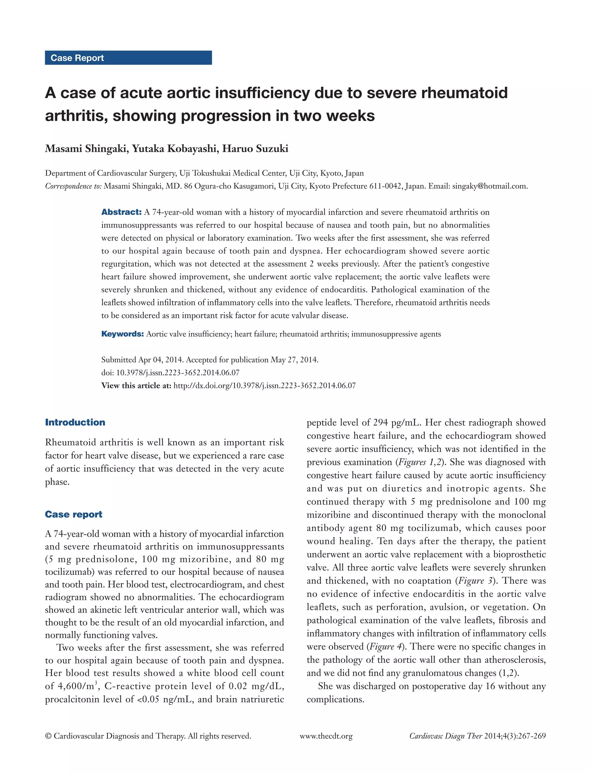

A 74-year-old woman with a history of myocardial infarction and severe rheumatoid arthritis was referred to the hospital twice within two weeks for nausea, tooth pain, and dyspnea. During the first visit, examinations showed no abnormalities. However, during the second visit two weeks later, echocardiography revealed severe aortic insufficiency that was not present previously. She underwent aortic valve replacement, which showed the aortic valve leaflets were severely shrunken and thickened with infiltration of inflammatory cells. This case report describes an rare instance of acute aortic insufficiency progressing within two weeks due to severe rheumatoid arthritis.