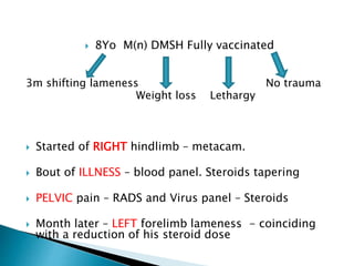
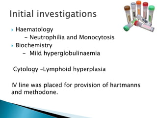
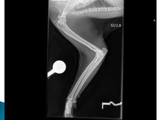
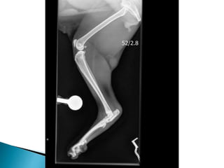
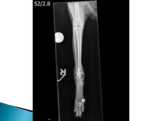
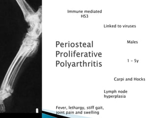
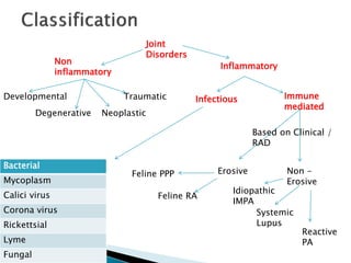
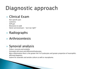
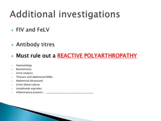
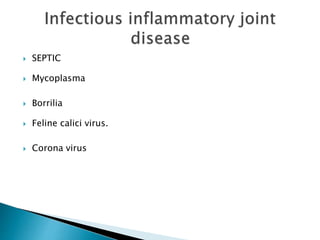
Glynn Woods
 8Yo M(n) DMSH Fully vaccinated
3m shifting lameness No trauma
Weight loss Lethargy
 Started of RIGHT hindlimb – metacam.
 Bout of ILLNESS – blood panel. Steroids tapering
 PELVIC pain – RADS and Virus panel – Steroids
 Month later – LEFT forelimb lameness - coinciding
with a reduction of his steroid dose
 Deteriorated significantly, in comparison to referring vet letter
 37.3 degrees, 186bpm and 36rpm
 Dull, lethargic and unwilling to move
 Painful in several limbs perceivable more so in his L fore
 When he did stand his hind leg gait was very stiff
 Joint effusion
 Popliteal lymph nodes were palpable
 No other significant findings on clinical exam
 Haematology
- Neutrophilia and Monocytosis
 Biochemistry
- Mild hyperglobulinaemia
Cytology –Lymphoid hyperplasia
IV line was placed for provision of hartmanns
and methodone.
.
 Viral Testing - negative
 Joint Culture
 Cytology
 Radiographs ***
 Ultrasound Abdomen
Immune mediated
HS3
Linked to viruses
Males
Carpi and Hocks
1 – 5y
Fever, lethargy, stiff gait,
joint pain and swelling
Lymph node
hyperplasia
Joint
Disorders
Non
inflammatory
Inflammatory
Developmental
Degenerative Neoplastic
Traumatic Infectious Immune
mediated
Erosive Non -
Erosive
Idiopathic
IMPA
Reactive
PA
Systemic
Lupus
Feline PPP
Feline RA
Bacterial
Mycoplasm
Calici virus
Corona virus
Rickettsial
Lyme
Fungal
Based on Clinical /
RAD
 Clinical Exam
- Non specific pain
- Lameness
- Stiff gait
- Reluctance to walk
- Owner percieved pain – “Just nae right!”
 Radiographs
 Arthrocentesis
 Synovial analysis
- Colour, viscosity and turbidity
- Estimated cell count and differential leukocytes
- More inflammation there is the greater the # of Leukocytes and greater proportion of neutrophils.
- Affirmed diagnosis.
- Submit for anaerobic and aerobic culture as well as mycoplasma.
 FIV and FeLV
 Antibody titres
 Must rule out a REACTIVE POLYARTHROPATHY
 Haematology
 Biochemistry
 Urine analysis
 Thoracis and abdomenal RADs
 Abdominal ultrasound
 Urine/blood culture
 Lymphnode aspirates
 Inflammatory proteins - ______________________________________
 SEPTIC
 Mycoplasma
 Borrilia
 Feline calici virus.
 Corona virus
 Highly prevalent in dogs. But considered RARE in cats
 Non erosive IMPA is caused by deposition of immune complexes in the synovial
membrane and the ensuing inflammatory response. Can occur primary (idiopathic),
secondary to stimulation (reactive) or as a feature of SLE.
 Reactive accounts for 25% pyenephritis, pneumonia, toxiplasmosisa dn enteritis, felv,
fip, and CBA.
 Rule out! Is so important.
 Not responding to doxy – then glucocorticoid warranted.
 Can use prednisone prednisolone, ciclosporin, leflunimide, chlorambucil,
methotrexate...
 Uncommon immune mediated disorders can result in erosive damage .Periosteal proliferative PA marked
periarticular . Second is a deforming arthritis resembling rhuematoid arthritis in humans.
 Both deemed Chronic Progressive PA.
 Postulations that it was caused by Feline syncitum virus in genetically predisposed cats. But because that virus
is so far and wide spread its hard to associate in papers.
 FPPPA. Male cats(intact or not), any age, fever lethargy, a stuff gait, joint pain, carpus and hock. Edema of skin
and soft tissues. LN hyperplasia. RAD changes not noticable for first 10-12 weeks. Over time it worsens the
periosteal changes. Synovial biopsy initally reeals a neutrophillic synoviitis and tendonitis but with time and
chronicity plasma cells and lymphocytes increase.
 Feline rheumatoid arthritis insidious, over weeks and months. Middle aged cats and siamese over represetned.
RARE. Fever and systemic disease do not occur so these cats are rarely investgate before severe disease
pathway. RADs – subchondral central and marginal erosiions and periarticular sof ttissue swelling.
 Pred 2mg.kg q 12h PO 3-4 days
 Same for 4 weeks
 Assess clinical response and perform fluid cytology
 Have they resolved?
 Tapering dose, evaluating clinical response and performing synovial cytology before
each reduction
NAW
AYE
 Treatment:
- Doxycyline Synulox Prednisolone Famotidine
 Check-up appointment
- Doing great!
- Blood glucose
OA which is common in cats
 Infectious > immune
mediated in cats.
 Suspect Calici virus
 Don’t forget mycoplasma
 Rule out reactive or infectious
causes.
 Don’t be scared of these
cases!
Grand Rounds Presentation

Grand Rounds Presentation

  • 1.
  • 2.
     8Yo M(n)DMSH Fully vaccinated 3m shifting lameness No trauma Weight loss Lethargy  Started of RIGHT hindlimb – metacam.  Bout of ILLNESS – blood panel. Steroids tapering  PELVIC pain – RADS and Virus panel – Steroids  Month later – LEFT forelimb lameness - coinciding with a reduction of his steroid dose
  • 3.
     Deteriorated significantly,in comparison to referring vet letter  37.3 degrees, 186bpm and 36rpm  Dull, lethargic and unwilling to move  Painful in several limbs perceivable more so in his L fore  When he did stand his hind leg gait was very stiff  Joint effusion  Popliteal lymph nodes were palpable  No other significant findings on clinical exam
  • 4.
     Haematology - Neutrophiliaand Monocytosis  Biochemistry - Mild hyperglobulinaemia Cytology –Lymphoid hyperplasia IV line was placed for provision of hartmanns and methodone. .
  • 5.
     Viral Testing- negative  Joint Culture  Cytology  Radiographs ***  Ultrasound Abdomen
  • 9.
    Immune mediated HS3 Linked toviruses Males Carpi and Hocks 1 – 5y Fever, lethargy, stiff gait, joint pain and swelling Lymph node hyperplasia
  • 10.
    Joint Disorders Non inflammatory Inflammatory Developmental Degenerative Neoplastic Traumatic InfectiousImmune mediated Erosive Non - Erosive Idiopathic IMPA Reactive PA Systemic Lupus Feline PPP Feline RA Bacterial Mycoplasm Calici virus Corona virus Rickettsial Lyme Fungal Based on Clinical / RAD
  • 11.
     Clinical Exam -Non specific pain - Lameness - Stiff gait - Reluctance to walk - Owner percieved pain – “Just nae right!”  Radiographs  Arthrocentesis  Synovial analysis - Colour, viscosity and turbidity - Estimated cell count and differential leukocytes - More inflammation there is the greater the # of Leukocytes and greater proportion of neutrophils. - Affirmed diagnosis. - Submit for anaerobic and aerobic culture as well as mycoplasma.
  • 12.
     FIV andFeLV  Antibody titres  Must rule out a REACTIVE POLYARTHROPATHY  Haematology  Biochemistry  Urine analysis  Thoracis and abdomenal RADs  Abdominal ultrasound  Urine/blood culture  Lymphnode aspirates  Inflammatory proteins - ______________________________________
  • 13.
     SEPTIC  Mycoplasma Borrilia  Feline calici virus.  Corona virus
  • 14.
     Highly prevalentin dogs. But considered RARE in cats  Non erosive IMPA is caused by deposition of immune complexes in the synovial membrane and the ensuing inflammatory response. Can occur primary (idiopathic), secondary to stimulation (reactive) or as a feature of SLE.  Reactive accounts for 25% pyenephritis, pneumonia, toxiplasmosisa dn enteritis, felv, fip, and CBA.  Rule out! Is so important.  Not responding to doxy – then glucocorticoid warranted.  Can use prednisone prednisolone, ciclosporin, leflunimide, chlorambucil, methotrexate...
  • 15.
     Uncommon immunemediated disorders can result in erosive damage .Periosteal proliferative PA marked periarticular . Second is a deforming arthritis resembling rhuematoid arthritis in humans.  Both deemed Chronic Progressive PA.  Postulations that it was caused by Feline syncitum virus in genetically predisposed cats. But because that virus is so far and wide spread its hard to associate in papers.  FPPPA. Male cats(intact or not), any age, fever lethargy, a stuff gait, joint pain, carpus and hock. Edema of skin and soft tissues. LN hyperplasia. RAD changes not noticable for first 10-12 weeks. Over time it worsens the periosteal changes. Synovial biopsy initally reeals a neutrophillic synoviitis and tendonitis but with time and chronicity plasma cells and lymphocytes increase.  Feline rheumatoid arthritis insidious, over weeks and months. Middle aged cats and siamese over represetned. RARE. Fever and systemic disease do not occur so these cats are rarely investgate before severe disease pathway. RADs – subchondral central and marginal erosiions and periarticular sof ttissue swelling.
  • 16.
     Pred 2mg.kgq 12h PO 3-4 days  Same for 4 weeks  Assess clinical response and perform fluid cytology  Have they resolved?  Tapering dose, evaluating clinical response and performing synovial cytology before each reduction NAW AYE
  • 17.
     Treatment: - DoxycylineSynulox Prednisolone Famotidine  Check-up appointment - Doing great! - Blood glucose
  • 18.
    OA which iscommon in cats  Infectious > immune mediated in cats.  Suspect Calici virus  Don’t forget mycoplasma  Rule out reactive or infectious causes.  Don’t be scared of these cases!

Editor's Notes

  • #3 8Yo M(n) DMSH Fully vaccinated 3 month history of shifting lameness Weight loss Lethargy. No illness or trauma. In essence his clinical history was very unspecific. With each bout of apparent lameness Koshka was taken to vets and treatment or investigations to some degree were performed. His initially pain relief was prescribed and he was seen to improve (MAY 2014). One month later he presented to the practice lethargic with a reduced appetite and so a SENOIR CAT PROFILE WAS RAN – showing no abnormalities. He was prescribed steroids at this stage and seemed to improve. However when weaned off signs quickly returned. Owner felt Koshka was very sensative over pelvis. Radiographs were performed but no obvious changes were noted. At this point the vets clearly were worried of some underlying disease process and standard virus panel was performed with results being –ive for FeLV , FIV and FCV. Another dose of steroids were prescribed, Convenia for leukocytes in urine 1 month later – Left forelimb lameness returned and this coincided with a reduction in his steroid dose to 0.25mg/Kg. At this point it made me think was this just coincidence of was there progression in the disease to the point that where steroids once were effective, they were now failing to reduce inflammation. This is the point at which cat was referred.
  • #4 When he was presented to us, he had deteriorated significantly which makes sense from the history because he had been off his steroids. He was dull, lethargic and unwilling to move. T was at low end of normal – 37.1 degrees P was strong, with no deficits and sat at 186bpm R was 32. Lameness examination was difficult as he was painful in several limbs. I seemed to receive a bigger response most obviously in his left fore limb as well as his hindlimb gait being stiff. On palpation (and even visually) there was significant effusion found in both hocks and both elbows along with mild swelling in his stifles. All of his limbs were extremely painful and he wasn’t too keen on being handled. His popliteal lymph nodes were palpable and around 1.5cm in diameter. Otherwise his clinical exam appeared normal. So I think it’s important to take a step back from the patient at this point and thinking generally to get things straight. We have a middle age cat that is sore on what conceivably from the history is all legs, but shifting. It has responded to steroid therapy with no other relevant signs of systemic disease. It’s pretty safe to say we have some form of inflammatory joint disease but as to the cause? Who knows?
  • #5  Cytology from FNA's of popliteal lymph node revealed small and intermediate sized lymphoid cells, with small numbers of lymphoblasts. The cytology was consistent with reactive lymphoid hyperplasia.
  • #6 FHV, FCV negative on swabs. FeLV, FIV negative FCo antibody negative Calicivirus negative on synovial sample also. Joint culture was sent away for anarobic and aerobic culture Mycoplasma culture was also requested alongwith specific PCR for individual strains.
  • #10 He had smooth recovery from anaesthesia and seemed much more comfortable on his methadone. Cytology and Culture both were negative. Hocks and elbows to have increased WBC, predominately neutrophilic inflammation (91- 94%). Mild toxic change to the neutrophils. The right and left stifle: Low Neutrophils (< 1 x10e9/l). The majority of cells present were large mononuclear cells (85 - 87%). Immune mediated hypersensitivity type III reaction. Run through the types… I = hay fever idea – IgE released. II = IMHA like whereby antibody binds to cell surface antigen III = Antibody binds to cell suface antogen however these are solvent in blood and not attached to cells and so can settle out in various locations throughout the body. Periosteal proliferative polyarthritis occurs most often in male cats (intact and castrated), but female cats can also be affected. Cats of any age can develop this polyarthritis, but it is most common in young adults. Affected cats experience fever, lethargy, a stiff gait, and joint pain and swelling, particularly of the carpus and hock. There is sometimes concurrent edema of the skin and soft tissues overlying the joints. Lymph node hyperplasia may cause severe regional lymphadenopathy. Synovial fluid analysis reveals a moderate-to-severe neutrophilic pleocytosis and culture is negative. Papers form the 1980’s actually found that 100% of cats with chronic progressive arthritis showed FSFV or antibodies to it. But is it just incidental because this virus in particular is found in normal cats also. It has been suggested that infection with FeLV or FIV-induced immunosuppression allows FeSFV to multiply in the joints of predisposed individuals. However in papers that I came across, surprisingly there isn't an increased incidence of FeLV or FIV in affected cats. It occurs more commonly in young, entire or castrated, male cats less than 5 years of age. Radiographic changes may not be evident for the first 10–12 weeks of illness and then early findings include periarticular soft tissue swelling and mild periosteal proliferation. This matache Koshka to a T being that after roughly two months of steroid effectiveness. An abnormally coarse trabecular pattern of the bones of affected joints. Over time, the periosteal proliferation worsens and extends beyond the confines of the joint, which may lead to ankylosis. Periarticular osteophytes, subchondral cyst-like lesions and periarticular erosions are evident. Calcification and erosion of bone may occur at the attachment of ligaments and tendons, causing painful lesions (enthesopathies).
  • #11 Non inflammatory are most common in cats and dogs overall OA was once thought to be rare in cats. However we now know that it is actually quite common in cats. Truth is 50 – 90% of cats have been shown to have age related primary cartilage degeneration or OA secondary to joint deformities. The difficulty arises in that majority of cats are minimally symptomatic. Lameness is rare with OA, but alterations in behaviour and lifestyle can be the biggest give away. Examples that owners often note include; Cat not wanting to move. Not attempting stairs. Cautious when moving off and on sofa. Not able to jump through the kitchen window. Inflammatory – infectious or immune mediated. These can either infect one limb – a monoarthritis – or multiple - polyarthritis. Infectious causes are a much more common cause of Polyarthritis in cats than immune mediated. Can be by direct infection of synovium but occasionally systemic infections/inflammation elsewhere in body give rise to circulating complexes that are deposited in joints resulting in a sterile PA known as Reactive Polyarthropathy. This contrasts heavily with the dog whereby it’s deemed common in ______________ Clinical manifestations are more acute with inflammatory disease than OA. Screamingly obvious if patient is monoarthritic but the appearance is often hard to notice in poly arthritis as the patient adopts a gait that is referred to as ‘walking on eggshells’
  • #12 Clin exam – non spec pain, lameness a stiff gait reluctance to walk or owner perceived pain should be evaluated with a careful physical and ortho exam. OA can lead to such few gross changes that joints feel normal on palpation. Inflammatory arthritis on the other hand lends itself to being painful to the touch. Although inflammatory joint disease exists it’s critical to be aware that inflammatory joint disease is much less common than Cat Bite Abscess, pancreatitis, viral infection – keeping Poly Arthritis at the back of our mind. It will be missed unless clinicians maintain a high index of suspicion. RADS important to determine erosive or non erosive. We need to be aware of incidental OA changes. Osteoarthritis causes osteophytes production, periarticular and meniscal mineralization. There is NO synovial effusion, punched out, subchondral, periarticular lesions or joint subluxation which is associated with immune mediated change. Arthrocentesis. Synovial fluid collection and analysis is required to confirm a diagnosis of inflammatory arthritis. GA recommended, slkin preperared. 25Guage 5/8 needle. Only 1 – 3 drops needed and standard FNA mode. Reluctance by vets probably results in significant under diagnosis of inflammatory joint disease in cats. 5 joints at least! Even in cats with apparent monoarthritis since one joint can be more inflamed than others. Radial carpal joint Elbo joint in partial flexion and just infront of the olecranon caudal to the epicondyle of humerus. Shoulder – cat in lateral recumbency with limbs parallel to table then a tiny flexion + enter just cranial to glenohumeral ligament. Hock just above tibial tarsal bone. Lateral approach to hock or caudal are more tricky as have to watch for branching of the saphenous. Bacterial arthritis is more likely to infect bigger joints like stifle and elbow in dogs but in cats it tends to be in the distal limbs. CAN ANYONE THINK WHY? Synovial analysis Normal is very viscous colourless and clear. Reduced viscosity suggests inflammation. <3000 cells per micro litre, 95% monocuclear cells, neutrophils roughly 10%. This applies still to OA. In circumstances of greater inflammation greater number of cells and increased proportion of neutrophils. Submitted for anaroebic and aerobic and swabs submitted for particular mycoplasma. .
  • #13 FIV and FeLV since immuno-suppression may be an important predisposition for infectious arthritis and FeLV has been implicated in the development of Pereosteal Proliferative PA. Antibody titres to help rule out any rickettsial pathogen, borrelia or toxoplasmosis How do we rule out a reactive polyarthropathy (25%)– basically by performing a complete patient screen. Reactive PA is a non erosive form – think back to our branching. Lymph node aspirates aid identification of infection or underlying neoplasia
  • #14 Cat bite abscess, pasturella, one joint due to paw swinging. Haematogenous spread is rare but seen with omphlabitis in kittens. Septic arthritis patients are normally febrile, lethargic and painful. Dx cytology of synovium, cultured from synovium, blood or urine from a cat with appropriate clinical signs. Increase neutrophils, bacterial evident with toxic change to neutrophils. Antibiotics given ASAP. Broad spec Amoxi clav. Quinolones enrofloxacin are good against pastuella. Retinal toxicity is a real worry with use of enrofloxacins in cats and so it’s appropriate to reach for pradaflox, especially in cases of coliform agent. If no response in 2-3 weeks then surgery for debridement and flush. Mycoplasma are normal flora of conjunctiva membranes, pharynx, resp, urogenital and generally considered non pathogenic. Gatea and felis been associated with erosive and non erosive PA They are deemed opportunistic and therefore primarily arise in immunocomprimised patients. It’s therefore critical to test for FIV and FeLV and lymphoma. Not only for treatment approach but as it will give a more reliable prognostic indicator. Reports of transient infections in healthy cats but full blown PA in compromised cats. Lymes disease in cats isn’t as well reported. Cats sero convert but show no clinical signs suggesting some form of resistance in the progression of infection. Can use the dog point of care assays against the C6 antibody. PCr is difficult because organisms low in numbers. Suspect treat with Abs with doxy good tissue distribution and will also address anaplasma and ehrlichia or bartonella. Feline calici virus. We all know the common symptoms; sneezing, fever, nasal discharge and nasal ulceration that we attribute to cat flu. But following discussion with Laura and further reading it’s very well documented that some cats actually acutely develop synovitis and lameness either separately or together with the typical signs of FCV. Can happen following vaccination - 80% of 123 vaccine reactions were associated with lameness and fever! PA is likely in kittens 5-7 days after vaccination . Lameness and fever normally resolve. No reports of it causing Polyarthritis in adults. Buprenorhine can be used to treat it in the interim. FIP – Dry or wet form caused by increased number of circulating antibody complexes.
  • #15 All cats should be vigorously investigated for direct infectious causes, systemic infections or neoplasia that could be responsible for a reactive PA. Before arriving at a primary IMPA. It is good practice to treat with doxy for 14 days and gauge response before making diagnosis of primary immune mediated disease. Most papers recommend the use of prednisolone at immunosuppressive doses. Prednisolone is preferred in comparison to prednisone because higher concentration of the active moiety is reached in circulation. In most cases reactive PA aren’t diagnosed because vets tend to believe the primary disease is actually causing the fever and unwillingness to walk. Drug induced Reactive – TMS, Phenobarbital, erythropoetin, penicillin, cephalexin and vaccines. Treat underlying cause. Even in cases whereby Primary IMPA is diagnosed it often turns out not to be the case – In one study I read with 6 cats; 4 responded to tapering preds, two didn’t and were euthanised. One of the PM’d cats had renal corynbacteria cultured and subclinical pyenephritis. 2/4 of the responders were FeLV + so a reactive poly arthropathy was more likely.
  • #17 2mg/kg 4 weeks > 1 mg/Kg 4 weeks > 0.5 mg/Kg 4 weeks
  • #18 The mainstay of treatment for progressive feline polyarthritis is immunosuppressive therapy (prednisolone+/- cyclophosphamide, chlormabucil, azathioprine or cyclosporine). The long term prognosis is variable, with 50% improving on glucocorticoids and analgesics alone. I have advised a recheck in two weeks time to ascertain response. If you have any further questions, please do not hesitate to contact me at any time. Thank you for referring this very interesting case to the Internal Medicine Service. He is a beautiful boy and has been a pleasure to treat.
  • #19 With exception of OA which is common in cats other causes of feline joint disease are considered rare – very rare. PA is more likely to be infectious than immune mediated in cats. Suspect Calici virus Synovial fluid culture for mycoplasma should always be requested in a cat. Doxycycline is there to be used but don’t over do it. Very important if you are going to start patient on immunosuppressive dose of steroids. Immune mediated PA causing periosteal proliferation is recognised in cats