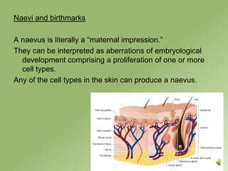
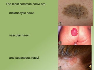
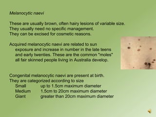
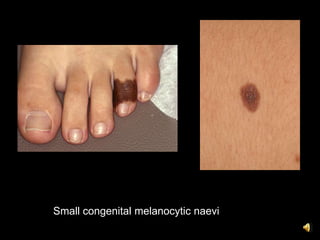
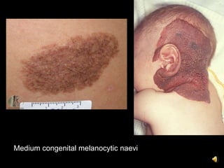
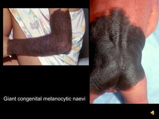
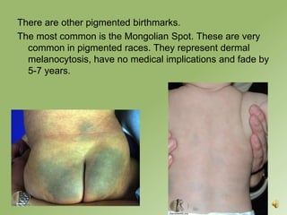
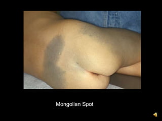
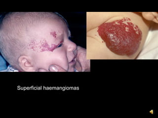
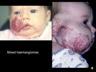
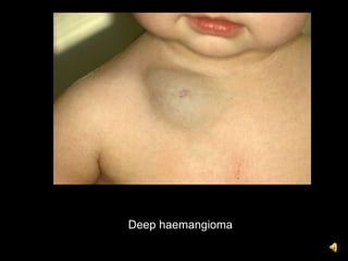
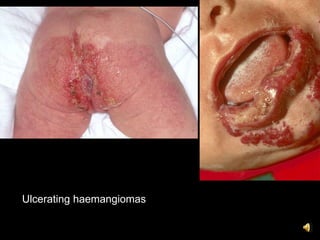
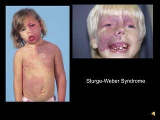
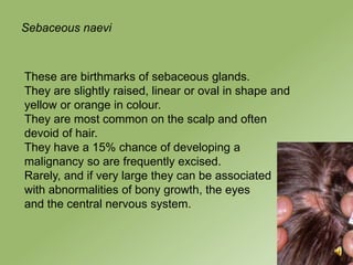
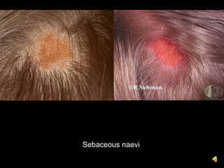
Birthmarks and Naevi

Dr Daniel Hewitt
Dermatologist
Skin and Cancer Foundation Westmead
Objectives

To understand that there are many different types of
  birthmarks and naevi.
To appreciate the main clinical characteristics of the most
  common types.
Paediatric dermatology

Many skin conditions affect children.
Atopic dermatitis and viral exanthems have been
  discussed. Most inflammatory dermatoses can affect
  both adults and children.
Here we will only discuss the more common naevi and
  birthmarks.
Naevi and birthmarks

A naevus is literally a “maternal impression.”
They can be interpreted as aberrations of embryological
  development comprising a proliferation of one or more
  cell types.
Any of the cell types in the skin can produce a naevus.
The most common naevi are

  melanocytic naevi


  vascular naevi


  and sebaceous naevi
Melanocytic naevi

These are usually brown, often hairy lesions of variable size.
They usually need no specific management.
They can be excised for cosmetic reasons.

Acquired melanocytic naevi are related to sun
  exposure and increase in number in the late teens
  and early twenties. These are the common "moles"
  all fair skinned people living in Australia develop.


Congenital melanocytic naevi are present at birth.
They are categorized according to size
  Small        up to 1.5cm maximum diameter
  Medium       1.5cm to 20cm maximum diameter
  Giant        greater than 20cm maximum diameter
Small congenital melanocytic naevi
Medium congenital melanocytic naevi
Giant congenital melanocytic naevi
There are other pigmented birthmarks.
The most common is the Mongolian Spot. These are very
  common in pigmented races. They represent dermal
  melanocytosis, have no medical implications and fade by
  5-7 years.
Mongolian Spot
Vascular birthmarks

There are two main types – infantile haemangiomas and
    vascular malformations.
It is very important to understand the difference in these as
    the natural histories and managements are completely
    different.
Infantile haemangiomas

These are benign tumours of vascular endothelium,
   characterized by a proliferative and an involutional
   phase.
Most often they can be observed with no specific
   management as they involute.
Infantile haemangiomas can be superficial (“strawberry
   birthmark,”) mixed or deep (“cavernous.”)
Superficial haemangiomas
Mixed haemangiomas
Deep haemangioma
Generally haemangiomas proliferate over 6-10 months and
  involute over 5-7 years.
However, complications of haemangiomas include
  Ulceration
  Ocular compromise
  Airway compromise
  High output cardiac failure
  Disseminated intravascular coagulation
  Systemic involvement
  Scarring

Such occurences may necessitate treatment with vascular
  laser, topical steroids, systemic steroids or systemic
  propranolol
Ulcerating haemangiomas
Vascular malformations

These are a developmental anomaly, predominantly
  capillary in origin, but sometimes derived from venous,
  arterial or lymphatic vessels. They are present at birth
  and persist throughout life.

Vascular laser is often used to treat these early as they
  generally get thicker and more prominent over time.

They are commonly known as “port wine stains.”
Capillary vascular malformations
and the thickening that may
develop in adult life
Capillary vascular malformations can be associated with
  other abnormalities.
The most well known syndrome is
Sturge-Weber Syndrome. This is a facial vascular
  malformation involving at least a portion of V1 in
  association with leptomeningeal angiomatosis and often
  glaucoma.
Sturge-Weber Syndrome
Sebaceous naevi


These are birthmarks of sebaceous glands.
They are slightly raised, linear or oval in shape and
yellow or orange in colour.
They are most common on the scalp and often
devoid of hair.
They have a 15% chance of developing a
malignancy so are frequently excised.
Rarely, and if very large they can be associated
with abnormalities of bony growth, the eyes
and the central nervous system.
Sebaceous naevi
Conclusion


There are many different types of birthmarks and naevi.
This is a very brief introduction to the most common.

These lesions are usually harmless, but occasionally
there can be serious complications which require specific
management.

9. birthmarks and naevi

  • 1.
    Birthmarks and Naevi DrDaniel Hewitt Dermatologist Skin and Cancer Foundation Westmead
  • 2.
    Objectives To understand thatthere are many different types of birthmarks and naevi. To appreciate the main clinical characteristics of the most common types.
  • 3.
    Paediatric dermatology Many skinconditions affect children. Atopic dermatitis and viral exanthems have been discussed. Most inflammatory dermatoses can affect both adults and children. Here we will only discuss the more common naevi and birthmarks.
  • 4.
    Naevi and birthmarks Anaevus is literally a “maternal impression.” They can be interpreted as aberrations of embryological development comprising a proliferation of one or more cell types. Any of the cell types in the skin can produce a naevus.
  • 5.
    The most commonnaevi are melanocytic naevi vascular naevi and sebaceous naevi
  • 6.
    Melanocytic naevi These areusually brown, often hairy lesions of variable size. They usually need no specific management. They can be excised for cosmetic reasons. Acquired melanocytic naevi are related to sun exposure and increase in number in the late teens and early twenties. These are the common "moles" all fair skinned people living in Australia develop. Congenital melanocytic naevi are present at birth. They are categorized according to size Small up to 1.5cm maximum diameter Medium 1.5cm to 20cm maximum diameter Giant greater than 20cm maximum diameter
  • 7.
  • 8.
  • 9.
  • 10.
    There are otherpigmented birthmarks. The most common is the Mongolian Spot. These are very common in pigmented races. They represent dermal melanocytosis, have no medical implications and fade by 5-7 years.
  • 11.
  • 12.
    Vascular birthmarks There aretwo main types – infantile haemangiomas and vascular malformations. It is very important to understand the difference in these as the natural histories and managements are completely different.
  • 13.
    Infantile haemangiomas These arebenign tumours of vascular endothelium, characterized by a proliferative and an involutional phase. Most often they can be observed with no specific management as they involute. Infantile haemangiomas can be superficial (“strawberry birthmark,”) mixed or deep (“cavernous.”)
  • 14.
  • 15.
  • 16.
  • 17.
    Generally haemangiomas proliferateover 6-10 months and involute over 5-7 years. However, complications of haemangiomas include Ulceration Ocular compromise Airway compromise High output cardiac failure Disseminated intravascular coagulation Systemic involvement Scarring Such occurences may necessitate treatment with vascular laser, topical steroids, systemic steroids or systemic propranolol
  • 18.
  • 19.
    Vascular malformations These area developmental anomaly, predominantly capillary in origin, but sometimes derived from venous, arterial or lymphatic vessels. They are present at birth and persist throughout life. Vascular laser is often used to treat these early as they generally get thicker and more prominent over time. They are commonly known as “port wine stains.”
  • 20.
    Capillary vascular malformations andthe thickening that may develop in adult life
  • 21.
    Capillary vascular malformationscan be associated with other abnormalities. The most well known syndrome is Sturge-Weber Syndrome. This is a facial vascular malformation involving at least a portion of V1 in association with leptomeningeal angiomatosis and often glaucoma.
  • 22.
  • 23.
    Sebaceous naevi These arebirthmarks of sebaceous glands. They are slightly raised, linear or oval in shape and yellow or orange in colour. They are most common on the scalp and often devoid of hair. They have a 15% chance of developing a malignancy so are frequently excised. Rarely, and if very large they can be associated with abnormalities of bony growth, the eyes and the central nervous system.
  • 24.
  • 25.
    Conclusion There are manydifferent types of birthmarks and naevi. This is a very brief introduction to the most common. These lesions are usually harmless, but occasionally there can be serious complications which require specific management.