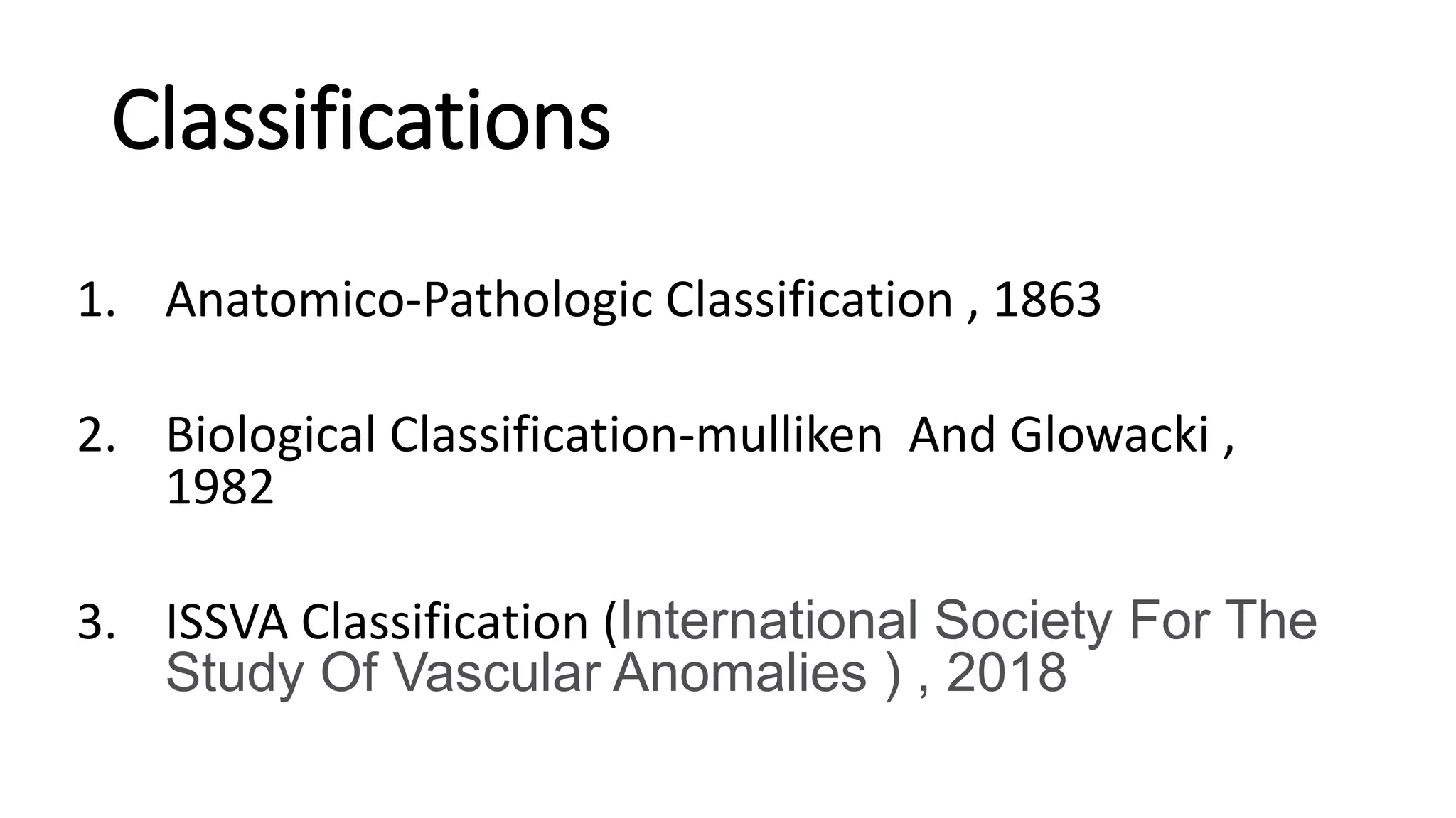
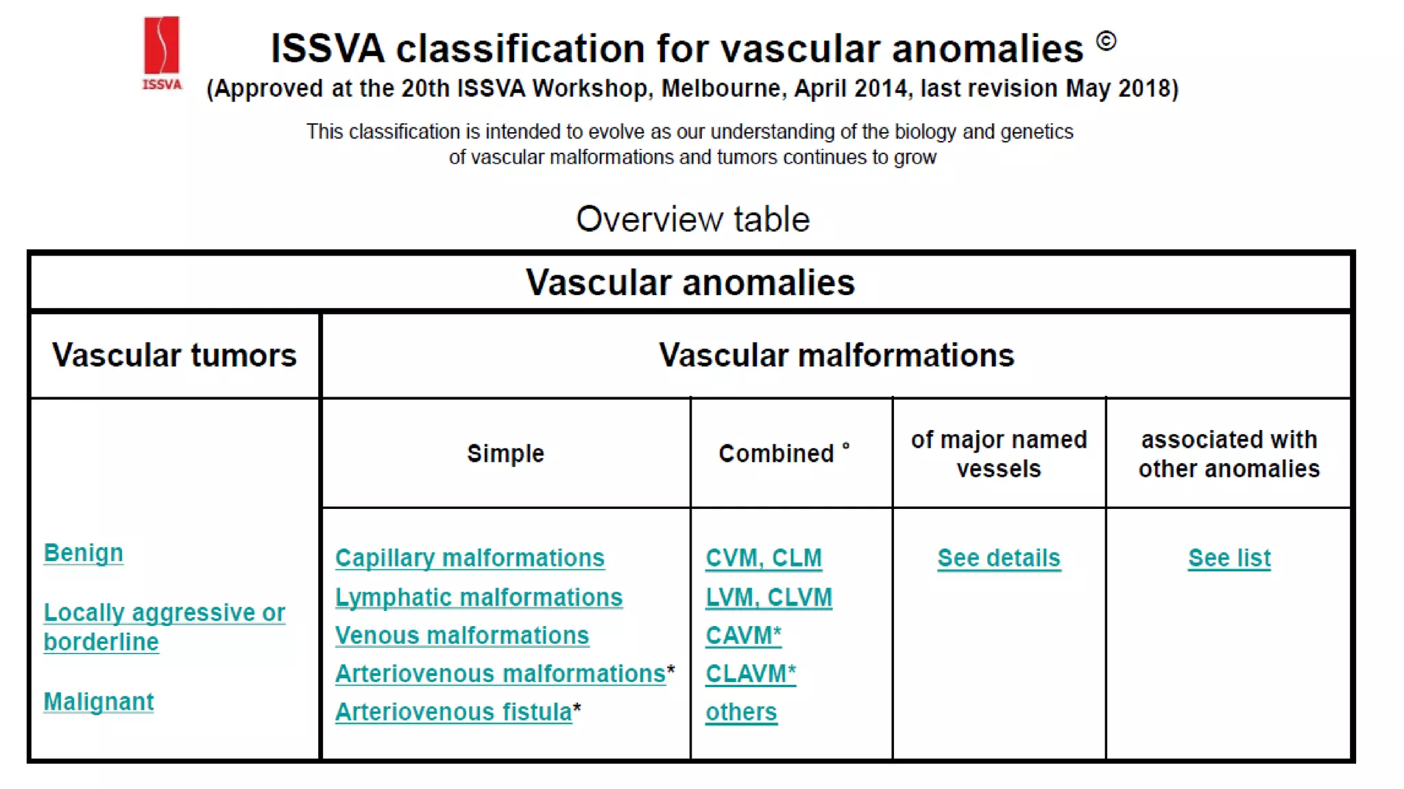
Vascular Malformation
BY
DR. KULDEEP PRAJAPATI R3 S/D
DR. JAY CHAUDHARI R3 S/D
 References
Rutherford's Vascular
Surgery And Endovascular
Therapy
S.K.Bhattacharya
Love And Bailey
Sabiston
Definitions
 The Term Vascular Malformation Is Used To Describe Malformed
Vessels Resulting From Arrested Development During Various Stages
Of Embryogenesis.
 It Is Present At Birth, Although Not Always Immediately Identifiable.
 Vms May Involve One Or All Three Circulatory Systems -Arterial,
Venous, And Lymphatic.
 Either As A Predominant Form (E.G., Venous Malformation) Or
As A Mixed Condition (E.G., Hemolymphatic Malformation).
 They Continue To Grow Regardless Of Type.
Classifications
1. Anatomico-Pathologic Classification , 1863
2. Biological Classification-mulliken And Glowacki ,
1982
3. ISSVA Classification (International Society For The
Study Of Vascular Anomalies ) , 2018
Anatomicopathological Classification
A) Angioma
B) Lymphangioma
1. Angioma Simplex: System Composed Of Capillaries.
2. Angioma Cavernosum: A Replacement Of Normal Vasculature With Large
Channels.
3. Angioma Racemosum: Tissue Consisted Of Markedly Dilated Interconnected
Vessels.
Biologic Classification
by Mulliken and Glowacki
 On Basis Of Cell Kinetics , There Are two Major Types Of
Vascular Anomalies.
1. Hemangioma : With Endothelial Hyperplasia.
2. Vascular Malformation : With Normal Endothelial Turnover.
Vascular Malformations
• Capillary (Cm)
• Lymphatic (Lm)
• Venous (VM)
• Arterial (Am)
 Combined
• Arteriovenous (Avm)
• Capillary-lymphatic (CLM)
• Capillary Venous (CVM)
• Lymphatico-venous (LVM)
• Capillary-lymphatico-venous
Low flow
High flow
 Most VM Are Sporadic, Autosomal Dominant .
 The Endothelial-specific Angiopoietin Receptor Tie2/Tek, Located On
9p21, Was Identified To Cause Familial Mucocutaneous VMs.
Embryology
 When the embryo begins its exponential growth, an equivalent
growth of the vascular system is required to deliver nutrition and
oxygen necessary for the proper development of the fetus.
 Undifferentiated capillary plexus becomes the initial
vasculature; subsequently, some capillaries undergo regression
and coalescence with others to form a reticular structure.
 This reticular structure is efficient to meet the embryo's needs during
the early, rapid-growth stage of embryogenesis.
 The clinical behavior of the vascular defect depends on the stage at
which arrested development occurs.
 Blood Vessel Develop
• 1st Stage : Endothelial Stage (Endothelium Lined Lakes Form)
• 2nd Stage : Retiform Stage (Capillary Communication Develops Between
These Lakes)
• Final Stage : Maturation Stage (Channel With Muscular Linings Are Artery
And Without It Are Veins.)
Abnormal Development At Any Of These Stage Result In Vascular
Malformation
Etiology
 A Number Of Genetic And Chromosomal Abnormalities Have Been
Implicated In The Development Of Cvms.
 These Have Been Associated With
(1) Exposure To Damaging Chemical Compounds During The First Trimester Of
Pregnancy;
(2) Infections Such As Rubella, Cytomegalic Inclusion Disease, Herpesvirus,
And Toxoplasmosis;
(3) Thalidomide And Other Drugs Such As Aminopterin, Cyclophosphamide,
Quinine, Anticonvulsant Drugs, Cortisone, And Corticotropin;
(4) Alcohol, Tobacco, And Cocaine Abuse.
(5) Maternal Diseases And Exposures Including Goiter, Diabetes Mellitus,
Thyroid Disease, Tuberculosis, Hypoxia, Carbon Monoxide And Lead Poisoning
Have Also Been Identified As Potential Causes Of Cvms.
Haemangioma
Haemangioma
• Hemangioma Is The Most Common
Tumor Of Infancy, Appears In
Neonates, Usually Within The First 2
Weeks.
• The word comes from Greek heame
“blood” , angio “vessel” , -oma “tumour”.
• Its developmental malformation of blood vessels rather than true
tumour.
• Its benign , self involuting tumour of endothelial cells lining of blood
vessel.
• Exhibit rapid growth upto 6-8 months follow by regression in 5-9
years.
Progression
• The natural history of
Hemangioma is characterized
by three phases:
1. Proliferating Phase
2. Involuting Phase
3. Involuted Phase
 Proliferating Phase
• Hemangioma grows rapidly during the first 6 to 8 months of
infancy, the skin becomes raised, bosselated, and a vivid crimson
color.
• Palpation at this stage reveals a tense and noncompressible mass.
 Involuting Phase
• Hemangioma reaches its peak before the first year; and for a time
thereafter, growth is proportionate to that of the child.
• The first signs of the involuting phase appear as the crimson color
fades to a dull purplish color, the skin gradually pales, and the tumor
feels less tense.
• The involuting phase continues until the child is 5 to 10 years of age.
Involuted Phase
• Regression is complete in 50% of children by age 5 years and in 70% by
age 7 years, with continued improvement until age 10 to 12 years.
• The hemangioma may persist in the form of residual tumor, loose skin,
telangiectasias, or scarring.
• Hemangiomas can be superficial, deep, or visceral in location.
Superficial lesions
• often exhibit the classic crimson color of the so-called strawberry
hemangioma.
Deep lesions
• Are those within the deep dermis or subcutaneous tissues often present
as pale blue or purple masses that may be confused with venous
malformations.
 Visceral lesions
• Are not apparent on physical examination, and in 50% of
cases, there are no accompanying cutaneous hemangiomas.
• In this setting, their presence may only be suggested by
physiologic findings such as hepatomegaly, congestive heart
failure, or stridor.
Gross appearance CT Angio of Hemangioma
Capillary Hemangioma
 Types
I. Salmon pink patch
II. Port wine stain
III. Strawberry hemangioma
IV. Spider naevus
Salmon pink patch
Pink patch over forehead in midline , over occiput.
Reselbles “stork bite”
Present Since birth ,
disappear by 1 year of age.
Strawberry Angioma
 Composed of immature vasoformative tissue,
 Involve skin and sub. Cut. Tissue most.
 Can occur any part of body .
 Most common head and neck .
 Submucous variety more prone to haemorrhage.
 Growth :
Normal at birth > red mark between 1-3 week >increase upto 1 year of life ,
form strawberry like mass protruding from skin > cease growth upto 7-8
years > then regress .
• Surface : irregular , pitted epithlium , smooth , small area of ulceration
covered with scab .
• Soft ,compressible
• Sign of emptying present
• Not pulsatile .
• Treatment :
1. wait upto 7-8 years
2. Excision with or without skin graft.
3. Conservative : apply co2 snow
: injection of sclerosant , hypertonic saline
: Laser therapy using Nd:YAG laser
Spider naevus
• Acquired condition
• Seen in chronic liver disease , pregnancy , menarche .
• Central red spot with radiating , linear , fine blood vessels like legs of
spider .
• Sign of emptying present.
• Treament : pulsed light or pulsed dye laser.
Spider naevus
Campbell de morgan spots
In elderly , over sunexposed area
AVF at dermal capillary level.
Venous hemangioma
 Congenital Multiplicity Of Venous Channel Having Varying Caliber.
 Present Since Birth
 No Tendency To Involute
 Gradually Increase In Size
 Sites : Skin Commonly Face , Ear , Lip
: Sub Cu. Tissue
: Mucous Membrane of Tongue , Mouth , Lips
: Internal Organ : Liver , Kidney , Brain Etc
Venous hemangioma
 Clinical feature
Swelling raised from surface
Bluish discoloration
Soft , fluctuant swelling
Emptying sing present ( except in thrombosis )
Not pulsatile
Phlebolith can be palpated
Arterial hemangioma
• This is type of congenital arterio-venous fistula .
• Arterialisation of veins (veins become dilated , tortuous , thick walled )
• Example : cirsoid aneurysm in scalp
Diagnosis :
Diffuse swelling : soft or cystic
Pulsatile
Compressible
Feels like bag of worms
When in relation to bone it may show bone erosion , osteoporosis
Arterial hemangioma
 Complications of haemangioma
 Atrophy of overlying skin
 Ulceration
 Haemorrhage
 Calcification
 Thrombosis
 Infection
 Pressure effect : bone erosion
 Malignant change : hemangioendothelioma ,
hemangiosarcoma
(I) Destortion
• Periorbital Haemangioma
• Blocks Visual Axis
• Ocular Proptosis
( II ) Destruction
( III ) Obstruction
• In Subglottic Haemangioma
• Hoarseness Of Voice, Stridor
• Manifests Before 12 Weeks Of Age
• Tracheostomy May Be Necessary
(Iv) Hemodynamics
• Cardiac Failure
• Respiratory Failure
• Renal Failure
• Neurologic Consequences
• Gastrointestinal Bleed
 DIAGNOSTIC IMAGING
• Hemangiomas are readily distinguished from other tumors by:
Ultrasonography (USG)
Computerized tomography (CT)
Magnetic resonance imaging (MRI)
Arteriography
Treatment
 Conservative
1. Wait and watch : eg. Salmon patch , strawberry hemangioma
2. Disguise blemish by cosmetic : port wine stain
3. Sclerotherapy : injection of hot water , hypertonic saline etc
4. Steroid therapy : repeated inj. Of hydrocortisone into the lesion
5. Cryotherapy : application of carbon dioxide snow.
(A) Intralesional steroids
• for small ulcerated lesions
• Triamcinolone = 3 to 5 mg / kg injection
• 3 to 5 injections at 6 week intervals
(B) Systemic steroids
• Prednisolone 2 to 3 mg / kg per day for two to three weeks
• Should see response in 7 to 10 days
• if good response 1 or 0.75 milligram per kilogram per day for 4 to 6 weeks
(C) Interferon Alpha Therapy
• For Endangering Lesion
• Failure Of Response To Steroids
• Complications Of Steroids
• Contraindication For Steroids
• Recombinant Interferon Alpha 2a Or 2b
• 2 To 3 Millions Unit Per Metre Square
• 6 To 12 Months Of Therapy
Complications
 febrile reaction
 Neutropenia
 Anemia
Other modalities
1) Embolic Therapy
• For Hepatic Haemangioma
2) Laser Therapy
• Flashlamp Pulsed Dye Laser
• 577-585 Nanometer
• Penetration 0.75 -1.2 Mm
• Best For Telangiectasia
3) Continuous Wave Co2 Laser
•4) Intra Lesional Nd-YAG Laser Or Use Diode
Laser
Surgery
Indication
1) Not Responding To Pharmacotherapy
2) Ulceration
3) For Obstructing Effect
Surgery
If lesionis localised
• Excision followed by skin grafting
• Some excision preceded by sclerosant to reduce size of lesion
If lesionis extensive
• Ligation of feeding blood vessel
• Theraupetic embolization of feeding artery
Radiotherapy
• Hemagioma is radio sensitive
• Radium mould may help
• S/E : skin necrosis , pigmentation , ulceration , haemorrhage
Vascular malformation
Vascular malformations
single vessel forms
• capillary
• arterial
• venous
• lymphatic
Combined
It can be
Slow flow
Fastflow (any malformation with an Arterial component)
Capillary malformation
Clinical features
• Usually evident at birth
• Previously called port Wine stain
• Dermatomal distribution
• Hypertrophy of underlying
soft tissues and bone.
• It can present as Sturge-Weber syndrome
(Facial Capillary Malformation + ipsilateral ocular +
leptomeningeal vascular anomalies)
Radiologic Investigations
-MRI
-Angiography –parenchymal blush
Management
Cosmetics
Lasers -tunable flashlamp pulsed dye laser (585nanometer )
- Useful only for face
-Less effect in Asian skin
-More complications in dark skin
Surgery
• Excision
• excision and skin grafting
Pulsed dye laser
Lymphatic malformations
• Macrocystic Lymphatic malformations
(outdated terms include -Lymphangioma cavernosum,
-Cystic hygroma,
-Lymphangioma cysticum)
• Usually presents as multiple cysts of >2 cm and are
commonly found in the posterior triangle of the neck,
and in the cervical area just below the angle of the
mandible.
Clinical features
• Affects
cervicofacial region,axilla,mediastinum
• Localized ,painless, non-pulsatile
swelling with no bruit or thrill,
• Having a rubbery compressible
consistency, and
• covered by normal appearing skin
unless hemorrhage or
communication with venous
malformations produce a blue
discolouration.
• Skeletal hypertrophy and distortion in 80% of cervicofacial Lymphatic
malformations occur by 10 years of age.
• Lymphatic malformations never regresses.
Radiologic features
• CT scan-low density cystic lesions
• MRI is the best imaging tool for conclusively
diagnosing lymphatic malformations,
enabling precise anatomic extension of the
lesion and cervical spaces involved
Management
Antibiotics
- complaints of injury followed by pain over swelling.It s/o bleeding into
the cysts
- In those situations antibiotics are given to prevent secondary
infection.
Sclerotherapy-
-intralesional Sodium dodecyl sulfate(SDS) ,ethanol,bleomycin
-intralesional OK 432
(Lyophilised mixture of attenuated streptococcus pyogenes)
• Sclerotherapy is the mainstay of treatment if it is smaller(<1 cm)
• If it is larger (>1 cm)
Lasers –Argon,Nd-Yag for tongue lesions
Surgery
• Tracheostomy –when tumor is pressing larynx
• Excision
Venous malformations
• Most common vascular anomaly
• Not obvious at birth
Pathogenesis
• Thin walled dilated channels
• Smooth muscle decreased
• intralesional clotting causes phleboliths .
It can associated with certain syndromes
• Turner syndrome –intestines and feet
• familial multiple glomangioma
• familial cutaneous cavernous
• Blue rubber bleb naevus syndrome
Clinical features
• Bluish
• Soft ,Compressible
• Solitary or multiple
• Usually grow with the age
• Size increases on dependency
• Localised Pain and phleboliths
• areas involved
-head and neck
-extremities
-Bowel
Radiological features
• MRI
-intravascular contrast materials
-Phleboliths-signal voids
-MR venography for assessment of deep veins
Management
Compressive stockings ± Aspirin
Anticoagulants (LMWH)
Sclerotherapy
i)Small lesions:1% SDS
ii)Large lesions-absolute ethanol
with/without lipiodol or
zein(corn protein)
Surgery-
• For limbs-
Excision/ Excision and skin graft
• Excision and skin grafting
 Combination(sclerotherapy +surgery)
Arteriovenous malformations
• AVMs are High-flow vascular anomalies composed of a complex
primitive network of arterial and venous channels (a nidus)—a
network that bypasses the normal capillary bed.
• Arterial inflow is diverted via these shunts into draining veins,
resulting in local and occasionally systemic hemodynamic
disturbances such as tissue ischemia and venous hypertension.
• Symptoms And Signs Of Arteriovenous Shunting, Including
- Local Hyperthermia,
-A Thrill, Bruit, And/Or
-Visibly Dilated Veins.
• Ischemic Changes And Ulceration Of The Skin Can Develop Distal To
The Shunt, Often With Intractable Pain And Intermittent Bleeding.
• Distal Gangrene Is Likely If Arterial Insufficiency Is Severe, And High-
output Cardiac Failure Is Unavoidable If A Large AVM Is not Treated .
• Usually small to start with
• Rapid enlargement after trauma/puberty
Radiological features
• CT and MRI shows extent
Nidus may not be seen
• Angiography to show dilated channels
SCHOBINGER STAGING SYSTEM
Management
Observation
-Cardiac evaluation
Embolisation-Using coils or beads
-Usually done 24-72 hrs before resection
during this blood flow is lesser
-Injection into nidus
-Obliterate abnormal vascular channels
-Promote ischemia
-Ultimate scarring
For Embolisation
-Liquids
Ethanol
N-butyl cynao acrylate
-Onyx(ethylene vinyl alcohol co-polymer)
-Solids or Coils
Pva (polyvinyl alcohol) particles
• If there is obstruction of inflow vessels >72 hrs
-Collateralisation of vessels
-Expansion of malformation
-Aggravation of symptoms
• Usually AVM is treated whenever endangering signs and symptoms
arise, such as
-ischemic pain,
-ulceration,
-bleeding, or
-increased cardiac output (Stages III and IV ).
Resection
Sclerotherapy -with ethanol
• When there is a simple vascular malformation with one feeding
artery,the nidus,ine drainage vein
• Treatment-Sclerotherapy with percutaneously administered
sclerosant is indicated.
• If vascular malformation with several feeding arteries and drainage
veins-
Sclerotherapy begins with a flow control procedure.
(occluded by means of a balloon catheter with or without use of coils or
N-butyl cyanoacrylate(NBCA) to decrease the number of drainage veins)
To achieve sclerosant stasis.Addisional sclerotherapy to the nidus is
then performed.
Arteriovenous fistula (AVF)
• Arteriovenous fistula (AVF) is the abnormal communication between
an artery and vein.
• AVFs can be either congenital or acquired.
• William Hunter first recognised it being different from traumatic
aneurysm.
Etiology
• Idiopathic
• Congenital
• Traumatic
• Infections
• Autoimmune
• Neoplastic
• Degenerative
• Iatrogenic
Etiology
• Congenital- Developmental errors
• Traumatic:
-Penetrating
-Iatrogenic- medical diagnostic or interventional procedures
(eg, angiography) or even after operative procedures that have
caused inadvertent trauma to the artery and vein.
• Erosion of an aneurysm into a neighboring vein
• Surgically created- Cimino shunt
Symptoms/Presentation
• History of trauma – gunshot wound
– stab wound
– blunt trauma and fractures
• Medical diagnostic or interventional procedures
• Operative procedures.
Presentation
• Swelling
• Dilated veins
• Unequal limb length and girth
• Skin ulceration.
• Pain and tenderness
• Functional impairment of limbs or joints from mass effect.
• Gangrene from prolonged tissue ischemia.
• Visceral AVMs can present with hematuria, hematemesis,
hemoptysis, or melena.
• Rarely, patients present initially with signs of congestive heart
failure(eg, dyspnea, lower limb edema)
Clinical Features
• High pulse rate
• Cardiac failure
• Overgrowth of limb
• Ischemia distally
• The Branham sign -slowing of the heart rate upon compression
proximal to the AVM.
Diagnosis
• Blood gas analysis in an arteriovenous fistula (AVF) reveals a higher
oxygen saturation in the venous blood immediately distal to the fistula
• Hemodynamic assessment with flow directed balloon catheter
(Swan-Ganz catheter) reveals high cardiac output and low peripheral
vascular resistance (PVR)
• Consumptive coagulopathy
– Low platelets,
– Elevated prothrombin time (PT) and partial thromboplastin time (PTT)
- Elevated clotting time
– Low fibrinogen
Imaging Studies
• Plain xray films may demonstrate soft tissue masses or abnormalities
within bony structures.
• Duplex scanning
– reversal of flow in the artery distal to the AVF(the steal phenomenon)
– proximal flow augmentation in mixed arteriovenous malformations
• Angiography
• CT angiography
• Digital subtraction angiography (DSA)
• Magnetic resonance angiography (MRA)
• Contrast angiography
– Early filling of veins
– hypertrophied and tortuous arteries proximal to the malformation,
– varicose and dilated veins distal to the fistula
• Nuclear scan-Radiolabeled studies can determine the shunt fraction,
which is the proportion of blood being shunted through the fistulous
tract.
Management
• Most AVMs can be observed through their typical phases of
development until they involute.
Congenital usually not operable
- Observe
- Non surgical management
Acquired surgically curable
• Corticosteroids (injected intralesionally or administered systemically)
• Interferon alfa
• Laser ablation
• Elastic support
• Alcohol sclerotherapy
Minimally invasive endovascular techniques.
• Embolisation
• A covered stent graft is deployed in the artery, thus covering the site of
communication between the artery and vein.
Arteriovenous Fistula Operative Therapy
• Division of fistula with reconstruction of involved artey+-vein
• Excision
• Quadruple ligation with bypass.
• Amputation
-Those patients severely afflicted with malformations who are not
candidates for local extirpation may be candidates for amputation and
rehabilitation with a limb prosthesis.
Thank You

Vascular malformation

  • 1.
    Vascular Malformation BY DR. KULDEEPPRAJAPATI R3 S/D DR. JAY CHAUDHARI R3 S/D  References Rutherford's Vascular Surgery And Endovascular Therapy S.K.Bhattacharya Love And Bailey Sabiston
  • 2.
    Definitions  The TermVascular Malformation Is Used To Describe Malformed Vessels Resulting From Arrested Development During Various Stages Of Embryogenesis.  It Is Present At Birth, Although Not Always Immediately Identifiable.  Vms May Involve One Or All Three Circulatory Systems -Arterial, Venous, And Lymphatic.
  • 3.
     Either AsA Predominant Form (E.G., Venous Malformation) Or As A Mixed Condition (E.G., Hemolymphatic Malformation).  They Continue To Grow Regardless Of Type.
  • 4.
    Classifications 1. Anatomico-Pathologic Classification, 1863 2. Biological Classification-mulliken And Glowacki , 1982 3. ISSVA Classification (International Society For The Study Of Vascular Anomalies ) , 2018
  • 5.
    Anatomicopathological Classification A) Angioma B)Lymphangioma 1. Angioma Simplex: System Composed Of Capillaries. 2. Angioma Cavernosum: A Replacement Of Normal Vasculature With Large Channels. 3. Angioma Racemosum: Tissue Consisted Of Markedly Dilated Interconnected Vessels.
  • 6.
    Biologic Classification by Mullikenand Glowacki  On Basis Of Cell Kinetics , There Are two Major Types Of Vascular Anomalies. 1. Hemangioma : With Endothelial Hyperplasia. 2. Vascular Malformation : With Normal Endothelial Turnover.
  • 7.
    Vascular Malformations • Capillary(Cm) • Lymphatic (Lm) • Venous (VM) • Arterial (Am)  Combined • Arteriovenous (Avm) • Capillary-lymphatic (CLM) • Capillary Venous (CVM) • Lymphatico-venous (LVM) • Capillary-lymphatico-venous Low flow High flow
  • 9.
     Most VMAre Sporadic, Autosomal Dominant .  The Endothelial-specific Angiopoietin Receptor Tie2/Tek, Located On 9p21, Was Identified To Cause Familial Mucocutaneous VMs.
  • 10.
    Embryology  When theembryo begins its exponential growth, an equivalent growth of the vascular system is required to deliver nutrition and oxygen necessary for the proper development of the fetus.  Undifferentiated capillary plexus becomes the initial vasculature; subsequently, some capillaries undergo regression and coalescence with others to form a reticular structure.
  • 11.
     This reticularstructure is efficient to meet the embryo's needs during the early, rapid-growth stage of embryogenesis.  The clinical behavior of the vascular defect depends on the stage at which arrested development occurs.
  • 12.
     Blood VesselDevelop • 1st Stage : Endothelial Stage (Endothelium Lined Lakes Form) • 2nd Stage : Retiform Stage (Capillary Communication Develops Between These Lakes) • Final Stage : Maturation Stage (Channel With Muscular Linings Are Artery And Without It Are Veins.) Abnormal Development At Any Of These Stage Result In Vascular Malformation
  • 13.
    Etiology  A NumberOf Genetic And Chromosomal Abnormalities Have Been Implicated In The Development Of Cvms.  These Have Been Associated With (1) Exposure To Damaging Chemical Compounds During The First Trimester Of Pregnancy; (2) Infections Such As Rubella, Cytomegalic Inclusion Disease, Herpesvirus, And Toxoplasmosis;
  • 14.
    (3) Thalidomide AndOther Drugs Such As Aminopterin, Cyclophosphamide, Quinine, Anticonvulsant Drugs, Cortisone, And Corticotropin; (4) Alcohol, Tobacco, And Cocaine Abuse. (5) Maternal Diseases And Exposures Including Goiter, Diabetes Mellitus, Thyroid Disease, Tuberculosis, Hypoxia, Carbon Monoxide And Lead Poisoning Have Also Been Identified As Potential Causes Of Cvms.
  • 15.
  • 16.
    Haemangioma • Hemangioma IsThe Most Common Tumor Of Infancy, Appears In Neonates, Usually Within The First 2 Weeks. • The word comes from Greek heame “blood” , angio “vessel” , -oma “tumour”.
  • 17.
    • Its developmentalmalformation of blood vessels rather than true tumour. • Its benign , self involuting tumour of endothelial cells lining of blood vessel. • Exhibit rapid growth upto 6-8 months follow by regression in 5-9 years.
  • 18.
    Progression • The naturalhistory of Hemangioma is characterized by three phases: 1. Proliferating Phase 2. Involuting Phase 3. Involuted Phase
  • 19.
     Proliferating Phase •Hemangioma grows rapidly during the first 6 to 8 months of infancy, the skin becomes raised, bosselated, and a vivid crimson color. • Palpation at this stage reveals a tense and noncompressible mass.  Involuting Phase • Hemangioma reaches its peak before the first year; and for a time thereafter, growth is proportionate to that of the child.
  • 20.
    • The firstsigns of the involuting phase appear as the crimson color fades to a dull purplish color, the skin gradually pales, and the tumor feels less tense. • The involuting phase continues until the child is 5 to 10 years of age. Involuted Phase • Regression is complete in 50% of children by age 5 years and in 70% by age 7 years, with continued improvement until age 10 to 12 years. • The hemangioma may persist in the form of residual tumor, loose skin, telangiectasias, or scarring.
  • 21.
    • Hemangiomas canbe superficial, deep, or visceral in location. Superficial lesions • often exhibit the classic crimson color of the so-called strawberry hemangioma. Deep lesions • Are those within the deep dermis or subcutaneous tissues often present as pale blue or purple masses that may be confused with venous malformations.
  • 23.
     Visceral lesions •Are not apparent on physical examination, and in 50% of cases, there are no accompanying cutaneous hemangiomas. • In this setting, their presence may only be suggested by physiologic findings such as hepatomegaly, congestive heart failure, or stridor.
  • 24.
    Gross appearance CTAngio of Hemangioma
  • 25.
    Capillary Hemangioma  Types I.Salmon pink patch II. Port wine stain III. Strawberry hemangioma IV. Spider naevus Salmon pink patch Pink patch over forehead in midline , over occiput. Reselbles “stork bite” Present Since birth , disappear by 1 year of age.
  • 26.
    Strawberry Angioma  Composedof immature vasoformative tissue,  Involve skin and sub. Cut. Tissue most.  Can occur any part of body .  Most common head and neck .  Submucous variety more prone to haemorrhage.  Growth : Normal at birth > red mark between 1-3 week >increase upto 1 year of life , form strawberry like mass protruding from skin > cease growth upto 7-8 years > then regress .
  • 27.
    • Surface :irregular , pitted epithlium , smooth , small area of ulceration covered with scab . • Soft ,compressible • Sign of emptying present • Not pulsatile . • Treatment : 1. wait upto 7-8 years 2. Excision with or without skin graft. 3. Conservative : apply co2 snow : injection of sclerosant , hypertonic saline : Laser therapy using Nd:YAG laser
  • 28.
    Spider naevus • Acquiredcondition • Seen in chronic liver disease , pregnancy , menarche . • Central red spot with radiating , linear , fine blood vessels like legs of spider . • Sign of emptying present. • Treament : pulsed light or pulsed dye laser.
  • 29.
  • 30.
    Campbell de morganspots In elderly , over sunexposed area AVF at dermal capillary level.
  • 31.
    Venous hemangioma  CongenitalMultiplicity Of Venous Channel Having Varying Caliber.  Present Since Birth  No Tendency To Involute  Gradually Increase In Size  Sites : Skin Commonly Face , Ear , Lip : Sub Cu. Tissue : Mucous Membrane of Tongue , Mouth , Lips : Internal Organ : Liver , Kidney , Brain Etc
  • 32.
  • 33.
     Clinical feature Swellingraised from surface Bluish discoloration Soft , fluctuant swelling Emptying sing present ( except in thrombosis ) Not pulsatile Phlebolith can be palpated
  • 34.
    Arterial hemangioma • Thisis type of congenital arterio-venous fistula . • Arterialisation of veins (veins become dilated , tortuous , thick walled ) • Example : cirsoid aneurysm in scalp Diagnosis : Diffuse swelling : soft or cystic Pulsatile Compressible Feels like bag of worms When in relation to bone it may show bone erosion , osteoporosis
  • 35.
  • 36.
     Complications ofhaemangioma  Atrophy of overlying skin  Ulceration  Haemorrhage  Calcification  Thrombosis  Infection  Pressure effect : bone erosion  Malignant change : hemangioendothelioma , hemangiosarcoma
  • 37.
    (I) Destortion • PeriorbitalHaemangioma • Blocks Visual Axis • Ocular Proptosis ( II ) Destruction
  • 38.
    ( III )Obstruction • In Subglottic Haemangioma • Hoarseness Of Voice, Stridor • Manifests Before 12 Weeks Of Age • Tracheostomy May Be Necessary (Iv) Hemodynamics • Cardiac Failure • Respiratory Failure • Renal Failure • Neurologic Consequences • Gastrointestinal Bleed
  • 39.
     DIAGNOSTIC IMAGING •Hemangiomas are readily distinguished from other tumors by: Ultrasonography (USG) Computerized tomography (CT) Magnetic resonance imaging (MRI) Arteriography
  • 40.
    Treatment  Conservative 1. Waitand watch : eg. Salmon patch , strawberry hemangioma 2. Disguise blemish by cosmetic : port wine stain 3. Sclerotherapy : injection of hot water , hypertonic saline etc 4. Steroid therapy : repeated inj. Of hydrocortisone into the lesion 5. Cryotherapy : application of carbon dioxide snow.
  • 41.
    (A) Intralesional steroids •for small ulcerated lesions • Triamcinolone = 3 to 5 mg / kg injection • 3 to 5 injections at 6 week intervals (B) Systemic steroids • Prednisolone 2 to 3 mg / kg per day for two to three weeks • Should see response in 7 to 10 days • if good response 1 or 0.75 milligram per kilogram per day for 4 to 6 weeks
  • 42.
    (C) Interferon AlphaTherapy • For Endangering Lesion • Failure Of Response To Steroids • Complications Of Steroids • Contraindication For Steroids • Recombinant Interferon Alpha 2a Or 2b • 2 To 3 Millions Unit Per Metre Square • 6 To 12 Months Of Therapy Complications  febrile reaction  Neutropenia  Anemia
  • 43.
    Other modalities 1) EmbolicTherapy • For Hepatic Haemangioma 2) Laser Therapy • Flashlamp Pulsed Dye Laser • 577-585 Nanometer • Penetration 0.75 -1.2 Mm • Best For Telangiectasia
  • 44.
  • 46.
    •4) Intra LesionalNd-YAG Laser Or Use Diode Laser
  • 49.
    Surgery Indication 1) Not RespondingTo Pharmacotherapy 2) Ulceration 3) For Obstructing Effect
  • 50.
    Surgery If lesionis localised •Excision followed by skin grafting • Some excision preceded by sclerosant to reduce size of lesion If lesionis extensive • Ligation of feeding blood vessel • Theraupetic embolization of feeding artery Radiotherapy • Hemagioma is radio sensitive • Radium mould may help • S/E : skin necrosis , pigmentation , ulceration , haemorrhage
  • 51.
  • 52.
    Vascular malformations single vesselforms • capillary • arterial • venous • lymphatic Combined It can be Slow flow Fastflow (any malformation with an Arterial component)
  • 53.
  • 54.
    Capillary malformation Clinical features •Usually evident at birth • Previously called port Wine stain • Dermatomal distribution • Hypertrophy of underlying soft tissues and bone.
  • 55.
    • It canpresent as Sturge-Weber syndrome (Facial Capillary Malformation + ipsilateral ocular + leptomeningeal vascular anomalies) Radiologic Investigations -MRI -Angiography –parenchymal blush
  • 56.
    Management Cosmetics Lasers -tunable flashlamppulsed dye laser (585nanometer ) - Useful only for face -Less effect in Asian skin -More complications in dark skin Surgery • Excision • excision and skin grafting
  • 57.
  • 58.
    Lymphatic malformations • MacrocysticLymphatic malformations (outdated terms include -Lymphangioma cavernosum, -Cystic hygroma, -Lymphangioma cysticum) • Usually presents as multiple cysts of >2 cm and are commonly found in the posterior triangle of the neck, and in the cervical area just below the angle of the mandible.
  • 59.
    Clinical features • Affects cervicofacialregion,axilla,mediastinum • Localized ,painless, non-pulsatile swelling with no bruit or thrill, • Having a rubbery compressible consistency, and • covered by normal appearing skin unless hemorrhage or communication with venous malformations produce a blue discolouration.
  • 60.
    • Skeletal hypertrophyand distortion in 80% of cervicofacial Lymphatic malformations occur by 10 years of age. • Lymphatic malformations never regresses.
  • 61.
    Radiologic features • CTscan-low density cystic lesions • MRI is the best imaging tool for conclusively diagnosing lymphatic malformations, enabling precise anatomic extension of the lesion and cervical spaces involved
  • 62.
    Management Antibiotics - complaints ofinjury followed by pain over swelling.It s/o bleeding into the cysts - In those situations antibiotics are given to prevent secondary infection. Sclerotherapy- -intralesional Sodium dodecyl sulfate(SDS) ,ethanol,bleomycin -intralesional OK 432 (Lyophilised mixture of attenuated streptococcus pyogenes) • Sclerotherapy is the mainstay of treatment if it is smaller(<1 cm)
  • 63.
    • If itis larger (>1 cm) Lasers –Argon,Nd-Yag for tongue lesions Surgery • Tracheostomy –when tumor is pressing larynx • Excision
  • 64.
  • 65.
    Venous malformations • Mostcommon vascular anomaly • Not obvious at birth Pathogenesis • Thin walled dilated channels • Smooth muscle decreased • intralesional clotting causes phleboliths .
  • 66.
    It can associatedwith certain syndromes • Turner syndrome –intestines and feet • familial multiple glomangioma • familial cutaneous cavernous • Blue rubber bleb naevus syndrome
  • 67.
    Clinical features • Bluish •Soft ,Compressible • Solitary or multiple • Usually grow with the age • Size increases on dependency • Localised Pain and phleboliths • areas involved -head and neck -extremities -Bowel
  • 68.
    Radiological features • MRI -intravascularcontrast materials -Phleboliths-signal voids -MR venography for assessment of deep veins
  • 69.
    Management Compressive stockings ±Aspirin Anticoagulants (LMWH) Sclerotherapy i)Small lesions:1% SDS ii)Large lesions-absolute ethanol with/without lipiodol or zein(corn protein)
  • 70.
    Surgery- • For limbs- Excision/Excision and skin graft
  • 71.
    • Excision andskin grafting  Combination(sclerotherapy +surgery)
  • 72.
  • 73.
    • AVMs areHigh-flow vascular anomalies composed of a complex primitive network of arterial and venous channels (a nidus)—a network that bypasses the normal capillary bed. • Arterial inflow is diverted via these shunts into draining veins, resulting in local and occasionally systemic hemodynamic disturbances such as tissue ischemia and venous hypertension. • Symptoms And Signs Of Arteriovenous Shunting, Including - Local Hyperthermia, -A Thrill, Bruit, And/Or -Visibly Dilated Veins.
  • 74.
    • Ischemic ChangesAnd Ulceration Of The Skin Can Develop Distal To The Shunt, Often With Intractable Pain And Intermittent Bleeding. • Distal Gangrene Is Likely If Arterial Insufficiency Is Severe, And High- output Cardiac Failure Is Unavoidable If A Large AVM Is not Treated .
  • 75.
    • Usually smallto start with • Rapid enlargement after trauma/puberty Radiological features • CT and MRI shows extent Nidus may not be seen • Angiography to show dilated channels
  • 76.
  • 77.
    Management Observation -Cardiac evaluation Embolisation-Using coilsor beads -Usually done 24-72 hrs before resection during this blood flow is lesser -Injection into nidus -Obliterate abnormal vascular channels -Promote ischemia -Ultimate scarring
  • 78.
    For Embolisation -Liquids Ethanol N-butyl cynaoacrylate -Onyx(ethylene vinyl alcohol co-polymer) -Solids or Coils Pva (polyvinyl alcohol) particles
  • 79.
    • If thereis obstruction of inflow vessels >72 hrs -Collateralisation of vessels -Expansion of malformation -Aggravation of symptoms • Usually AVM is treated whenever endangering signs and symptoms arise, such as -ischemic pain, -ulceration, -bleeding, or -increased cardiac output (Stages III and IV ).
  • 80.
    Resection Sclerotherapy -with ethanol •When there is a simple vascular malformation with one feeding artery,the nidus,ine drainage vein • Treatment-Sclerotherapy with percutaneously administered sclerosant is indicated.
  • 81.
    • If vascularmalformation with several feeding arteries and drainage veins- Sclerotherapy begins with a flow control procedure. (occluded by means of a balloon catheter with or without use of coils or N-butyl cyanoacrylate(NBCA) to decrease the number of drainage veins) To achieve sclerosant stasis.Addisional sclerotherapy to the nidus is then performed.
  • 82.
    Arteriovenous fistula (AVF) •Arteriovenous fistula (AVF) is the abnormal communication between an artery and vein. • AVFs can be either congenital or acquired. • William Hunter first recognised it being different from traumatic aneurysm.
  • 83.
    Etiology • Idiopathic • Congenital •Traumatic • Infections • Autoimmune • Neoplastic • Degenerative • Iatrogenic
  • 84.
    Etiology • Congenital- Developmentalerrors • Traumatic: -Penetrating -Iatrogenic- medical diagnostic or interventional procedures (eg, angiography) or even after operative procedures that have caused inadvertent trauma to the artery and vein. • Erosion of an aneurysm into a neighboring vein • Surgically created- Cimino shunt
  • 85.
    Symptoms/Presentation • History oftrauma – gunshot wound – stab wound – blunt trauma and fractures • Medical diagnostic or interventional procedures • Operative procedures. Presentation • Swelling • Dilated veins • Unequal limb length and girth • Skin ulceration. • Pain and tenderness
  • 86.
    • Functional impairmentof limbs or joints from mass effect. • Gangrene from prolonged tissue ischemia. • Visceral AVMs can present with hematuria, hematemesis, hemoptysis, or melena. • Rarely, patients present initially with signs of congestive heart failure(eg, dyspnea, lower limb edema)
  • 87.
    Clinical Features • Highpulse rate • Cardiac failure • Overgrowth of limb • Ischemia distally • The Branham sign -slowing of the heart rate upon compression proximal to the AVM.
  • 88.
    Diagnosis • Blood gasanalysis in an arteriovenous fistula (AVF) reveals a higher oxygen saturation in the venous blood immediately distal to the fistula • Hemodynamic assessment with flow directed balloon catheter (Swan-Ganz catheter) reveals high cardiac output and low peripheral vascular resistance (PVR)
  • 89.
    • Consumptive coagulopathy –Low platelets, – Elevated prothrombin time (PT) and partial thromboplastin time (PTT) - Elevated clotting time – Low fibrinogen
  • 90.
    Imaging Studies • Plainxray films may demonstrate soft tissue masses or abnormalities within bony structures. • Duplex scanning – reversal of flow in the artery distal to the AVF(the steal phenomenon) – proximal flow augmentation in mixed arteriovenous malformations • Angiography • CT angiography • Digital subtraction angiography (DSA) • Magnetic resonance angiography (MRA)
  • 91.
    • Contrast angiography –Early filling of veins – hypertrophied and tortuous arteries proximal to the malformation, – varicose and dilated veins distal to the fistula • Nuclear scan-Radiolabeled studies can determine the shunt fraction, which is the proportion of blood being shunted through the fistulous tract.
  • 92.
    Management • Most AVMscan be observed through their typical phases of development until they involute. Congenital usually not operable - Observe - Non surgical management Acquired surgically curable • Corticosteroids (injected intralesionally or administered systemically) • Interferon alfa • Laser ablation • Elastic support • Alcohol sclerotherapy
  • 93.
    Minimally invasive endovasculartechniques. • Embolisation • A covered stent graft is deployed in the artery, thus covering the site of communication between the artery and vein. Arteriovenous Fistula Operative Therapy • Division of fistula with reconstruction of involved artey+-vein • Excision • Quadruple ligation with bypass. • Amputation -Those patients severely afflicted with malformations who are not candidates for local extirpation may be candidates for amputation and rehabilitation with a limb prosthesis.
  • 95.

Editor's Notes