
Recommended
Recommended
More Related Content
Similar to 8. Endocrine lecture eight (1) (1).pptx endocrinologist
Similar to 8. Endocrine lecture eight (1) (1).pptx endocrinologist (20)
More from bwalyakangwa582
More from bwalyakangwa582 (6)
Recently uploaded
Recently uploaded (20)
8. Endocrine lecture eight (1) (1).pptx endocrinologist
- 1. Lecture 8: Endocrine By Dr S B Phiri Insulin, Glucagon, and Diabetes Mellitus THE PANCREAS
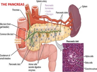
- 2. PANCREAS • The pancreas, in addition to its digestive functions, secretes two important hormones, insulin and glucagon, that are crucial for normal regulation of glucose, lipid, and protein metabolism. • Other minor hormones whose function are not well established include amylin, somatostatin, and pancreatic polypeptide, Physiologic Anatomy of the Pancreas. • The pancreas is composed of two major types of tissues, – the acini, which secrete digestive juices into the duodenum, – the islets of Langerhans, which secrete insulin and glucagon directly into the blood. • The islets contain three major types of cells, alpha, beta, and delta cells, which are distinguished from one another by their morphological and staining characteristics. • The beta cells, constituting about 60 per cent of all the cells of the islets, lie mainly in the middle of each islet and secrete insulin and amylin, a hormone that is often secreted in parallel with insulin, although its function is unclear. • The alpha cells, about 25 per cent of the total, secrete glucagon. • The delta cells, about 10 per cent of the total, secrete somatostatin. • In addition, at least one other type of cell, the PP cell, is present in small numbers in the islets and secretes a hormone of uncertain function called pancreatic polypeptide. • The close interrelations among these cell types in the islets of Langerhans allow cell-to-cell communication and direct control of secretion of some of the hormones by the other hormones. – For instance, insulin inhibits glucagon secretion, amylin inhibits insulin secretion, and somatostatin inhibits the secretion of both insulin and glucagon.
- 3. Insulin Is a Hormone Associated with Energy Abundance • That is, when there is great abundance of energy-giving foods in the diet, especially excess amounts of carbohydrates, insulin is secreted in great quantity. • In turn, the insulin plays an important role in storing the excess energy. – In the case of excess carbohydrates, it causes them to be stored as glycogen mainly in the liver and muscles. – Also, all the excess carbohydrates that cannot be stored as glycogen are converted under the stimulus of insulin into fats and stored in the adipose tissue. – In the case of proteins, insulin has a direct effect in promoting amino acid uptake by cells and conversion of these amino acids into protein. – In addition, it inhibits the breakdown of the proteins that are already in the cells. 3
- 4. Insulin Chemistry and Synthesis • Insulin is a small protein; human insulin has a molecular weight of 5808. It is composed of two amino acid chains, connected to each other by disulfide linkages. • When the two amino acid chains are split apart, the functional activity of the insulin molecule is lost. • Insulin is synthesized in the beta cells to form initially a preproinsulin. • This initial preprohormone with molecular weight of about 11,500, is cleaved in the endoplasmic reticulum to form a proinsulin with a molecular weight of about 9000; • Most of proinsulin is further cleaved in the Golgi apparatus to form insulin and peptide fragments before being packaged in the secretory granules. • When insulin is secreted into the blood, it circulates almost entirely in an unbound form; – it has a plasma half-life that averages only about 6 minutes, so that it is mainly cleared from the circulation within 10 to 15 minutes. • Except for that portion of the insulin that combines with receptors in the target cells, the remainder is degraded by the enzyme insulinase mainly in the liver, to a lesser extent in the kidneys and muscles, and slightly in most other tissues. • This rapid removal from the plasma is important for rapid control of metabolic functions. Human insulin molecule 4
- 5. Mechanisms of Insulin Secretion 5 Basic mechanisms of glucose stimulation of insulin secretion by beta cells of the pancreas. GLUT, glucose transporter BETA CELL
- 6. • The beta cells have a large number of glucose transporters (GLUT-2) that permit a rate of glucose influx that is proportional to the blood concentration in the physiologic range. • Once inside the cells, glucose is phosphorylated to glucose-6-phosphate by glucokinase. • This step appears to be the rate limiting for glucose metabolism in the beta cell and is considered the major mechanism for glucose sensing and adjustment of the amount of secreted insulin to the blood glucose levels. • The glucose-6-phosphate is subsequently oxidized to form adenosine triphosphate (ATP), which inhibits the ATP-sensitive potassium channels of the cell. • Closure of the potassium channels depolarizes the cell membrane, thereby opening voltage-gated calcium channels, which are sensitive to changes in membrane voltage. • This produces an influx of calcium that stimulates fusion of the docked insulin-containing vesicles with the cell membrane and secretion of insulin into the extracellular fluid by exocytosis. • Other nutrients, such as certain amino acids, can also be metabolized by the beta cells to increase intracellular ATP levels and stimulate insulin secretion. • Some hormones, such as glucagon and gastric inhibitory peptide, as well as acetylcholine increase intracellular calcium levels through other signaling pathways and enhance the effect of glucose, although they do not have major effects on insulin secretion in the absence of glucose. • Other hormones, including somatostatin and norepinephrine (by activating a-adrenergic receptors), inhibit exocytosis of insulin. • Sulfonylurea drugs stimulate insulin secretion by binding to the ATP-sensitive potassium channels and blocking their activity. – This results in a depolarizing effect that triggers insulin secretion, making these drugs very useful in stimulating insulin secretion in patients with type II diabetes, 6
- 7. Control of Insulin Secretion • Increased blood glucose (>100mg/dL) stimulates insulin secretion • Some amino acids stimulate insulin secretion – Lysine and arginine are the most potent – They strongly potentiate the glucose stimulus for insulin secretion – insulin in turn promotes transport of amino acids into the tissue cells as well as intracellular formation of protein • Gastrointestinal Hormones. A mixture of several important gastrointestinal hormones-gastrin, secretin, cholecystokinin, and gastric inhibitory peptide (which seems to be the most potent) - causes a moderate increase in insulin secretion. – These hormones are released in the gastrointestinal tract after a person eats a meal. – They then cause an “anticipatory” increase in blood insulin in preparation for the glucose and amino acids to be absorbed from the meal. – These gastrointestinal hormones generally act the same way as amino acids to increase the sensitivity of insulin response to increased blood glucose.
- 8. Other Hormones and the Autonomic Nervous System. • Other hormones that either directly increase insulin secretion or potentiate the glucose stimulus for insulin secretion include glucagon, growth hormone, cortisol, and, to a lesser extent, progesterone and estrogen. • The importance of the stimulatory effects of these hormones is that prolonged secretion of any one of them in large quantities can occasionally lead to exhaustion of the beta cells of the islets of Langerhans and thereby increase the risk for developing diabetes mellitus. • Indeed, diabetes often occurs in people who are maintained on high pharmacological doses of some of these hormones. • Diabetes is particularly common in giants or acromegalic people with growth hormone- secreting tumors, or in people whose adrenal glands secrete excess glucocorticoids. • Under some conditions, stimulation of the parasympathetic nerves to the pancreas can increase insulin secretion. 8
- 9. Activation of Target Cell Receptors by Insulin and the Resulting Cellular Effects • To initiate its effects on target cells, insulin first binds with and activates a membrane receptor protein. • It is the activated receptor, not the insulin, that causes the subsequent effects. • The insulin receptor is a combination of four subunits held together by disulfide linkages: – two alpha subunits that lie entirely outside the cell membrane and – two beta subunits that penetrate through the membrane, protruding into the cell cytoplasm. • The insulin binds with the alpha subunits on the outside of the cell, but because of the linkages with the beta subunits, the portions of the beta subunits protruding into the cell become autophosphorylated. • Thus, the insulin receptor is an example of an enzyme-linked receptor. • Autophosphorylation of the beta subunits of the receptor activates a local tyrosine kinase, which in turn causes phosphorylation of multiple other intracellular enzymes including a group called insulin-receptor substrates (IRS). • Different types of IRS (e.g. IRS-1, IRS-2, IRS-3) are expressed in different tissues. The net effect is to activate some of these enzymes while inactivating others. • In this way, insulin directs the intracellular metabolic machinery to produce the desired effects on carbohydrate, fat, and protein metabolism. 9
- 10. The end effects of insulin stimulation are the following: • Within seconds after insulin binds with its membrane receptors, the membranes of about 80 per cent of the body’s cells markedly increase their uptake of glucose. – This is especially true of muscle cells and adipose cells but is not true of most neurons in the brain. – The increased glucose transported into the cells is immediately phosphorylated and becomes a substrate for all the usual carbohydrate metabolic functions. – The increased glucose transport is believed to result from translocation of multiple intracellular vesicles to the cell membranes; these vesicles carry in their own membranes multiple molecules of glucose transport proteins, which bind with the cell membrane and facilitate glucose uptake into the cells. – When insulin is no longer available, these vesicles separate from the cell membrane within about 3 to 5 minutes and move back to the cell interior to be used again and again as needed. • The cell membrane becomes more permeable to many of the amino acids, potassium ions, and phosphate ions, causing increased transport of these substances into the cell. • Slower effects occur during the next 10 to 15minutes to change the activity levels of many more intracellular metabolic enzymes. These effects result mainly from the changed states of phosphorylation of the enzymes. • Much slower effects continue to occur for hours and even several days. They result from changed rates of translation of messenger RNAs at the ribosomes to form new proteins and still slower effects from changed rates of transcription of DNA in the cell nucleus
- 11. Effect of Insulin on Carbohydrate Metabolism • Immediately after a high-carbohydrate meal, the glucose that is absorbed into the blood causes rapid secretion of insulin. • The insulin in turn causes rapid uptake, storage, and use of glucose by almost all tissues of the body, but especially by the muscles, adipose tissue, and liver. Insulin Promotes Muscle Glucose Uptake and Metabolism • During much of the day, muscle tissue depends not on glucose for its energy but on fatty acids. – The principal reason for this is that the normal resting muscle membrane is only slightly permeable to glucose, except when the muscle fiber is stimulated by insulin; – Between meals, the amount of insulin that is secreted is too small to promote significant amounts of glucose entry into the muscle cells. • However, under two conditions the muscles do use large amounts of glucose. – One of these is during moderate or heavy exercise. • This usage of glucose does not require large amounts of insulin, because exercising muscle fibers become more permeable to glucose even in the absence of insulin because of the contraction process itself. – The second condition for muscle usage of large amounts of glucose is during the few hours after a meal. • At this time the blood glucose concentration is high and the pancreas is secreting large quantities of insulin. The extra insulin causes rapid transport of glucose into the muscle cells. • This causes the muscle cell during this period to use glucose preferentially over fatty acids, as we discuss later. 11 Storage of Glycogen in Muscle. • If the muscles are not exercising after a meal and yet glucose is transported into the muscle cells in abundance, then most of the glucose is stored in the form of muscle glycogen instead of being used for energy. • The glycogen can later be used for energy by the muscle. • It is especially useful for short periods of extreme energy use by the muscles and to provide spurts of anaerobic energy.
- 12. Insulin Promotes Liver Uptake, Storage, and Use of Glucose • Insulin causes most of the glucose absorbed after a meal to be stored almost immediately in the liver in the form of glycogen. • Then, between meals, when food is not available and the blood glucose concentration begins to fall, insulin secretion decreases rapidly and the liver glycogen is split back into glucose, which is released back into the blood to keep the glucose concentration from falling too low. • The mechanism by which insulin causes glucose uptake and storage in the liver includes several almost simultaneous steps: – Insulin inactivates liver phosphorylase, the principal enzyme that causes liver glycogen to split into glucose. – Insulin causes enhanced uptake of glucose from the blood by the liver cells. • It does this by increasing the activity of the enzyme glucokinase, which is one of the enzymes that causes the initial phosphorylation of glucose after it diffuses into the liver cells. – Insulin also increases the activities of the enzymes that promote glycogen synthesis, including especially glycogen synthase, • The net effect of all these actions is to increase the amount of glycogen in the liver. The glycogen can increase to a total of about 5 to 6 per cent of the liver mass, which is equivalent to almost 100 grams of stored glycogen in the whole liver.
- 13. Glucose Is Released from the Liver Between Meals. • When the blood glucose level begins to fall to a low level between meals, several events transpire that cause the liver to release glucose back into the circulating blood: – The decreasing blood glucose causes the pancreas to decrease its insulin secretion. – The lack of insulin then reverses all the effects listed earlier for glycogen storage, essentially stopping further synthesis of glycogen in the liver and preventing further uptake of glucose by the liver from the blood. – The lack of insulin (along with increase of glucagon, which is discussed later) activates the enzyme phosphorylase, which causes the splitting of glycogen into glucose phosphate. – The enzyme glucose 6- phosphatase, which had been inhibited by insulin, now becomes activated by the insulin lack and causes the phosphate radical to split away from the glucose; this allows the free glucose to diffuse back into the blood. • Thus, the liver removes glucose from the blood when it is present in excess after a meal and returns it to the blood when the blood glucose concentration falls between meals.
- 14. Insulin Promotes Conversion of Excess Glucose into Fatty Acids and Inhibits Gluconeogenesis in the Liver. • When the quantity of glucose entering the liver cells is more than can be stored as glycogen or can be used for local hepatocyte metabolism, insulin promotes the conversion of all this excess glucose into fatty acids. • These fatty acids are subsequently packaged as triglycerides in very-low-density lipoproteins and transported in this form by way of the blood to the adipose tissue and deposited as fat. • Insulin also inhibits gluconeogenesis. – It does this mainly by decreasing the quantities and activities of the liver enzymes required for gluconeogenesis. – Also insulin decreases the release of amino acids from muscle and other extrahepatic tissues and in turn the availability of these necessary precursors required for gluconeogenesis.
- 15. Lack of Effect of Insulin on Glucose Uptake and Usage by the Brain • The brain is quite different from most other tissues of the body in that insulin has little effect on uptake or use of glucose. • Instead, the brain cells are permeable to glucose and can use glucose without the intermediation of insulin. • The brain cells normally use only glucose for energy. • Therefore, it is essential that the blood glucose level always be maintained above a critical level. • When the blood glucose falls too low, symptoms of hypoglycemic shock develop. Effect of Insulin on Carbohydrate Metabolism in Other Cells • Insulin increases glucose transport into and glucose usage by most other cells of the body (with the exception of the brain cells) • The transport of glucose into adipose cells mainly provides substrate for the glycerol portion of the fat molecule. • Therefore, in this indirect way, insulin promotes deposition of fat in these cells.
- 16. Effect of Insulin on Fat Metabolism Insulin Promotes Fat Synthesis and Storage • Insulin has several effects that lead to fat storage in adipose tissue. • First, insulin increases the utilization of glucose by most of the body’s tissues, which automatically decreases the utilization of fat, thus functioning as a fat sparer. • Insulin also promotes fatty acid synthesis. – especially true when more carbohydrates are ingested than can be used for immediate energy, thus providing the substrate for fat synthesis. • Almost all this synthesis occurs in the liver cells, and the fatty acids are then transported from the liver by way of the blood lipoproteins to the adipose cells to be stored.
- 17. The different factors that lead to increased fatty acid synthesis in the liver include the following: • Insulin increases the transport of glucose into the liver cells. After the liver glycogen concentration reaches 5 to 6 per cent, this in itself inhibits further glycogen synthesis. – Then all the additional glucose entering the liver cells becomes available to form fat. – The glucose is first split to pyruvate in the glycolytic pathway, and the pyruvate subsequently is converted to acetyl coenzyme A (acetyl-CoA), the substrate from which fatty acids are synthesized. • An excess of citrate and isocitrate ions is formed by the citric acid cycle when excess amounts of glucose are being used for energy. – These ions then have a direct effect in activating acetyl-CoA carboxylase, the enzyme required to carboxylate acetyl-CoA to form malonyl-CoA, the first stage of fatty acid synthesis. • Most of the fatty acids are then synthesized within the liver itself and used to form triglycerides, the usual form of storage fat. – They are released from the liver cells to the blood in the lipoproteins. – Insulin activates lipoprotein lipase in the capillary walls of the adipose tissue, which splits the triglycerides again into fatty acids, – a requirement for them to be absorbed into the adipose cells, where they are again converted to triglycerides and stored.
- 18. Role of Insulin in Storage of Fat in the Adipose Cells. Insulin has two other essential effects that are required for fat storage in adipose cells: • Insulin inhibits the action of hormone-sensitive lipase. This is the enzyme that causes hydrolysis of the triglycerides already stored in the fat cells. – Therefore, the release of fatty acids from the adipose tissue into the circulating blood is inhibited. • Insulin promotes glucose transport through the cell membrane into the fat cells in exactly the same ways that it promotes glucose transport into muscle cells. – some of this glucose is then used to synthesize minute amounts of fatty acids, – but more important, it also forms large quantities of a-glycerol phosphate which supplies the glycerol that combines with fatty acids to form the triglycerides that are the storage form of fat in adipose cells. • Therefore, when insulin is not available, even storage of the large amounts of fatty acids transported from the liver in the lipoproteins is almost blocked.
- 19. Insulin Deficiency Increases Use of Fat for Energy All aspects of fat breakdown and use for providing energy are greatly enhanced in the absence of insulin. This occurs even normally between meals when secretion of insulin is minimal, but it becomes extreme in diabetes mellitus when secretion of insulin is almost zero. The resulting effects are as follows. • Insulin Deficiency Causes Lipolysis of Storage Fat and Release of Free Fatty Acids. – In the absence of insulin, all the effects of insulin noted earlier that cause storage of fat are reversed. – The most important effect is that the enzyme hormone-sensitive lipase in the fat cells becomes strongly activated. – This causes hydrolysis of the stored triglycerides, releasing large quantities of fatty acids and glycerol into the circulating blood. – Consequently, the plasma concentration of free fatty acids begins to rise within minutes. – This free fatty acid then becomes the main energy substrate used by essentially all tissues of the body besides the brain. • Insulin Deficiency Increases Plasma Cholesterol and Phospholipid Concentrations. • The excess of fatty acids in the plasma also promotes liver conversion of some of the fatty acids into phospholipids and cholesterol. These two substances, along with excess triglycerides formed at the same time in the liver, are then discharged into the blood in the lipoproteins. • Occasionally the plasma lipoproteins increase as much as threefold in the absence of insulin, giving a high total concentration of plasma. • This high lipid concentration—especially the high concentration of cholesterol—promotes the development of atherosclerosis in people with serious diabetes.
- 20. Excess Usage of Fats During Insulin Lack Causes Ketosis and Acidosis. • Insulin lack also causes excessive amounts of acetoacetic acid to be formed in the liver cells. • This results from the following effect: – In the absence of insulin the carnitine transport mechanism becomes increasingly activated. – In the mitochondria, beta oxidation of the fatty acids then proceeds very rapidly, releasing extreme amounts of acetyl-CoA. – A large part of this excess acetyl-CoA is then condensed to form acetoacetic acid, which in turn is released into the circulating blood. – Most of this passes to the peripheral cells, where it is again converted into acetyl-CoA and used for energy in the usual manner. • At the same time, the absence of insulin also depresses the utilization of acetoacetic acid in the peripheral tissues. – Thus, so much acetoacetic acid is released from the liver that it cannot all be metabolized by the tissues. – Some of the acetoacetic acid is also converted into b-hydroxybutyric acid and acetone. • These two substances, along with the acetoacetic acid, are called ketone bodies, and their presence in large quantities in the body fluids is called ketosis. • The acetoacetic acid and the b-hydroxybutyric acid can cause severe acidosis and coma, which often leads to death.
- 21. Effect of Insulin on Protein Metabolism and on Growth Insulin Promotes Protein Synthesis and Storage. During the few hours after a meal when excess quantities of nutrients are available in the circulating blood, not only carbohydrates and fats but proteins as well are stored in the tissues; insulin is required for this to occur. Some of the effects of insulin are. • Insulin stimulates transport of many of the amino acids into the cells. – Among the amino acids most strongly transported are valine, leucine, isoleucine, tyrosine, and phenylalanine. – Thus, insulin shares with growth hormone the capability of increasing the uptake of amino acids into cells. • Insulin increases the translation of messenger • Over a longer period of time, insulin also increases the rate of transcription of selected DNA genetic sequences • Insulin inhibits the catabolism of proteins • In the liver, insulin depresses the rate of gluconeogenesis In summary, insulin promotes protein formation and prevents the degradation of proteins Insulin lack causes protein depletion and increased plasma amino acids Insulin and Growth Hormone Interact Synergistically to Promote Growth. • the two hormones function synergistically to promote growth, each performing a specific function that is separate from that of the other
Editor's Notes
- SUR; Drug Sulfonylurea