This document summarizes research on PMMA/Ag2O acrylic bone cement coated with collagen to improve biomineralization, biocompatibility, and antibacterial properties. Collagen was electrodeposited on cement samples containing 0.5%, 1%, and 2% silver oxide. FTIR analysis showed collagen deposition depended on silver content. In vitro tests in simulated body fluid revealed silver ion release and mineralization of hydroxyapatite on surfaces over 3 weeks. Antimicrobial tests showed inhibition of E. coli and S. aureus growth, with effectiveness depending on silver content.
This document summarizes a study that investigated improving osseointegration of alumina/zirconia ceramic implants through fluoride surface treatment. Specifically, it discusses:
1) Fabricating alumina/zirconia composites and modifying their surface with SnF2 and NaBF4 treatments. Characterization showed the treatments incorporated fluoride onto the surface.
2) Evaluating the treatments in vitro with human fibroblast cultures, which showed better cell proliferation and layer formation on SnF2-treated surfaces.
3) Testing the treatments in vivo with rabbit femur implants, which revealed the SnF2 treatment resulted in better bone-implant contact after 6 weeks.
SIMONA CAVALU_Bioactivity evaluation of new silver doped bone cementSimona Cavalu
This document summarizes a study that evaluated the bioactivity and antimicrobial properties of two types of silver-doped bone cement composites. Electrochemical measurements and SEM/FTIR analysis were used to analyze the composites after incubation in simulated body fluid for up to 14 days. The results showed that both composites released silver and calcium ions into the fluid over time. One composite released silver ions more rapidly initially but both developed a hydroxyapatite-like surface layer indicating bioactivity. The layer formed more intensely on the surface of the composite also containing antibiotics. The study demonstrates the antimicrobial activity and bone bonding properties of the silver-doped composites.
SIMONA CAVALU_Microstructure and bioactivity of acrylic bone cementsSimona Cavalu
Polymer-ceramic composites based on polymethyl methacrylate are widely used in orthopaedics as suture materials and
fixation devices due to their biocompatibility and ability to support bony growth (osteoconductive) and also bone bioactive
(to form a calcium phosphate layer on its surface). In this study are compared the microstructure, bioactivity and
biocompatibility of two different types of biocomposites: BIOLOS3®and ANTIBIOTIC SIMPLEX®. They are investigated in
vitro in simulated body fluid using electrochemical measurements, SEM microscopy and ATR-FTIR spectroscopy in order to
evaluate the properties of the surface layer.
This document describes a study investigating bioceramic nanoparticles doped with magnesium and silver for use as implant materials. The nanoparticles were synthesized using a quick alkali-mediated sol gel method and had the composition xMgO-yAg2O-(40Àx)CaO-(20.5Ày)Na2O-35.5SiO2-4P2O5 with varying molar percentages of MgO and Ag2O. Characterization techniques were used to analyze the structure, morphology, degradation behavior, and antimicrobial properties of the samples. In vitro tests examined the samples' ability to support cell growth, deliver antibiotics in a controlled manner, and stimulate bone regeneration. The goal was to
Cavalu Simona_PMMA/Mg3Al2(SiO4)3 BONE CEMENTS: PRODUCTION, STRUCTURAL CHARACT...Simona Cavalu
The document discusses the production and characterization of acrylic bone cements reinforced with 5% magnesium aluminosilicate (Mg3Al2(SiO4)3). Structural analysis using FTIR and XRD showed the inclusion of the Mg3Al2(SiO4)3 crystalline phase within the PMMA polymer matrix. SEM images revealed the additive created a porous structure in PMMA, facilitating hydroxyapatite formation in vitro. Biocompatibility tests with human fibroblasts showed cell attachment and proliferation on specimens with and without the magnesium aluminosilicate; no cytotoxic effects were observed. The study aims to improve the mechanical and bioactive properties of acrylic bone cements.
The document discusses optimizing cell adhesion on magnesium-based implant materials through pre-incubation under cell culture conditions. It finds that pre-incubating Mg and Mg10Gd1Nd alloy samples in cell culture medium for just 6 hours is enough to form a natural protective surface layer suitable for cell culture by decreasing corrosion rates and increasing cell viability compared to untreated samples. Longer pre-incubation times further optimize cell adhesion and proliferation but do not significantly improve results enough to justify the increased contamination risk of longer experiments.
This study investigated how dental pulp cells and osteoblasts respond to gold substrates with different micropattern features. The researchers designed masks to create dot, line, hole, and hexagon patterns of varying sizes on gold films sputtered onto silicon wafers. Characterization with profilometry, AFM, and EDAX showed that film thickness and surface roughness increased with thicker films. After coating the patterns with an amino group and depositing cells, fluorescence imaging indicated the micropatterns were non-cytotoxic and osteoblasts attached normally. Future work will examine how cell behavior differs across the pattern features.
In this presentation, the chemical composition of rigid polyurethane foams was studied using FTIR microscopy and diffuse reflectance Fourier transform spectroscopy (DRIFTS) FTIR.
For more information, go to ssi.shimadzu.com and follow Shiamdzu on Twitter @ShimadzuSSI. Thanks for viewing!
Bioactive glass-ceramics used as a replacement material for bone tissues because
of its bioactivity, compatibility, and abilities to form a crystallized hydroxyapatite
(HA) bonding layer which has a similarity in composition and structure to the
inorganic component of bones mineral phase. The major objectives of this research is
estimating the effects of using MgO as an additive on the property of bioactive glass
ceramic in 45SiO2, 24.5 Na2O, 6 P2O5, (24.5-x)CaO, X MgO where X= 0,1,3 and 5
wt.%. Bioactive glass ceramic was made by traditional glasses melting techniques at
1200°C for 2hr. Compressed sample sintered at (850)°C, the X-ray diffraction test
confirmed the formations of (Na2CaSi3O8) and (Na2Ca2Si3O9) phases in the
structure of pure bioglass-ceramic and with (1wt.%) MgO, while (Na2CaSiO4) and
CaMg(SiO3)2 phases were present in the structure of bioglass-ceramic with
(3wt.%)MgO, and CaMg(SiO3)2 phase in the structure of bioglass-ceramic with
(5wt.%) MgO. Results of mechanical and physical studies showed that increasing
(MgO wt.%) in glass structure led to improving the bending strength, compressive
strength, microhardness, and density associating with decreasing in porosity. FTIR
confirmed the formations of the Si-O-Mg bond. The biological test was done by
immersion the sample in (SBF) simulated body fluid for (21 days). XRD, SEM and pH
tests revealed that appetite layers made on the samples surface, giving an evidence of
their bioactivity. As general the results of the present study demonstrate that partial
replacements of CaO by MgO in 45S5 glass ceramic improve the mechanical
properties without altering the bioactivity suggesting that glass ceramic was suitable
for the biomedical applications
Duddeck edi journal-1-2015_surface-analysis_datapro_cl
This document summarizes the interim results of a study examining the surface quality of 65 different implant systems from 37 manufacturers using scanning electron microscopy. Some key findings include:
- The Integra implant showed organic residue predominantly near the thread edges, which was identified as carbon in elemental analysis and may originate from the polyethylene packaging.
- Implant surfaces are analyzed to examine technical precision, possible impurities, and surface microstructure which influences osseointegration. Methods include qualitative and quantitative elemental analysis as well as 3D reconstruction of surface topography.
- Implant systems use various surface treatments like blasting, etching, coating, and anodization to optimize osseointegration,
1. The study compares silver nanoparticles coated and uncoated NiO–Fe2O3–CaO–SiO2–P2O5 ferromagnetic bioactive ceramics.
2. XRD and TEM analysis show the growth of hydroxyapatite layers on both coated and uncoated samples immersed in simulated body fluid.
3. Coated samples demonstrate better antimicrobial properties, degradation, and drug delivery compared to uncoated samples. The presence of silver nanoparticles improves these properties.
1) The study investigated the effects of enamel matrix derivative (EMD) in liquid formulation versus gel formulation on osteoblast and periodontal ligament cell proliferation and differentiation.
2) Results showed that both EMD formulations increased cell proliferation, growth factor expression, and calcium deposition similarly.
3) Future research is needed to evaluate EMD liquid formulation combined with bone grafts for periodontal regeneration compared to the gel formulation.
This document describes research into modifying titanium surfaces with nanoporous coatings to enhance protein adsorption for improved biocompatibility in orthopedic and dental implants. The researchers fabricated titanium surfaces with chemical etching and anodization to create nanoporous, silver-doped nanoporous, and gold-doped nanoporous coatings. Electrochemical impedance spectroscopy was used to analyze the stability and adsorption of bovine serum albumin protein on the modified surfaces. Scanning electron microscopy and energy-dispersive X-ray spectroscopy characterized the surface morphologies and compositions. The results showed that the nanoporous coatings enhanced protein adsorption and corrosion resistance, indicating improved biocompatibility for implant applications.
1) The study evaluated and compared the adhesion of two self-etch adhesive systems (OptiBond XTR and Bond Force) on normal and sclerotic dentin using scanning electron microscopy (SEM) and transmission electron microscopy (TEM).
2) SEM examination showed that on normal dentin, both adhesives formed hybrid layers of varying thickness, with OptiBond XTR penetrating dentinal tubules and Bond Force not penetrating. On sclerotic dentin, hybrid layers were thinner but OptiBond XTR still penetrated tubules.
3) TEM examination revealed that on normal dentin, Bond Force penetrated tubules as well, but not on sclerotic dentin. Hybrid
This document discusses a study on the responses of rabbit bone marrow cells to a biomimetic zeolite coating on nanoporous titanium. The researchers prepared zeolite-coated titanium samples and characterized their surface properties. They then cultured rabbit bone marrow-derived mesenchymal stem cells on the coated and uncoated titanium samples. The cells showed higher viability, proliferation, attachment and alkaline phosphatase activity on the zeolite-coated surfaces over 21 days, indicating the zeolite coating improved biocompatibility. Additionally, more bone-like nodule formation was observed on the zeolite-coated surfaces, suggesting potential for use as cancellous bone substitutes. In conclusion, the zeolite-coated nanoporous
Bioactive materials are revolutionizing oral health care and the quest for newer materials is never ending especially in the field of dental science. Research on biomaterials intensely involves interdisciplinary contributions from several major areas and requires extensive knowledge of medical science, materials science, biochemistry, biomedical engineering and clinical science. They are broadly used in the field of conservative dentistry and periodontics for regeneration, repair and reconstruction by acting directly on the vital tissue inducing its healing and repair through induction of various growth factors and different cells. This article reviews on the properties and clinical application of newer bioactive materials in endodontics, with a primary focus on the biocompatibility and tissue response to these materials.
This document outlines an investigation into protein adsorption on surfaces modified with self-assembled monolayers of amido amino acids. The study aims to modify surfaces with histidine, leucine, serine, and tryptophan and examine protein adsorption of IgG, albumin, and fibrinogen using quartz crystal microbalance biosensors. The document details the experimental procedures for surface sterilization and activation, optimization of self-assembled monolayer formation, and characterization of modified surfaces and protein adsorption.
This document summarizes bioactive materials and their bonding mechanisms with living tissues. It defines two types of bioactivity - osteoproductive and osteoconductive - based on whether the material elicits intracellular and extracellular responses. Bioactive glasses, glass-ceramics, and calcium phosphate ceramics are discussed. The mechanisms of bioactive bonding are explained as an 11-stage process where a hydroxycarbonate apatite layer forms on the implant surface, enabling bonding with tissues through both chemical and biological interactions.
This document summarizes 4 articles related to dental implants. The first article discusses a case study of one-piece implant design and concludes that it eliminates structural weaknesses of two-piece implants and increases success rates of immediately loaded implants with high insertion torque. The second article discusses factors affecting dental implant success including biocompatibility, tissue interactions, osteointegration and surface treatments. The third article assesses bone quality for implants and categorizes bone quality into 4 types. The fourth article provides an overview of corrosion aspects of titanium and its alloys used in dental implants.
Antimicrobial Coatings: The Research and Regulatory PerspectiveApril Bright
Coatings have long been considered an avenue for infection prevention in orthopedic procedures. These coatings, some of which utilize silver, have largely not been commercialized because regulators seek greater evidence of their safety, creating a long, expensive road for device companies. Announcements in the last half of 2018 and early 2019 indicate that companies continue to push to get them on the market and that productive conversations are taking place with regulators. This session began with a history of antimicrobial coatings followed by a look at recent research and technology.
Temporal relationship between sucrose –associated changes inDr. Roshni Maurya
The study evaluated the timing of effects of sucrose on the biochemical and microbiological composition of dental biofilm and its relationship to enamel demineralization. The findings showed that dental biofilm exposed 8 times per day to 20% sucrose solution significantly increased enamel mineral loss over time compared to the negative control. Sucrose increased levels of mutans streptococci and lactobacilli in dental biofilm and the concentration of insoluble extracellular polymeric substances compared to the negative control and glucose+fructose solution. While changes in biofilm composition occurred earlier, significant enamel demineralization only occurred after 7 days of biofilm accumulation.
This document summarizes the use of biphasic calcium phosphate (BCP) ceramics for bone regeneration and tissue engineering applications. BCP ceramics have proven biocompatibility and osteoconductivity. More recently, some studies have also shown their osteoinductive potential. For tissue engineering, BCP ceramics are promising scaffolds that can support stem cell behavior to favor bone regeneration. The document discusses the physicochemical properties of BCP ceramics that create a microenvironment favorable for bone formation, including chemical composition, resorption rate, porosity, and surface characteristics. BCP ceramics seeded with mesenchymal stem cells have been used to regenerate large bone defects in animal studies.
This document summarizes research on modifying the surface of titanium alloys through radio frequency magnetron sputtering of titanium dioxide (TiO2) thin films at different thicknesses. TiO2 films with thicknesses of 200, 300, and 500nm were deposited on commercially pure titanium (CPTi) and Ti-6Al-4V alloy substrates heated to 100°C using argon gas under low pressure. Field emission scanning electron microscopy, optical microscopy, and Vickers microhardness tests were performed to analyze surface morphology, microstructure, and hardness at each film thickness. Preliminary results indicate hardness increases and corrosion resistance improves for coated samples compared to uncoated, especially for the 300nm film thickness.
Joint Replacement: The Current and Future Impact of CoatingsApril Bright
The control of surface properties to reduce wear and corrosion and improve biocompatibility is of particular interest today as device companies—and surgeons, payors and patients—seek to extend the life of knee and hip implants. In this session, device companies shared research on their joint replacement coatings and materials, covering pros, cons and the future of their technology.
This document discusses applications of composite materials in the biomedical field. It describes how composites consist of two or more materials with distinct properties and are used in biomedical applications like implants, surgical devices, and prosthetics. Specific applications discussed include using carbon fiber composites for orthopedic implants like hip replacements and bone plates, fiber-reinforced dental composites, composite scaffolds for tissue engineering, and composite materials for diagnostic devices and cancer treatment. Important considerations for biomedical composites include biocompatibility, mechanical properties, degradation properties, and aesthetics.
This document summarizes research on the adsorption behavior of hyaluronidase enzyme onto silver nanoparticles and polymethyl methacrylate (PMMA) bone substitute surfaces. Surface-enhanced Raman scattering (SERS) and infrared (IR) spectroscopy techniques were used to analyze the adsorption. SERS spectra of hyaluronidase adsorbed to silver nanoparticles showed well-resolved Raman signals and indicated oxygen adsorption from functional groups. IR analysis of PMMA incubated with hyaluronidase showed shifts in amide band positions, suggesting changes to secondary structure upon adsorption. Band deconvolution and fitting of amide peaks revealed decreases in α-helix and β-sheet content for adsorbed hyaluronidase, with increases
This document summarizes a study that compares the microstructure, bioactivity, and biocompatibility of two types of acrylic bone cements: BIOLOS3® and ANTIBIOTIC SIMPLEX®. Electrochemical measurements, SEM imaging, and FTIR spectroscopy were used to analyze the materials after incubation in simulated body fluid for up to 34 days. The results showed that ANTIBIOTIC SIMPLEX® developed a more intense mineralization layer on its surface, as evidenced by greater calcium phosphate formation and water uptake. Hemolysis testing also indicated ANTIBIOTIC SIMPLEX® had better blood compatibility. In conclusion, the antibiotic-containing cement demonstrated superior in vitro bioactivity and bi
SIMONA CAVALU_Adsorption behavior of hyaluronidase onto silverSimona Cavalu
Surface enhanced Raman scattering (SERS) and ATR-FTIR spectroscopy was applied in this work, in order to get
information about the adsorption behavior of the title macromolecule with respect to different surfaces. In this case, the
silver substrate can be considered as artificial substrate and the investigations regarding the mechanisms of adsorption can
be useful in order to elucidate the active site properties of this enzyme. Our purpose is to study the adsorption mechanism
of hyaluronidase onto silver nanoparticles and PMMA (polymethyl methacrylate) substrates as well as qualitative and
quantitative aspects regarding perturbations of protein secondary structure (α-helix, β-sheet and unordered structures) upon
adsorption, using deconvolution techniques.
SIMONA CAVALU_ Bone Cement silver joam2007Simona Cavalu
This study evaluated two types of silver-doped bone cement composites for their properties and ability to release silver ions with antibacterial effects. Electrochemical measurements showed both composites reduced calcium ion concentration in simulated body fluid over 14 days, with one composite releasing more silver ions within 5 hours. Scanning electron microscopy images revealed both composites developed hydroxyapatite crystal structures on their surfaces after 14 days, indicating bioactivity. Infrared spectroscopy confirmed mineral deposition and showed one composite absorbed more water over time. The antibiotic-containing composite showed greater mineralization and formation of a calcium phosphate layer, suggesting it may be more suitable for applications requiring bone growth.
SIMONA CAVALU_Biomaterials and regenerative biomedicineSimona Cavalu
This document appears to be listing the authors Iulian Vasile ANTONIAC, Simona CAVALU, and Stefan Ioan VOICU and their affiliation with Biomaterials and Regenerative Biomedicine. It seems to be citing their work but provides no other contextual information about the topic, findings, or purpose of their work within that field.
The knowledge of the key factors involved in ethiopathogenesis of the gallstones disease requires chemical, structural and elemental composition analysis
This document summarizes research on the adsorption behavior of hyaluronidase enzyme onto silver nanoparticles and polymethyl methacrylate (PMMA) bone substitute surfaces. Surface-enhanced Raman scattering (SERS) and infrared (IR) spectroscopy techniques were used to analyze the adsorption. SERS spectra of hyaluronidase adsorbed to silver nanoparticles showed well-resolved Raman signals and suggested oxygen adsorption from functional groups. IR analysis of PMMA incubated with hyaluronidase showed shifts in amide band positions, indicating changes to secondary structure upon adsorption. Band deconvolution and fitting of amide peaks revealed decreases in α-helix and β-sheet content for adsorbed hyaluronidase, with increases
Simona cavalu_ Adherence Properties of Acrylic Bone Cement to AluminaSimona Cavalu
The aim of this study is to investigate the adherence properties of acrylic cement based on PMMA to alumina
ceramics. These ceramics are suitable for orthopedic and dental applications, as bioinert components in prosthetic
surgery. The surface of alumina specimens were subjected to a special treatment based on acid etched followed by
two dierent uoride treatments: SnF2 and NaBF4, respectively.
SIMONA CAVALU_Raman and surface-enhanced Raman spectroscopy of tempyo spin la...Simona Cavalu
Tempyo labelled ovalbumin at different pH values was prepared and investigated using Raman and SERS spectroscopy.
Raman spectra of tempyo labelled ovalbumin in the pH range from 6.7 to 11 were compared to those of the corresponding free
ovalbumin. In the basic pH range from 6.7 to 11 the molecular conformation was found to be unaffected by the tempyo
presence. Adsorption versatility to the colloidal Ag particles of pure- and tempyo labelled ovalbumin was also found to be
unchanged in this basic pH range. As the SERS binding site of protein the a-helix conformation is favourable.
SIMONA CAVALU_Surface Modification of Alumina/ Zirconia Ceramics Upon Differe...Simona Cavalu
The aim of this study was to prepare and to characterize the structure of Al2O3–3YSZ composites with 5% TiO2 addition
as well as the surface modification upon treatments with SnF2 and NaBF4, respectively. SEM micrographs showed the
controlled densification of the composites as an effect of 3YSZ and TiO2 addition to alumina matrix. By FTIR and XRD,
the characteristics of Al-O and Zr-O vibrations, respectively, the diffractions lines related to a-corundum and zirconia in
tetragonal phase were discussed. Qualitative and quantitative results obtained by XPS and ATR FTIR demonstrated that the
proposed materials are more sensitive to SnF2 than to NaBF4 treatment.
SIMONA CAVALU_Correlation between structural properties and in vivo biocompat...Simona Cavalu
The document summarizes research on the structural properties and biocompatibility of alumina/zirconia bioceramics. X-ray diffraction and Fourier transform infrared spectroscopy showed that titanium dioxide addition modified the structure of alumina/zirconia composites while retaining the tetragonal phase of zirconia. Animal studies found that both 90% alumina-10% zirconia and 90% alumina-10% zirconia with 5% titanium dioxide composites integrated well with bone over 6 weeks without inflammatory responses. Calcium/phosphorus ratios from scanning electron microscopy indicated improved osseointegration with titanium dioxide addition.
SIMONA CAVALU_Alternative approaches using animal model for implant biomaterialsSimona Cavalu
This document summarizes an animal study testing new dental and orthopedic implant materials. It discusses the motivation for animal testing prior to human clinical trials. It describes the materials to be tested - dense alumina-zirconia composites. Small animal models (Wistar rats) and larger animal models (rabbits) are proposed. The surgical procedures, monitoring, and analysis methods are outlined, including radiography, histology, and SEM/EDX to examine the bone-implant interface. Preliminary conclusions suggest the materials show good biocompatibility in animals and that surface coatings could improve biological response. The study involves collaboration between universities in Romania and Turkey.
Experimental study of gas flux characteristics in a co2 selective silica base...IAEME Publication
1. The document describes an experimental study of gas flux characteristics in a CO2 selective silica-based ceramic membrane.
2. A fresh ceramic membrane was prepared using a sol-gel dip coating technique and tested for separating CO2, O2, CH4, and N2 gases.
3. Results showed the membrane had high CO2 flux and CO2/N2 selectivity, confirming its potential for CO2 capture from flue gases. The gas flux increased linearly with pressure.
This document describes research into developing a new green resist material for microfabrication processes using animal-derived biopolymers. Bovine serum albumin (BSA) and fish gelatin were tested as potential water-based resists. BSA films of 5-10% concentration were successfully spin coated on silicon and silica wafers, with thicker films produced at lower spin speeds. Ellipsometry measurements determined film thicknesses ranging from 70-270 nm for BSA and 6-15 nm for 1.5% fish gelatin. Exposure to plasma etching showed BSA films were etched at rates of 4-16 nm/min, with thicker BSA films unexpectedly etching faster. While BSA showed potential as
— Processes based on immobilized enzymes have been studied extensively in the last few decades and today are also applied to the safeguard of environmental parameters. In this work, zeolite composite flat membranes with different chemical composition, transition metal, and microporous structures were prepared using in situ and secondary growth crystallization synthesis methods in/on stainless steel porous disks. All zeolite materials were been used in catalase adsorption to analyze the zeolite behavior andthe effect of chemical composition and structure on interaction with the enzyme. This study shows that the electrostatic type of interaction seems to be of the utmost importance in influencing immobilization, while the zeolite Brönsted acidity of the support is the subordinate parameter, which differentiates the adsorption performances of different zeolite structures (that distinct for chemical composition of the framework). Moreover, it permits to conclude that transition metal-containing membranes adsorb a higher percentage of the enzyme with respect to no-exchanged membranes and that, for all materials synthesized, the amount of catalase adsorbed onto the zeolite crystals and membranes increases with the temperature.
SIMONA CAVALU_Comparison between nanostructured aluminosilicate systems with ...Simona Cavalu
The document compares the biocompatibility of aluminosilicate samples containing iron and either dysprosium or yttrium. Scanning electron microscopy images show morphological changes on the sample surfaces after incubation in a collagen solution. Attenuated total reflectance Fourier-transform infrared spectroscopy analysis and deconvolution of the amide I region indicate qualitative and quantitative differences in the secondary structure of adsorbed collagen compared to native collagen. Specifically, collagen adsorbed to the yttrium aluminosilicate sample showed a more pronounced modification of secondary structure, indicating lower biocompatibility. Cyclic voltammetry further supported the quantitative investigations by showing enhanced current intensity and decreased oxidation potential of collagen adsorbed to the electrode surface
This document presents a novel method for quantifying surface modifications of glass fibre-reinforced polyester composites exposed to UV radiation. The method involves staining the composites with methylene blue dye after UV exposure. Colour analysis of digital images of the stained composites is then used to assess degradation, as degradation leads to increased dye adsorption and darker staining. The colour changes correlated well with FTIR spectroscopy and microscopy results. The method is presented as being easier, faster and cheaper than traditional analysis techniques for evaluating UV degradation of such composites.
In this study, kinetics of demineralization of chitin extraction from snail shells was
investigated. Chitin was extracted from snail shells by demineralizing the
deproteinized shells in 1.2 M HCl solution. Prior to demineralization, the raw snail
shells were deproteinized using 1 M NaOH solution to remove proteins and organic
matter present in the shells. The product was dried before the demineralization
process was carried out. The results showed that based on the R2 values obtained for
each of the shrinking core models considered which include; fluid film diffusion
(FFD), ash layer diffusion (ALD), and chemical reaction control (CRC), it was noted
that the CRC model was prevalent for all the various range of particle sizes analyzed
(6.3 – 4.75 mm, 4.75 – 2 mm, 2 – 1 mm, and 600 – 300 μm). The surface morphologies
and the Fourier Transform Infra-Red (FTIR) bands of the extracted chitin were
similar to previous studies
This document discusses a study on poly(vinyl chloride)/layered silicate nanocomposites. The study analyzed the effect of organoclay type (PGV or 1.31PS) and content on the electrical, antibacterial, and oxygen barrier properties of PVC compounds. Key findings include:
1) Dielectric properties (permittivity and loss) of PVC/1.31PS nanocomposites decreased with increasing organoclay content up to 2.5 phr, indicating improved electrical insulation properties, while PVC/PGV increased with content.
2) PVC/1.31PS (2.5 phr) showed significant resistance to Gram-positive and Gram
Structural Stability of Nano-Crystalline Aluminum-Glass CompositesIOSRJAP
Effects of particle size of the material in nanometer on solid state, structural properties AluminumGlass based samples are reported in this study. The particle size of 25µm was used for both Aluminum and Glass powders respectively. The samples were prepared in powdered form of various Al-Glass proportions. The weight of Aluminum ranges between 20 – 60 percent in the composites. Results showed from X-ray diffraction analyses that Aluminum addition and the determined particle size greatly influence the structural stability of the samples. It was revealed that samples were structurally stable in the range of Aluminum composition and the observed size of crystals, measured Width at Middle of Maximum Height and structural parameters were found in nanometers. The Al-Glass materials were noted to crystallize with FCC and BCC lattice structures.
The document describes an experimental study of the tribological properties of commercially pure titanium with different microstructures and coatings. The results show that titanium with an ultrafine-grained structure produced through severe plastic deformation has lower friction coefficient values and higher load-bearing capacity compared to coarse-grained titanium. Titanium samples coated with TiC using ion plasma spraying or TiO2 using microarc oxidation also exhibited lower friction coefficients than uncoated samples. The study provides data on friction coefficients and shear strengths of coated and uncoated titanium with different grain sizes.
Effect of Temperature on Wear Rate of Si-Epoxy- Eglass Polymer composite Mate...IDES Editor
With the increase use of polymer composites in
erosive work environment, it has become extremely important
to investigate their erosion characteristics. The hybrid
composite consisting of a Epoxy resin reinforced with Eglass
fiber and Si particles was developed. The effect of temperature
on wear rate applied to Si – Epoxy – Eglass polymer composite
has been studied. It has been found that the wear rate increases
as the temperature of carrier media (air) is increased.
Temperature of carrier media is one of the significant factor
influencing the erosion wear. The results were compared with
the plain epoxy composite material. Micrographs of eroded
surface are also presented to study the wear rate and Wear
behavior of composite.
A Study on the Strength of the Bacterial Concrete Embedded with Bacillus Mega...IRJET Journal
This document summarizes a study on how adding the bacteria Bacillus megaterium to concrete affects its strength properties. Concrete cylinders and prisms were cast with and without the bacteria. Testing found that adding the bacteria led to increases in both the split tensile strength and flexural strength of the concrete. Scanning electron microscope analysis indicated that pores in the concrete were partially filled by material growth from the addition of the bacteria. The maximum compressive strength was achieved with a bacteria cell concentration of 105 cells per ml of mixing water. Overall, the addition of B. megaterium was found to significantly improve the strength characteristics of concrete compared to conventional concrete without bacteria.
This document summarizes a research article that studied the effect of adding nano silicon dioxide (SiO2) particles to polylactic acid (PLA) films on some mechanical properties. PLA films were produced with 0%, 1%, 3%, and 5% nano SiO2 by weight using solution casting. Tests showed that adding 5% nano SiO2 increased the tensile strength of PLA films by 35% and the Young's modulus by 25% compared to pure PLA films. Fourier transform infrared spectroscopy and X-ray diffraction characterization indicated good dispersion of the nano SiO2 particles in the PLA matrix. The improved mechanical properties are due to the uniform distribution of nano SiO2 particles in the PLA and
Dry Sliding Wear Behaviour of Rheocat Al-5.7Si-2Cu-0.3Mg AlloyDr. Manal Abdullatif
In this study, the effect of improved microstructure of Al-5.7Si-2Cu-0.3Mg alloys by using semisolid process on hardness and dry sliding wear behaviour were investigated. The microstructures of conventional cast alloy were totally dendritic, while in rheocasting the dendrit-ic transfer to fine globular microstructures after using cooling slope casting. Tribological tests were carried out by using a pin-on-disc apparatus in dry sliding conditions. Wear tests were at low sliding speed 1ms-1 ,applied load at 50N and three different sliding distance (i.e., 1.8Km, 5.4Km and 9Km) respectively. An optical microscope and a scanning electron microscope were used to examine the micro-structure and to understand the wear mechanism on the worn surface of both samples. The results showed that, the wear resistance of rheocast alloy was improved and higher than that those produce by conventional casting. The volume loss of rheocast alloy show reduc-tion more than 18% at 1.8Km and 10% at 9Km compared to as-cast alloy. Moderate wear regimes were appeared in both alloys, accord-ing to the range of wear rate. The friction coefficient had increased due to increase in the contact point between pin and disc materials. The dominant wear mechanism for conventional and rheocasting alloys was adhesion wear and abrasive wear respectively.
1) The document investigates the effect of stacking sequence and hybridization on the tensile and flexural properties of composites made from basalt, jute, flax, and E-glass fibers reinforced with epoxy resin.
2) It was found that stacking sequence had little effect on tensile properties but a significant effect on flexural strength and modulus, with a sandwich-like sequence performing better.
3) Hybrid composites containing E-glass and basalt fibers had the highest specific tensile strength and modulus. E-glass/basalt performed better than E-glass/jute and E-glass/flax combinations in terms of strength to weight ratio.
IRJET- Structural Health Monitoring of Glass Fibre Reinforced Concrete BeamIRJET Journal
This document summarizes a study on monitoring the structural health of a glass fibre reinforced concrete beam using thermocouple sensors. Glass fibre mesh was used to reinforce concrete prisms instead of steel reinforcement. Thermocouple sensors embedded in the prisms measured the internal temperature at different loads. A relationship was derived between internal temperature and applied load. The temperature increased by 0.2°C for every load change. The study concluded that temperature monitoring can provide a warning of damage as temperature rises with increasing loads. Further research is needed to develop a more economical health monitoring system using thermocouple sensors.
The document summarizes research on the effect of adding nano silicon dioxide (SiO2) particles to polylactic acid (PLA) films on their mechanical properties. PLA films were produced with 0%, 1%, 3%, and 5% nano SiO2 via solution casting. Tests showed adding 5% nano SiO2 increased the tensile strength of PLA films by 35% and the Young's modulus by 25%. Characterization with FTIR and X-ray diffraction confirmed the nano SiO2 was well dispersed in the PLA matrix and showed interactions between the materials. The improved mechanical properties are due to good polymer-filler interactions from uniform nano SiO2 dispersion in the PLA matrix. The research
Preparation and properties of calcium-silicate filled resins for dental restoration. Part II: Micro-mechanical behaviour to primed mineral-depleted dentine.
Profeta AC.
Study of mechanical and morphological properties of glass fiber reinforced mo...eSAT Publishing House
IJRET : International Journal of Research in Engineering and Technology is an international peer reviewed, online journal published by eSAT Publishing House for the enhancement of research in various disciplines of Engineering and Technology. The aim and scope of the journal is to provide an academic medium and an important reference for the advancement and dissemination of research results that support high-level learning, teaching and research in the fields of Engineering and Technology. We bring together Scientists, Academician, Field Engineers, Scholars and Students of related fields of Engineering and Technology
Ceramic membrane coating with graphene oxide for tannery wastewater treatmenthunypink
The document summarizes a study on using a graphene oxide coated ceramic membrane for treating tannery wastewater. Key points:
- Tannery wastewater is highly contaminated and poses risks to the environment. A ceramic membrane was coated with graphene oxide using dip coating to treat this wastewater.
- Experiments were conducted at different pressures from 0.7 to 3 bar to evaluate the coated membrane's rejection of contaminants like total solids, salts, and conductivity.
- Results showed the coating improved the membrane's rejection performance. Rejection percentages increased for parameters like total solids, dissolved solids, suspended solids, salinity and conductivity compared to an uncoated membrane.
New evidences of key factors involved in “silent stones” ethiopathogenesis an...Simona Cavalu
The knowledge of the key factors involved in ethiopathogenesis of the gallstones disease requires chemical, structural and elemental composition analysis.
This document discusses Raman and surface enhanced Raman spectroscopy (SERS) experiments performed on bovine serum albumin and cytochrome c proteins labeled with 2,2,5,5-tetramethyl-3-pyrrolin-1-yloxy-3-carboxamide (tempyo) spin label at various pH values. The Raman and SERS spectra of the labeled proteins are compared to the unlabeled proteins. For bovine serum albumin, the SERS results suggest the α-helix conformation is favored at the binding site and the tempyo label does not significantly impact the vibrational structure. For cytochrome c, SERS enhancement of the porphyrin macrocycle bands indicates N-adsorption of
SIMONA CAVALU_IDENTIFICATION OF THE URINARY STONE COMPOSITION UPON EXTRACORPO...Simona Cavalu
Abstract. The study was carried out to investigate the composition and type of different urinary
stones upon application of extracorporeal shock wave lithotripsy. ATR FTIR spectra revealed the
marker bands of a mixed stone composition containing calcium oxalate monohydrate/ calcium
carbonate, respectively calcium oxalate monohydrate/cystine. The surface morphology of the samples
and elemental analysis was performed by SEM-EDAX confirming the presence of oxalate, carbonate
and cystine in the samples. Combination of FTIR spectroscopy and SEM-EDAX allowed quantitative
and qualitative evaluation of components, the spatial distribution and the percent of major and trace
elements present in a single sample.
SIMONA CAVALU_Raman and Surface Enhanced Raman Spectroscopy of 2,2,5,5-Tetram...Simona Cavalu
ABSTRACT: 2,2,5,5-Tetramethyl-3-pyrrolin-1-yloxy-3-carboxamide (tempyo) labeled bovine
serum albumin and cytochrome c at different pH values were prepared and
investigated using Raman–resonance Raman (RR) spectroscopy and surface enhanced
Raman scattering (SERS) spectroscopy. The Raman spectra of tempyo labeled proteins
in the pH 6.7–11 range were compared to those of the corresponding free species. The
SERS spectra were interpreted in terms of the structural changes of the tempyo labeled
proteins adsorbed on the silver colloidal surface. The tempyo spin label was found to be
inactive in the Raman–RR and SERS spectra of the proteins. The a-helix conformation
was concluded to be more favorable as the SERS binding site of bovine serumalbumin.
In the cytochrome c the enhancement of the bands assigned to the porphyrin macrocycle
stretching mode allowed the supposition of the N-adsorption onto the colloidal surface.
Test bank for karp s cell and molecular biology 9th edition by gerald karp.pdfrightmanforbloodline
Test bank for karp s cell and molecular biology 9th edition by gerald karp.pdf
DECLARATION OF HELSINKI - History and principlesanaghabharat01
This SlideShare presentation provides a comprehensive overview of the Declaration of Helsinki, a foundational document outlining ethical guidelines for conducting medical research involving human subjects.
10 Benefits an EPCR Software should Bring to EMS Organizations Traumasoft LLC
The benefits of an ePCR solution should extend to the whole EMS organization, not just certain groups of people or certain departments. It should provide more than just a form for entering and a database for storing information. It should also include a workflow of how information is communicated, used and stored across the entire organization.
Know the difference between Endodontics and Orthodontics.Gokuldas Hospital
Your smile is beautiful.
Let’s be honest. Maintaining that beautiful smile is not an easy task. It is more than brushing and flossing. Sometimes, you might encounter dental issues that need special dental care. These issues can range anywhere from misalignment of the jaw to pain in the root of teeth.
Breast cancer: Post menopausal endocrine therapyDr. Sumit KUMAR
Breast cancer in postmenopausal women with hormone receptor-positive (HR+) status is a common and complex condition that necessitates a multifaceted approach to management. HR+ breast cancer means that the cancer cells grow in response to hormones such as estrogen and progesterone. This subtype is prevalent among postmenopausal women and typically exhibits a more indolent course compared to other forms of breast cancer, which allows for a variety of treatment options.
Diagnosis and Staging
The diagnosis of HR+ breast cancer begins with clinical evaluation, imaging, and biopsy. Imaging modalities such as mammography, ultrasound, and MRI help in assessing the extent of the disease. Histopathological examination and immunohistochemical staining of the biopsy sample confirm the diagnosis and hormone receptor status by identifying the presence of estrogen receptors (ER) and progesterone receptors (PR) on the tumor cells.
Staging involves determining the size of the tumor (T), the involvement of regional lymph nodes (N), and the presence of distant metastasis (M). The American Joint Committee on Cancer (AJCC) staging system is commonly used. Accurate staging is critical as it guides treatment decisions.
Treatment Options
Endocrine Therapy
Endocrine therapy is the cornerstone of treatment for HR+ breast cancer in postmenopausal women. The primary goal is to reduce the levels of estrogen or block its effects on cancer cells. Commonly used agents include:
Selective Estrogen Receptor Modulators (SERMs): Tamoxifen is a SERM that binds to estrogen receptors, blocking estrogen from stimulating breast cancer cells. It is effective but may have side effects such as increased risk of endometrial cancer and thromboembolic events.
Aromatase Inhibitors (AIs): These drugs, including anastrozole, letrozole, and exemestane, lower estrogen levels by inhibiting the aromatase enzyme, which converts androgens to estrogen in peripheral tissues. AIs are generally preferred in postmenopausal women due to their efficacy and safety profile compared to tamoxifen.
Selective Estrogen Receptor Downregulators (SERDs): Fulvestrant is a SERD that degrades estrogen receptors and is used in cases where resistance to other endocrine therapies develops.
Combination Therapies
Combining endocrine therapy with other treatments enhances efficacy. Examples include:
Endocrine Therapy with CDK4/6 Inhibitors: Palbociclib, ribociclib, and abemaciclib are CDK4/6 inhibitors that, when combined with endocrine therapy, significantly improve progression-free survival in advanced HR+ breast cancer.
Endocrine Therapy with mTOR Inhibitors: Everolimus, an mTOR inhibitor, can be added to endocrine therapy for patients who have developed resistance to aromatase inhibitors.
Chemotherapy
Chemotherapy is generally reserved for patients with high-risk features, such as large tumor size, high-grade histology, or extensive lymph node involvement. Regimens often include anthracyclines and taxanes.
Nano-gold for Cancer Therapy chemistry investigatory projectSIVAVINAYAKPK
chemistry investigatory project
The development of nanogold-based cancer therapy could revolutionize oncology by providing a more targeted, less invasive treatment option. This project contributes to the growing body of research aimed at harnessing nanotechnology for medical applications, paving the way for future clinical trials and potential commercial applications.
Cancer remains one of the leading causes of death worldwide, prompting the need for innovative treatment methods. Nanotechnology offers promising new approaches, including the use of gold nanoparticles (nanogold) for targeted cancer therapy. Nanogold particles possess unique physical and chemical properties that make them suitable for drug delivery, imaging, and photothermal therapy.
Travel Clinic Cardiff: Health Advice for International TravelersNX Healthcare
Travel Clinic Cardiff offers comprehensive travel health services, including vaccinations, travel advice, and preventive care for international travelers. Our expert team ensures you are well-prepared and protected for your journey, providing personalized consultations tailored to your destination. Conveniently located in Cardiff, we help you travel with confidence and peace of mind. Visit us: www.nxhealthcare.co.uk
The skin is the largest organ and its health plays a vital role among the other sense organs. The skin concerns like acne breakout, psoriasis, or anything similar along the lines, finding a qualified and experienced dermatologist becomes paramount.
Histololgy of Female Reproductive System.pptxAyeshaZaid1
Dive into an in-depth exploration of the histological structure of female reproductive system with this comprehensive lecture. Presented by Dr. Ayesha Irfan, Assistant Professor of Anatomy, this presentation covers the Gross anatomy and functional histology of the female reproductive organs. Ideal for students, educators, and anyone interested in medical science, this lecture provides clear explanations, detailed diagrams, and valuable insights into female reproductive system. Enhance your knowledge and understanding of this essential aspect of human biology.
1. Digest Journal of Nanomaterials and Biostructures Vol. 6, No 2, April - June 2011, p. 779 - 790
BIOACTIVITY AND ANTIMICROBIAL PROPERTIES OF PMMA/Ag2O ACRYLIC BONE CEMENT COLLAGEN COATED
S. CAVALU*
Orthopedic acrylic cements have to fulfill several medical requirements, such as: low values of maximum cure temperature (to avoid thermal necrosis of the bone tissue, during the setting of the cement), moderate sitting time (so that cement does not cure too fast or too slow), high values of compressive strength (allowing the cured cement mantle to withstand the compressive loads involved by normal daily activities) [3]. A new biomaterials generation was born by combining bioactivity, biocompatibility and antibacterial properties of materials, either by antibiotic [4,5] or silver loading [6,7]. The use of antibiotic-loaded bone cement is a well-accepted adjunct in the treatment of infected joint arthroplasty and is gaining further application as a method of prophylaxis. The influence of antibiotic inclusion on cement mechanical properties, specifically fatigue, determines its resistance to crack formation and the long-term in vivo structural integrity , V. SIMONa, G. GOLLERb, I. AKINb University of Oradea, Faculty of Medicine and Pharmacy, 410068 Oradea, Romania, aBabes-Bolyai University, Faculty of Physics & Institute of Interdisciplinary Research in Bio-Nano-Sciences, 400084 Cluj-Napoca, Romania bIstanbul Technical University, Metalurgical and Materials Engineering Dept, 34469 Maslak Istanbul, Turkey PMMA/Ag2O bone cements with collagen coating were prepared in order to improve their biomineralisation, biocompatibitity and antibacterial properties as well. ATR FTIR spectroscopy analysis demonstrated that collagen electrodeposition on their surface depends on the silver content in samples. In vitro tests, performed in simulated body fluid (SBF) during three weeks of SBF incubation, revealed the dissolution profile of Ag+ from the acrylic matrix, the results being in concordance with antibacterial tests performed against Escherichia coli and Staphylococcus aureus. The mineralization process upon incubation was confirmed by the surface morphology analyses (SEM) combined with ATR FTIR, which evidenced the phosphate characteristic bands of hydroxyapatite, depending on the silver oxide content in the samples. (Received April 11, 2011; Accepted April 19, 2011) Keywords: Acrylic bone cement, Collagen, Antimicrobial activity, SEM, ATR FTIR 1. Introduction The development of a novel biomimetic implant based on polymer composites has yet to be justified as a valid and effective design for orthopedic implants. An important parameter that can help predict long-term behavior of the prostheses is the permanent displacement, or migration, of the implant relative to bone. This incidence of migration is one of the predominant causes of failure and is mainly due to micromotion, the lack of fixation stability at the bone-implant interface. It was demonstrated [1] the importance of material flexibility and its effects on stress shielding. Materials with a high flexibility tend to have less bone resorption in the calcar region and the stress level in bone is significantly lower. The mechanical environment of the host bone is altered by the implantation of a foreign material and consequently, bone remodeling occurs in order to adapt the bone structure to this new situation [2].
* Correspondsing author: scavalu@rdslink.ro
2. 780
of the cement. Several factors influence the choice of antibiotic to add to bone cement. The antibiotic must be able to withstand the exothermic temperature of polymerization, be available as a powder, have a low incidence of allergy, and be able to elute from the cement over an appropriate time period. Several bone cements containing antibiotics such as erythromycin, colystin, gentamicin and tobramycin have been commercially available in Europe for many years. On the other hand, silver based antimicrobials capture much attention not only because of the non toxicity of the active Ag+ to human cells [8,] but because of their novelty being a long lasting biocide with high temperature stability and low volatility. The antimicrobial efficacy of these composites depends on their ability to release the silver ions from these composites upon interaction with biological fluids. Hence, silver doped materials are use as an alternative (or complementary) to antibiotic loaded cements, silver being capable of killing over 650 forms of bacteria, viruses [9]. In the same times, a complete biological integration into the surrounding tissues is a critical step for clinical success of an implant. It has been previously demonstrated that biomimetic coatings consisting either of collagen type I and hydroxyapatite or mineralized collagen are suitable surfaces to enhance cell attachment and proliferation [10, 11]. Collagen comprises 90% of the extracellular matrix of bone and it occupies a key role in the interaction of osteoblasts and their environment. We have developed in this work a new strategy for orthopedic/dental implants based on both concepts improvement: bioactivity and antibacterial activity by incorporating different concentration of Ag2O in PMMA bone cement followed by collagen electrodeposition. The study is focused on scanning electron microscopic analysis before and after immersion in SBF and complementary ATR FTIR spectroscopy which evidenced the phosphate characteristic bands of hydroxyapatite, depending on the silver oxide content in the samples. 2. Experimental The PMMA/Ag2O samples were prepared using commercial PMMA based cements (BIOLOS3®) as starting material, by mixing the powder polymethyl methacrylate (copolymer) with Ag2O particles as antimicrobial agent in concentration of 0.5, 1 and 2% (wt/wt) with respect to the total powder amount and the polymerization was initiated by adding an appropriate volume of liquid monomer (methyl methacrylate). The curing parameters were registered according to the ASTM Standard [12]. Time and temperature were measured from the onset of the mixing powder with the liquid. Two determinations were performed for each sample formulation. The electrodeposition of soluble collagen type I, from calf skin (Sigma Chemicals) was performed for each specimen using a three electrode electrochemistry system, following the protocol described in our previous paper [13]. The first evidence of collagen layer on the surfaces was obtained by recording the ATR FTIR spectra comparing the coated and uncoated surfaces of the samples, using a Spectrum BX II Perkin Elmer spectrometer, equipped with MIRacle ATR accessory (ZnSe crystal), with a scanning speed of 32 cm-1 min-1 and the spectral width 2.0 cm-1. Deconvolution of amide I band of native collagen and adsorbed on the samples surfaces was performed in order to investigate the conformational changes of the protein during the coating process. The spectra were smoothed with a 9-point Savitsky–Golay smooth function to remove the white noise. The second derivative spectral analysis of amide I band was applied to locate positions and assign them to different functional groups. Before starting the fitting procedure, the obtained depths of the minima in the second derivative spectrum and subsequently, the calculated maximum intensities, were corrected for the interference of all neighbouring peaks. All second- derivative spectra, calculated with the derivative function of Opus software, were baseline- corrected, based on the method of Dong and Caughey [14] and area-normalized under the second derivative amide I region, 1700–1600 cm-1. Curve fitting was performed by setting the number of component bands found by second-derivative analysis with fixed bandwidth (12 cm-1) and Gaussian profile. The area under each peak was used to calculate the percentage of each component and, finally, to analyze the percentage of secondary structure component [15].
In vitro tests were carried out in Simulated body fluid (SBF), prepared according to Kokubo protocol [16]. Electrochemical measurements were performed in SBF in static conditions,
3. 781
upon incubation of the materials during three weeks (with and without collagen coating), using a CyberComm 6500 Multimeter (EUTECH Instruments) equipped with Ag+ ion selective electrodes and data aquisition software. The detection limit using this method is 10-6 M for Ag+ ions. The second evidence of collagen coating was revealed by SEM analysis on the surfaces, as well as the morphology before and after three weeks incubation in SBF. Microstructure characterization was made by using JEOL JSM 7000F Field Emission Scanning Electron Microscope. Antimicrobial activity of the specimens (cylindrical shape, 10 mm diameter and 3 mm high) was determined against the bacterial pathogens (Escherichia coli ATCC 2732 and Staphylococcus aureus ATCC 6538 P) by the agar disc diffusion assay, in Petri dishes (measuring 120 mm each side) containing 20 mL of nutrient agar with 105CFU/mL of bacteria. The plates were incubated at 370C for 24 h. The antibacterial activity was evaluated by measuring the zone of growth inhibition surrounding the discs. 3. Results and discussions In spite of its promising bioactivity, the previously reported collagen/calcium phosphate composite [17] is mechanically weak and consequently limited to non-loading applications. An alternative way to improve the mechanical properties of such composites is to apply a collagen layer and such designs are commonly used in medical devices as hip implants. Thus, the bioactive surface would accelerate bone growth and implant fixation, thereby shortening the healing period of the patient. The coating procedure by electrodeposition was already reported [13] using chronoamperometric method. Briefly, a three-electrode electrochemistry system was used by keeping the working electrode at the potential value -2V during 1800 seconds, the current during electrodeposition being monitored by a potentiostat. The current intensity dropped dramatically in the first few minutes of deposition, then remained relatively constant until ~ 20 min. The rapid decrease in intensity represents the capacitive charging of the electrode, followed by a bearing with approximately constant intensity, limited by the diffusion of collagen concentration. Collagen solution was prepared in a concentration of 1 mg/mL of electrolyte and pH was adjusted to 4.8-5.3.
ATR FTIR spectrometric measurement was carried out to further clarify the presence of collagen coating on the surface of each specimen containing different Ag2O concentration. The spectra recorded before and after collagen electrodeposition on PMMA- based bone cement are presented comparatively for each specimen in fig. 1-3 along with the corresponding spectra recorded after 20 days incubation in Simulated Body Fluid (SBF). The marker bands of PMMA are a sharp and intense peak at 1720 cm-1 due to the presence of ester carbonyl group ν(C=O) stretching vibration, a broad band at 1436 cm-1 due to δ(CH3) vibration mode, the peaks in the range 1265-1000 cm-1 assigned to O-C-O, C-CH3 and C-COO stretching vibrations, and the region between 980-800 cm-1 due to the bending of C-H bond [18, 19]. The PMMA characteristic vibrations are very similar for all the three specimens prepared in this study, containing respectively 0.5, 1 and 2% Ag2O. The ATR FTIR spectra recorded after electrodeposition confirms the presence of the collagen on the specimens surface throughout the characteristics of the distinct peaks of collagen: amide I at 1635/1638 cm-1 (C=O stretching), amide II at 1553/1556 cm-1 (N-H deformation) and amide III around 1240 cm-1 (combined N-H bending and C-N stretching). The relative intensity of these bands is very different with respect to each specimen showing that the collagen deposition depends on the silver concentration in the samples. In the same time, the characteristic bands of PMMA are obviously reduced in intensity as the collagen layer is formed on the surfaces. The results are in accordance with previous reported study regarding collagen self assembly coating by electrolysis on silicate cathode surface [20]. The bioactivity of the surface layer of the specimens was demonstrated by the spectra recorded after 21 days incubation in SBF as shown in Fig. 1c, 2c and 3c. A pronounced decreased in intensity of the bands related to the polymer matrix and to the collagen layer is observed when compare with the corresponding spectra before incubation. Especially for the amide I and II bands of collagen, a broadening and shift to lower wavenumbers was found. The surface mineralization was demonstrated by the strong band at 1015/1020 cm-1 which are the characteristics of the calcium phosphate in hydroxyapatite- type biomimetic coatings [10,21, 22]. The appearance of a new band
4. 782
as a shoulder at 1086/1082 cm-1 is also an indicative of the P-O antisymmetric vibration. Thus, as already reported [23], the cations as well as the anions in SBF can be readily and reversibly exchanged.
1800.017001600150014001300120011001000900800.0100203040506070809101119cm-1 17211436138112651238113910689738201800.017001600150014001300120011001000900800.0200406081012141618202250cm-1 17201638155614491382133912391191113410759801800.017001600150014001300120011001000900800.006010203040506070885cm-1 17211635154314421242119010861020958873831 Fig. 1 ATR FTIR spectra of the specimen with 0.5% Ag20 recorded before (a) and after collagen electrodeposition (b) and upon 20 days incubation in SBF (c). The hydrated collagen layer is not stable and it is progressively replaced by apatite during ageing in an aqueous media (maturation). The mechanism of this transformation is not yet well known but it involves a decrease of the amount of HPO42- ions and an increase of the calcium content of the mineral phase. Simultaneously OH- ions are included in the structure and water is excluded [24].
Specimen 1: 0.5% Ag20
a)
b)
c)
5. 783
1800.017001600150014001300120011001000900800.00.0010.010.020.030.040.050.060.070.076cm-1 1721148014351384126912381188114310659849658411800.017001600150014001300120011001000900800.00.0100.050.100.150.200.220cm-1 17201635154414451405133612301199107610299371800.017001600150014001300120011001000900800.000420.0100.0150.0200.0250.0300.0350417 cm-1 172016301541145113971246119810821015958876836 Fig. 2 ATR FTIR spectra of the specimen with 1% Ag20 recorded before (a) and after collagen electrodeposition (b) and upon 20 days incubation in SBF (c). The primer condition imposed to materials considered for biomedical applications is biocompatibility dictated by the manner in which their surface interact with blood constituents (erythrocytes, platelets) as well as the proteins [25, 26]. The type and amounts of adsorbed proteins mediate subsequent adhesion, proliferation and differentiation of cells as well as depositing of mineral phases. The behavior of a protein at an interface is likely to differ considerably from its behavior in the bulk.
Specimen 2: 1% Ag2O
b)
a)
c)
6. 784
1800.017001600150014001300120011001000900800.006020304050607080902cm-1 1721143613841266123611851139111610659819138361800.017001600150014001300120011001000900800.320051015202590cm-1 1719163215531453133912381203107910321800.017001600150014001300120011001000900800.0481015202530354028cm-1 17201630154414201240119010821020958880840 Fig. 3 ATR FTIR spectra of the specimen with 2% Ag20 recorded before (a) and after collagen electrodeposition (b) and upon 20 days incubation in SBF (c). Because of the different local environment at the interface, the protein may have the opportunity of adopting a more disordered state exposing its hydrophobic core to the aqueous phase, often called surface denaturation. Denaturation is a process by which hydrogen bonds, hydrophobic interactions and salt linkages are broken and the protein is unfolded. The denaturation of secondary structure involves also changes in ratio among the three common structures: α helix, β sheets or turns and unordered [14, 27]. FTIR spectroscopy can be used to study protein secondary structure in any state, i.e. aqueous, frozen, dried or even as an insoluble aggregate, and for this reason it is one of the most used techniques for studying stress induced alterations in protein conformation and for quantifying protein secondary structure. ATR-FTIR can provide important information leading to the development of novel biomaterials as replacements for damaged or diseased natural tissue. The spectral region of amide I (~1640 cm-1), amide II (~1540 cm-1) and amide III (~1240cm-1) are very sensitive to the conformational changes in the secondary structure of proteins. Computational techniques based on the second derivative spectra and deconvolution procedure is used for percentage evaluation of each secondary structure and also the perturbations upon the adsorption to different surfaces [28,29]. Collagen type I is the most abundant protein of the extracellular matrix, a fibrillar triple helical structure that forms gel networks in irregular connective tissue. Collagen is also proline- rich and self assembles into fibrils [30,31].
As a reference, the FTIR spectrum of native collagen is shown in Fig. 4a , pointing out the features characteristic of amide I, II and III which are the most intense vibrational modes. The present study is focused on the amide I behavior, which is due primarily to the stretching vibrations of the peptide carbonyl group. According to the literature, the intensity of amide I band
a)
b)
c)
Specimen 3: 2% Ag2O
7. 785
of collagen decreases markedly upon denaturation, and after deconvolution, four prominent components are present both in the native or denaturated protein spectrum [15,32]. That the relative intensities of these four peaks vary with the extent of collagen-fold or triple helix content speaks to the point that they are clearly conformationally dependent. Specific components within the fine structure of amide I adsorbed collagen is correlated with different states of hydrogen bonding associated with the local conformations of the alpha chain peptide backbones. This heterogeneity can arise either from intrinsic basicity differences in the strengths of the hydrogen bonds associated with the carbonyls [33]. Deconvolution of amide I band of native collagen and adsorbed to our specimens is shown in Fig. 4 (b,c,e) and the assignment of the components in Table 1 was made on the basis of the previous reported studies, along with the quantitative analysis. Curve fits to the amide I native collagen reveals four Gaussian components at 1630, 1644, 1665 and 1682 cm-1 representing helix-related hydrogen-bounded set of carbonyls. According to the literature, the highest frequency carbonyl absorption peak represents the weakest H-bonded system [34]. Beside the characteristic frequencies of α helix conformation, the peak located in the higher region, at 1682 cm-1, represent the formation of an antiparallel β-sheet structure (or turns). Both the intensity and the location of the characteristic peaks are modified upon adsorption. As a general behavior, one can observe a shift toward lower frequencies, a decrease in α helix total content and concomitant increase of turn percentage upon adsorption, as a consequence of denaturation.
1600161016201630164016501660167016801690-0.0000050.0000000.0000050.0000100.0000150.000020e) Wavenumber cm-1 Absorbance (a.u.) Fig.4. ATR FTIR spectrum of native collagen type I, from calf skin as received from Sigma Chemicals (a), deconvoluted amide I absorption band of native collagen (b) and adsorbed collagen to specimens with 0.5% Ag20 (c), 1% Ag20 (d) and 2% Ag20 (e) respectively.
The silver ion released in simulated body fluid was monitored during 21 days incubation of the three specimens. The results presented in fig. 5 indicate that the silver ion concentration in fluid increase with increasing silver oxide content in the sample. A small amount of silver was detected after the first ten hours of incubation, the Ag+ release showed only a marginal increase during the first 3 days followed by an abrupt rise after one week especially for composites containing higher silver oxide concentrations. As previousely reported in literature, concentrations of silver ions between 0.10 and 0.22 mM, released from Ag2O- doped bioactive glass, were found
180016001400120010008006000.0000.0250.0500.0750.1000.1250.1500.1750.2000.2250.250 1228Amide III Wavenumber cm-1 Absorbance (a.u.) 1640Amide I 1546Amide II a) 160016201640166016801700 Absorbance (a.u) Wavenumber (cm-1) d)
8. 786
to have bactericidal activity against gram- positive bacterium [35]. In table 2 are presented the results regarding the antibacterial efficiency of the Ag2O/PMMA specimens against gram positive (Staphylococcus aureus ATCC 6538 P) and gram negative (Escherichia coli ATCC 2732), respective the diameters of the inhibition areas read after 24 hours incubation at 37°C. The inhibition effect is significantly more intense in the case of Escherichia coli. The mechanism of the antimicrobial action of silver ions is not completely known. However, the effect of silver ions on bacteria is linked with its interaction with thiol group compounds found in the respiratory enzymes of the bacterial cells. Silver binds to the bacterial cell wall and cell membrane and inhibits the respiration process. In case of E-coli, silver acts by inhibiting the uptake of phosphate and releasing phosphate, mannitol, succinate, proline and glutamine from the E-coli cells. In addition, it was shown that Ag+ ions prevent DNA replication by binding to the polynucleotide molecules, hence resulting in bacterial death [36]. Table 1.
Collagen amide I
α helix
α helix
α helix
turns
ν(cm-1)
A (%)
ν(cm-1)
A (%)
ν(cm-1)
A (%)
ν(cm-1)
A (%)
native collagen
1630
28.3
1644
33.2
1665
34.7
1682
3.8
Specimen 1 0.5% Ag2O
1625
40.2
1641
25.5
1657
23.5
1670
10.8
Specimen 2 1% Ag2O
1619
4.2
1637
37.7
1657
43.5
1682
14.6
Specimen 3 2% Ag2O
1630
34.0
1640
44.0
1663
12.0
1673
10.0 02468105100100200300400500 21 days7 days10 days3 days10 hours Ag+ concentration (mM) 0.40.30.20.10.05 Specimen 1Specimen 2Specimen 3 Fig 5. Electrochemical measurements of Ag+ release from the specimens with different silver oxide content, during 21 days incubation in SBF.
9. 787
Table 2.
Bacterium
Diameter of inhibition area (mm)/ Specimen
0.5% Ag2O
1% Ag2O
2% Ag2O
Staphylococcus aureus ATCC 6538
10.2
12
13.5
Escherichia coli ATCC 2732
12.5
14.2
18
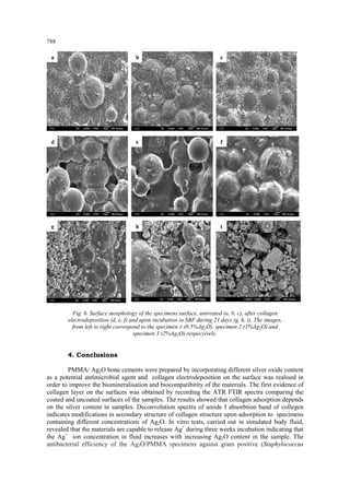
In order to study the morphological details of the samples surfaces, SEM analysis were performed before and after collagen electrodeposition and after 21 days incubation in SBF solution, and presented in fig. 6. Before any treatments (fig. 6 a, b, c) the surfaces presents the granular, regular shape of the PMMA bone cement and dispersed silver oxide particles. After electrodeposition, the images clearly illustrates the formation of collagen films on the sample surfaces (fig.6 d, e, f), covering uniformly the PMMA granules. According to the literature [15], once the protein has covered the surface of implants, host cells are no longer able to contact the underlying foreign-body material but only the protein–coated surface. The adsorbed protein layer- rather then the foreign material itself may stimulate or inhibit further biochemical processes. The bioactivity of the samples was demonstrated by the intense nucleation process of calcium containing crystals (fig. 6 g,h,i), the distribution of the aggregates being densely on the specimen 3 surface. The formation of hydroxyapatite crystals in this case was strongly influenced by the presence of collagen layer, as demonstrated by comparing with our previousely reported study on the bioactivity of silver/PMMA acrylic cements without collagen film [22,23].
10. 788
Fig. 6. Surface morphology of the specimens surface, untreated (a, b, c), after collagen electrodeposition (d, e, f) and upon incubation in SBF during 21 days (g, h, i). The images, from left to right correspond to the specimen 1 (0.5%Ag2O), specimen 2 (1%Ag2O) and specimen 3 (2%Ag2O) respecyively. 4. Conclusions
PMMA/ Ag2O bone cements were prepared by incorporating different silver oxide content as a potential antimicrobial agent and collagen electrodeposition on the surface was realised in order to improve the biomineralisation and biocompatibitity of the materials. The first evidence of collagen layer on the surfaces was obtained by recording the ATR FTIR spectra comparing the coated and uncoated surfaces of the samples. The results showed that collagen adsorption depends on the silver content in samples. Deconvolution spectra of amide I absorbtion band of collegen indicates modifications in secondary structure of collagen structure upon adsorption to specimens containing different concentrations of Ag2O. In vitro tests, carried out in simulated body fluid, revealed that the materials are capable to release Ag+ during three weeks incubation indicating that the Ag+ ion concentration in fluid increases with increasing Ag2O content in the sample. The antibacterial efficiency of the Ag2O/PMMA specimens against gram positive (Staphylococcus
a
b
c
d
e
f
g
h
i
11. 789
aureus ATCC 6538 P) and gram negative (Escherichia coli ATCC 2732), bacterium shows that he inhibition effect is significantly more intense in the case of Escherichia coli. The mineralization process upon 21days incubation time was also dependent on the Ag2O content in the samples. Moreover, the SEM micrographs show that formation of hydroxyapatite crystals was strongly influenced by the presence of collagen layer, but dependent on the silver oxide as well. Acknowledgements: This research was accomplished in the framework of Romania-Turkey Bilateral Cooperation, project nr. 385/2010. References [1] R. Huiskes, H. Weinans, B. VanRietbergen, Clin Orthop Res 274, 124 (1997). [2] F.K. Chang, J. L. Perez, J. A. Davidson, Journal of Biomedical Materials Research, 24(7), 873 (1990). [3] M. C. Rusu, D. L. Rusu, M. Rusu, JOAM –Symposia 1(6,) 1020 – 1026(2009) [4] Y.He, J.P. Trotignon, B.Loty, A. Tcharkhtchi, J. Verdu, J Biomed Mater Res 63, 800(2002). [5] H.Van de Belt, D.Neut, W. Schenk, J.R.Van Horn, H.C. Van der Mei, H. J. Busscher, Acta Orthop Scand 72, 557(2001). [6] R.Kumar, H.Munstedt, Biomaterials 26(14), 2081(2004). [7] M.Kawashita, S.Tsuneyama, F.Miyaji,T.Kokubo, K.Yamamoto, Biomaterials 21, 393 (2000). [8] J.Hardes, A. Streitburger, H. Ahrens, T. Nusselt, C. Gebert, W. Winkelmann, A.Battmann, G. Gosheger, Sarcoma, doi:10.1155/2007/26539 (2007) [9] A. Gupta, S. Silver, Nat Biotechnol 16, 888(1998) [10] Y. Fan, K. Duan, R. Wang, Biomaterials 26(14) 1623-1632( 2005) [11] S. Roessler, R. Born, D. Scharnweber, H. Worch, J Mater Sci Mater Med 12, 871-877(2001) [12] *** American Society for Testing and Materials (ASTM), Standard F451-99a, 2000 Annual Book of ASTM Standard, Vol. 13.01, 55 (2000). [13] S. Cavalu S., F. Banica , V. Simon , JOAM Symposia, 2, (1),140 (2010). [14] A. Dong, W.S. Caughey, Methods Enzymol. 232, 139 (1994). [15] S. Tunc, M.F. Maintz, G. Steiner, L. Vasquez, M.T. Pham, R. Salzer, Colloids Surf.B, 42, 219-225 (2005). [16] ] T. Kokubo, S. Ito, Z.T.Huang, T.Hayashi, S.Sakka, T.Kitsugi, T.Yamamuro, J Biomed Mater Res 24, 331(1990). [17] C. Du, F.Z. Cui, W. Zhang, Q.L. Feng, X.D. Zhu, K. De Groot, J. Biomed. Mater. Res. 50, 518-527(2000). [18] A. Balamurugan, S. Kannan, V. Selvaraj, S. Rajeswari, Trends Biomater Artif Organs 18(1) 41(2004). [19] I. Notinger, J.J. Blaker, V. Maquet, L.L. Hench, A. Boccaccini, Asian J. Physics 15(2) 221(2006). [20] J.C. Brodie, J. Merry, M.H. Grant, J. Mater. Sci: Mater Med 17,43-48 (2006). [21] S. Roessler, R. Born, D. Scharnweber, H. Worch, J Mater Sci Mater Med 12, 871-877(2001) [22] S. Cavalu, V. Simon, J. Optoelectron Adv. Mater. 8(4) 1520-1523(2006) [23] S.Cavalu, V.Simon, C. Albon, C. Hozan, JOAM 9(3) 693-697 (2007) [24] P. Sutandar, D.J.Ahn, E.I.Franses, Macromolecules 27,7316-7328 (1994) [25] W. Aken, Steen Dawis ed., Kluwer academic publishers, Norwell, Massachusetts, USA, (1989). [26] K. Vijayanand, D. K. Pattanayak, T. R. Rama Mohan, R. Banerjee, Trends Biomater. Artif. Organs,18 (2), 73-83 (2005). [27] A. Dong, J. D. Meyer, J.L Brown, M.C. Manning, J.F. Carpenter, Arch. Biochem. Biophys. 383, 148-155 (2000).
12. 790
[28] G. Damian, S. Cavalu, Asian Chem. Letters, 9(1-2), 3-9 (2005). [29] S. Cavalu, V. Simon, JOAM 9(11), 3297-3302 (2007). [30] L.J. Juszczak, J. Biol. Chem. 279(9), 7395-7404 (2004). [31] S. Leikin, V.A.Parsegian, W.H.Yang, G.E.Walrafen, Proc. Nat. Acad.Sci. USA, 94, 11312-11317 (1997). [32] K.J. Payne, A. Veis, Biopolymers, 27, 1749-1760 (1988). [33] J. D. Whittle, N.A. Bullett, R.D. Short, C.W. I. Douglas, A.P. Hollander, J. Davies, J. Mater. Chem., 2, 2726-2732 (2002). [34] K.J. Payne, A. Veis, Biopolymers, 27, 1749-1760 (1988). [35] M. Bellantone, H. D. Williams, L.L. Hench, Antimicrobial Agents and Chemotherapy 46(6), 1940-1945 (2002). [36] F. Heidarpour, W. A. Wan AB. Karim Ghani, F. R. Bin Ahmadun, S. Sobri, M. Zargar, M. R. Mozafari, Digest J Nanomat. Biostruct. 5(3), 797 (2010).