SlideShare a Scribd company logo
ORIGINAL RESEARCH
A Zebrafish Model of 5q-Syndrome Using CRISPR/Cas9 Targeting
RPS14 Reveals a p53-Independent and p53-Dependent Mechanism of
Erythroid Failure
Jason Ear a
, Jessica Hsueh a
, Melinda Nguyen a
, QingHua Zhang a
, Victoria Sung b
,
Rajesh Chopra c
, Kathleen M. Sakamoto d
, Shuo Lin a,*
a
Department of Molecular, Cell and Developmental Biology, University of California Los Angeles, Los Angeles, CA 90095, USA
b
Celgene Corporation, San Francisco 94158, USA
c
Celgene Corporation, Summit, NJ 07901, USA
d
Division of Hematology/Oncology, Department of Pediatrics, Stanford University School of Medicine, Stanford, CA 94304, USA
Received 28 January 2016; revised 21 February 2016; accepted 6 March 2016
Available online 2 April 2016
ABSTRACT
5q-syndrome is a distinct form of myelodysplastic syndrome (MDS) where a deletion on chromosome 5 is the underlying cause. MDS is
characterized by bone marrow failures, including macrocytic anemia. Genetic mapping and studies using various models support the
notion that ribosomal protein S14 (RPS14) is the candidate gene for the erythroid failure. Targeted disruption of RPS14 causes an increase
in p53 activity and p53-mediated apoptosis, similar to what is observed with other ribosomal proteins. However, due to the higher risk for
cancer development in patients with ribosome deficiency, targeting the p53 pathway is not a viable treatment option. To better understand
the pathology of RPS14 deficiency in 5q-deletion, we generated a zebrafish model harboring a mutation in the RPS14 gene. This model
mirrors the anemic phenotype seen in 5q-syndrome. Moreover, the anemia is due to a late-stage erythropoietic defect, where the
erythropoietic defect is initially p53-independent and then becomes p53-dependent. Finally, we demonstrate the versatility of this model
to test various pharmacological agents, such as RAP-011, L-leucine, and dexamethasone in order to identify molecules that can reverse the
anemic phenotype.
KEYWORDS: 5q-syndrome; RPS14; Ribosomopathy; Myelodysplastic syndrome
INTRODUCTION
Chromosome 5q-deletion syndrome, a.k.a. 5q-syndrome, is a
hematological disorder characterized as a distinct form of
myelodysplastic syndrome (MDS) where patients harbor a
deletion on chromosome 5. Approximately 10% to 20% of all
MDS cases are caused by chromosomal deletions. Patients
with 5q-syndrome have macrocytic anemia, and commonly
present themselves with other hematological phenotypes (i.e.,
thrombocytosis and megakaryocyte hyperplasia) (Boultwood
et al., 1994; Barlow et al., 2010b). Unlike MDS patients
without 5q-deletion, there is a relatively low rate of progres-
sion to acute myeloid leukemia (AML). Lenalidomide, a
thalidomide analog, has been shown to increase erythropoiesis
in patients with 5q-syndrome; however, in some cases, there is
a deleterious effect, which leads to thrombocytopenia and
neutropenia (List et al., 2005, 2006; Wei et al., 2013). The side
effects of lenalidomide treatment create a need for further
mechanistic studies on 5q-syndrome and for the identification
of novel therapeutics.* Corresponding author. Tel: þ1 310 267 4970, fax: þ1 310 267 4971.
E-mail address: shuolin@ucla.edu (S. Lin).
Available online at www.sciencedirect.com
ScienceDirect
Journal of Genetics and Genomics 43 (2016) 307e318
JGG
http://dx.doi.org/10.1016/j.jgg.2016.03.007
1673-8527/Copyright Ó 2016, Institute of Genetics and Developmental Biology, Chinese Academy of Sciences, and Genetics Society of China. Published by Elsevier
Limited and Science Press. All rights reserved.
A commonly deleted region (CDR) of 5q-syndrome has
been narrowed to a 1.5-Mb locus on chromosome 5
(Boultwood et al., 1994, 2002). Several genes in this CDR
have been suggested to play a role in the pathogenesis of the
disease, with haploinsufficiency of ribosomal protein S14
(RPS14) contributing to the anemic phenotype (Barlow et al.,
2010b; Boultwood, 2011). As a consequence of this genetic
aberration, expression of RPS14 is severely downregulated in
patients with 5q-deletion (Boultwood et al., 2007; Wu et al.,
2013). Knockdown of RPS14 using RNAi in CD34þ
he-
matopoietic progenitor cells severely affected erythroid dif-
ferentiation, similar to what is observed in patients (Ebert
et al., 2008). Furthermore, a small chromosomal deletion in
a mouse model containing a deletion of RPS14 was shown to
have macrocytic anemia (Barlow et al., 2010a). The anemic
correlation between RPS14 deficiency and 5q-deletion led to
the classification of 5q-syndrome as a ribosomopathy (a dis-
order due to aberrant ribosome function or biogenesis).
Haploinsufficiency of ribosomal proteins has been impli-
cated in several bone marrow failure disorders including
DiamondeBlackfan anemia (DBA), Dyskeratosis congenital
(DKC), ShwachmaneDiamond syndrome (SDS), and
Cartilage-hair hypoplasia (CHH) (Luzzatto and Karadimitris,
1998; Draptchinskaia et al., 1999; Ganapathi et al., 2007;
Thiel and Rauch, 2011). A deficiency in ribosomal proteins
has been shown to cause an increase in the activity of the
tumor suppressor, p53. Activation of the p53-pathway leads to
cell cycle arrest and apoptosis. While the exact mechanism of
p53 activation remains to be fully understood, a ribosomal
protein (RP)-mdm2-p53 checkpoint mechanism seems to be
involved (Boultwood, 2011; Miliani de Marval and Zhang,
2011). In a mouse model of 5q-syndrome, an elevated level
of p53 and cell death is observed. Furthermore, crossing the
model into a p53-null background reverses the degree of cell
death (Barlow et al., 2010a). While these studies may suggest
targeting p53 as a potential therapeutic strategy, caution is
warranted in this approach due to a risk of tumor development
when targeting this pathway. Inhibiting p53 is further ill-
advised when one considers the elevated risk in cancer
development in patients with ribosomopathies. Recently,
various studies have demonstrated that both p53-dependent
and p53-independent pathways play a role in the pathology
of the disease (Danilova et al., 2008; Narla et al., 2014).
Targeting the p53-independent pathway, therefore, offers a
more attractive therapeutic strategy.
In this study, through the use of CRISPR/Cas9 gene targeting,
we generated a zebrafish model of RPS14 deficiency in order to
mirrorthe anemic phenotype of patients with5q-sydrome. Using
this model, we demonstrate that a delay in erythroid maturation
leads to anemia in embryos deficient of RPS14 through a
p53-independent and a p53-dependent mechanism. This delay is
partially reversed upon treatment with RAP-011 (an activin
receptor type 2A ligand trap), L-leucine, as well as with
dexamethasone. In all, this model will serve as a useful model to
further investigate the mechanism underlying ribosome-
associated disorders and to test various drug candidates in an
intact animal model.
RESULTS
CRISPR/Cas9 targeting of RPS14
A deletion on chromosome 5, which contains the RPS14 gene,
is expected to lead to haploinsufficient levels of ribosomal
protein S14. The zebrafish RPS14 gene is located on chro-
mosome 21 and its encoded protein shares over 99% amino
acid identity to the human RPS14 (Fig. S1). To target the
RPS14 gene in zebrafish, we applied CRISPR/Cas9 gene-
editing technologies and generated a guide-RNA (gRNA)
targeting exon 2 of the gene, near the translational start site. To
facilitate validation of nuclease activity and screening, we
targeted an AvrI I restriction digest site on exon 2 of the gene
(Fig. 1A). Injection of the in vitro synthesized gRNA, along
with Cas9 mRNA, into the zebrafish embryos successfully
targeted the AvrI I restriction enzyme cut site, as indicated by
the retention of an upper band after restriction digest of
the PCR product with restriction endonuclease (Fig. 1B).
Interestingly, a population of the injected embryos displayed a
decrease in erythroid levels as revealed through the use of o-
dianisidine staining to label hemoglobin positive cells
throughout the entire embryo (Fig. 1C).
Injected embryos were raised to adulthood and mutants
were screened for transmission into germline (Fig. 1D).
Progeny from positive germline carriers were genotyped, and
mutations containing insertions as well as deletions were
revealed, thereby confirming the success targeting of RPS14 in
zebrafish (Fig. 1E).
Deficiency in RPS14 leads to morphological defects and
an increase in apoptosis
Three mutant lines were outcrossed and maintained for further
analysis (Fig. 1E and F). Two lines carried introduction of an
early stop codon (RPS14D4bp and RPS14DINS) and one line
(RPS14D21bp) carried an in-frame deletion (Fig. 1F).
RPS14DINS was incrossed and embryos carrying two
copies of the mutation displayed aplasia in the head at
24 hours post fertilization (hpf), and by 30 hpf, a decrease in
pigmentation and defective yolk extension was apparent
(Fig. 2A, A0
, and B). By 48 hpf, there was a slight recovery in
pigmentation; however, the embryos had a smaller head,
edema, and retained the defective yolk extension (Fig. 2C).
Overtime, the severity of the phenotypes increased and the
mutation became lethal around 6 to 7 days post fertilization
(dpf) (Fig. 2DeF). These morphologically abnormal embryos
were confirmed to be mutants for RPS14 through PCR vali-
dation (Fig. 3A). The RPS14D4bp mutants also displayed
similar morphological abnormalities to the RPS14DINS line
(Figs. 2, and S2); therefore, the RPS14DINS line was used for
the remainder of the work.
No observable defects were present in the RPS14D21bp
(Fig. S2). In fact, homozygous carriers of this mutation were
able to grow to adulthood and give rise to homozygous
progenies (Fig. S3). This suggests that the observed phenotype
308 J. Ear et al. / Journal of Genetics and Genomics 43 (2016) 307e318
in the RPS14DINS and RPS14D4bp is due to a null mutation
in the gene, and not through random mutation.
Next, to determine if the morphological defect is accom-
panied with an increase in levels of apoptosis, we performed
TUNEL staining on whole embryos. At all time points
analyzed, an increase in TUNEL staining positive cells was
observed in mutant embryos compared to age matched sibling
control (Fig. 2G). This falls in line with previous findings
RPS14
Exon 1 Exon 2
Translational start codon
Uninjected
F0
embryos
F1
embryos
F0
adults
F1
adults
U
ninjectedInjected Cas9 nuclease
Guide RNA
Exon 3
Exon 4
Exon 5
Avrl l cut site
48 hpf
gRNA/Cas9
A
B
D
E F
C
Fig. 1. Targeting RPS14 using CRISPR/Cas9.
A: Illustration depicting the RPS14 target site. B: In Cas9 mRNA and RPS14 gRNA injected embryos, region flanking the AvrI I restriction digest site was PCR
amplified followed by restriction digest of the PCR product with AvrI I. C: o-dianisidine staining of Cas9 mRNA and RPS14 gRNA injected embryos.
Approximately 50% of embryos displayed reduction in staining. D: Screening scheme used to identify RPS14 mutants. Injected embryos were raised to adulthood
and crossed for screening. Embryos (F1 embryos) were screened by restriction digest and individual clutches containing mutant carriers were raised to adulthood.
F1 adults were tail cut and screened for mutations in RPS14 through restriction digest. E: Genomic sequences of mutations found in RPS14. F: Predicted protein
sequence based on DNA sequence.
309J. Ear et al. / Journal of Genetics and Genomics 43 (2016) 307e318
demonstrating that a deficiency in RPS14 lead to an increase
in apoptosis. Interestingly, apoptosis was scattered throughout
the embryo and not apparent in regions of blood development.
To confirm that the morphologically abnormal embryos are
truly deficient for RSP14, transcript measurements for RPS14
were performed using quantitative real-time PCR (qPCR). At
both 24 and 48 hpf, the amount of RPS14 transcripts was
significantly downregulated compared to morphologically
normal sibling control embryos (Fig. 3B). This was further
confirmed by whole-mount in situ hybridization (WISH)
(Fig. 3C). Strong expression of RPS14 is normally observed
throughout the embryo at 24 and 48 hpf; however, in our
mutants we can see a severe reduction in expression levels.
Additionally, there were no changes in expression levels of
ribosomal protein RPL11 or RPS19 between mutants and
control groups (Fig. S4), indicating that the phenotype is
specifically due to loss of RPS14.
Finally, we confirmed that the morphological defects in our
mutants was due to a decrease in levels of RPS14 by per-
forming a rescue experiment using in vitro transcribed RPS14
24 hpf
48 hpf
4 dpf
48 hpf24 hpf
30 hpf
Sibling
Sibling
Sibling
Sibling Sibling Sibling
5 dpf
3 dpf
Sibling
3 dpf
Sibling
Mutant
Mutant
Mutant
Mutant Mutant Mutant
Mutant
Mutant
Mutant
Sibling
A
C
E
G
F
D
A′ B
Fig. 2. RPS14 deficient embryos display physical abnormalities as well as increase in apoptosis.
AeF: Morphological phenotype of RPS14 deficient embryos. At 24 hpf (A and A0
), aplasia (arrowhead) can be seen in the head region. By 30 hpf (B), there is a
delay in pigmentation. At 48 hpf (C) and 3 dpf (D), a smaller head and edema (arrowhead) are present. At 4 dpf (E), edema (arrowhead) is more apparent. At 5 dpf
(F), embryos still continue to have severe edema and smaller head, and display a high degree of lethality. Most embryos die between 5 and 6 dpf. G: In situ cell
death staining of morphologically abnormal embryos. Arrowheads point to the region of positive TUNEL staining.
310 J. Ear et al. / Journal of Genetics and Genomics 43 (2016) 307e318
mRNA. Upon rescue using RPS14 mRNA, mutants showed an
improvement in morphology (Fig. 3D). This suggests that a
deficiency in RPS14 leads to the morphological defects seen in
our mutant embryos.
Erythroid failure is observed upon RPS14 deficiency
Because ribosome deficiency typically leads to anemia in
patients, we sought to study the effects of RPS14 deficiency
on the development of red blood cells in our model. RPS14
mutants at 48 hpf and 3 dpf showed decreased levels of
o-dianisidine positive cells, suggesting a defect in the
production of mature erythroid cells in the embryos (Fig. 4A).
This decrease in o-dianisidine staining can be restored
when embryos were injected with RPS14 mRNA (Fig. S5),
suggesting that the anemic phenotype is due to a deficiency in
RPS14.
To better assess the erythroid defect, we crossed the RPS14
mutant line into the LCR2:EGFP transgenic fish line, which
specifically have all globin positive cells labeled with EGFP
(Ganis et al., 2012). We observed a similar degree in the amount
of EGFP positive cells between mutant and sibling control
embryos at 48 hpf and a slightly lower amount of EGFP positive
cells in mutant embryos at 3 dpf (Fig. 4B). Interestingly, sibling
control had two populations of EGFP expressing cells (high- and
low-EGFP), whereas mutants predominantly contained one
population of EGFP expressing cells with an intermediate level
of EGFP expression (Figs. 4C, D and S6). Analysis of the
cells using flow cytometry revealed no change in the size of the
cell (data not shown). The presence of EGFP positive cells
and lack of o-dianisidine staining suggest that a defect in the
terminal maturation of erythroid cells is impaired upon RPS14
deficiency.
To further characterize the blood defect in our model, we
performed WISH for various erythroid markers throughout
different stages of embryonic development. gata1 is one of the
earliest markers used to detect the specification of hemato-
poietic cells into the erythroid lineage (Detrich et al., 1995).
1.2
0.8
0.6
0.4
0.2
0
1.0
RPS 14ΔINS
WT
Sibling Mutant
M
arker(200
bp)
Relativefoldinduction
24 hpf
24 hpf
Sibling
Sibling Mutant
Mutant Mutant
Sibling
48 hpf
48 hpf
48 hpf
RPS 14
Sibling + RPS14 mRNA Mutant + RPS14 mRNA
* *A
C
D
B
Fig. 3. Deficiency in RPS14 is reversed by RPS14 mRNA injections.
A: PCR genotype of RPS14 mutant embryos. B: qPCR of transcript levels of RPS14. *, P < 0.05. C: WISH for RPS14 in mutants and wild-type sibling at 24 hpf
(left) or 48 hpf (right). D: Rescue of RPS14 deficient embryos with in vitro transcribed RPS14 mRNA. Arrowhead points to the rescue in head defect.
311J. Ear et al. / Journal of Genetics and Genomics 43 (2016) 307e318
Other markers of erythroid cells used in zebrafish include
globin transcripts such as hbbe1.1 and hbae3. There was no
observable difference in expression levels of gata1, hbbe1.1,
and hbae3 between mutant and sibling control embryos at
24 hpf, suggesting that RPS14 deficiency does not lead to
defects in the specification of erythroid cells (Fig. 5A).
Expression of gata1 was normally downregulated in the
zebrafish after 24 hpf. The expression level of gata1 in the
RPS14 mutants was downregulated in a similar fashion to
sibling control (Fig. 5B). In addition, there was a similar
expression level of global transcript at 48 hpf and only a
slightly lower level of expression at 3 dpf (Fig. 5B and C).
100
80
60
40
20
0
0 102
103
104
105
Cellcount(%ofmax.)
EGFP intensity
Merge
Sibling
Mutant
Mutant
Mutant
Mutant Mutant
Mutant Mutant
Mutant Mutant
EGFP DAPI
Sibling
Sibling Sibling Sibling
Sibling
Sibling Sibling
Sibling Sibling
Mutant
48 hpf
24 hpf
3 dpf 3 dpf
48 hpf Ventral view
Ventral view
3 dpfA
B
C D
Fig. 4. RPS14 deficiency leads to anemia in mutant embryos.
A: o-dianisidine staining for RPS14 between siblings or mutants at 48 hpf (left) or 3 dpf (right). Arrowheads point to the sites of weak o-dianisidine staining. B:
RPS14 mutation was bred into LCR:EGFP transgenic fish line. EGFP positive cells can be seen circulating throughout mutant embryos. At 24 and 48 hpf,
approximately equal number of EGFP positive cells can be seen. At 3 dpf, mutants display a slightly lower number of EGFP positive cells. C: Blood cells from
48 hpf embryos were smeared onto glass slides and immunostained for EGFP. Two populations of EGFP positive cells can be seen in sibling control (arrowhead,
EGFP-high and asterisk, EGFP-low). Mutants show a more intermediate phenotype of EGFP expression. D: EGFP positive cells from 48 hpf embryos were
analyzed by flow cytometry.
312 J. Ear et al. / Journal of Genetics and Genomics 43 (2016) 307e318
When the expression pattern of mpx was analyzed, we
observed a similar expression pattern between mutants and
sibling control, suggesting that the specification of primitive
myeloid cells is unaffected (Fig. S7). Overall, these data
suggest that hematopoiesis is affected in our model of RPS14
deficiency and that the anemia is due to a late-stage terminal
maturation of erythroid cells.
p53-activity is upregulated in embryos deficient for
RPS14
Haploinsufficiency of ribosome proteins has been shown to
cause elevated levels of p53 and cell cycle arrest (Danilova
et al., 2008). As expected, a mutation in RPS14 led to
increased levels of p53 and p53-target genes (Fig. 6A and B).
Furthermore, this increase was not restricted to hematopoietic
tissues, but throughout the entire embryo (Fig. 6A).
To determine if targeting the p53-pathway can alleviate the
anemic phenotype in our model, we injected morpholino
targeting p53 in our model (Fig. 6C). Targeting p53 led to a
slight morphological rescue in our model (Fig. 6D). However,
when we performed o-dianisidine staining on p53-morpholino
( p53-MO) injected embryos, no rescue was observed at
48 hpf (Fig. 6E). Interestingly, targeting p53 rescued anemic
phenotype at 3 dpf (Fig. 6F). Injection of p53-MO did not
show any observable changes to the erythroid levels in wild-
type sibling control embryos (Fig. S8). Taken together, these
findings suggest that upon RPS14 deficiency, both the p53-
independent and p53-dependent pathways are involved in
the anemic phenotype.
RAP-011 reverses the anemia associated with RPS14
In another model of ribosome deficiency, we demonstrated
that an elevated level of lft1 leads to decreased levels of
mature erythroid cells (Ear et al., 2015). Furthermore, the
effects of lft1 overexpression can be reversed upon treatment
with the activin receptor ligand-trap, RAP-011. In RPS14
deficiency, an elevated level of lft1 is also observed (Fig. 7A),
which is comparable to that seen in other models of
ribosomopathies (Ear et al., 2015). Interestingly, we observe
that lft1 is normally expressed in an asymmetric pattern in the
embryos in presumably hatching gland cells. Upon RPS14
deficiency, the asymmetric pattern is loss and lft1 is expressed
24 hpf
Sibling
24 hpf
Sibling
24 hpf
Sibling
48 hpf
Sibling
48 hpf
Sibling
3 dpf
Sibling
3 dpf
Sibling
48 hpf
Sibling
gata1 Mutant hbbe1.1 Mutant hbae3 Mutant
hbae3 Mutant
hbae3 Mutant
A
B
C
hbbe1.1 Mutant
hbbe1.1 Mutant
gata1 Mutant
Fig. 5. Expression of erythroid specific transcripts is unaffected in RPS14 mutants.
WISH shows that expression levels of gata1, hbbe1.1, and hbae3 in mutants are unaffected at 24 hpf (A) and 48 hpf (B). C: Slight reduction in hbbe1.1 and hbae3
expression appears at 3 dpf in mutant embryos.
313J. Ear et al. / Journal of Genetics and Genomics 43 (2016) 307e318
in a more bilateral pattern over the yolk (Fig. 7B). Using
RAP-011 in our model, we showed that it could partially
reverse the anemic phenotype associated with RSP14
deficiency (Fig. 7C).
Finally, we wanted to investigate if other treatments could
be used in our model. We found that treatment with either L-
leucine or dexamethasone could also partially rescue the
anemic phenotype in our model (Fig. 7D).
*
*
*
8
7
6
5
4
3
2
1
0
7
6
5
4
3
2
1
0
Relativefoldinduction
p53
p53
p21
p21
m
dm
2
m
dm
2
pum
a
bax
24 hpf
48 hpf
48 hpf
3 dpf
48 hpf
Mutant
Mutant
+ p53-MO
p53 p21
p21p53
Sibling
Sibling
Sibling
Sibling
+ p53-MO
Mutant
+ p53-MO
Mutant
+ p53-MO
Mutant Mutant Mutant
Mutant
Mutant
Sibling
Mutant
+ p53-MOMutant
Sibling Sibling
mdm2
mdm2
A
B
D
E
F
C
Fig. 6. p53-independent and p53-dependent mechanisms contribute to the anemic phenotype in RPS14 deficiency.
A: WISH shows that p53, p21, and mdm2 are ubiquitously expressed in RPS14 deficient embryos. B: Transcript measurement of p53 and p53-target genes ( p21,
mdm2, puma, bax) was preformed by qPCR. The value is represented as fold change over sibling control. C: qPCR was used to confirm knockdown of p53 and
p53-target genes in RPS14 mutants using p53-MO. *, P < 0.05. D: Slight morphological rescue (arrowhead) was observed in mutants with knockdown of p53.
E and F: o-dianisidine staining of mutants with p53-MO at 48 hpf (E) and 3 dpf (F). Arrowheads in F point to the rescue in o-dianisidine staining. Observed
phenotype: E, mutant ¼ 22/22, mutant þ p53-MO ¼ 19/20. F, mutant ¼ 20/20, mutant þ p53-MO ¼ 20/25.
314 J. Ear et al. / Journal of Genetics and Genomics 43 (2016) 307e318
2
1.5
1
0.5
0
lft1relativefoldinductiont
Sibling
M
utant
lft1
48 hpf
48 hpf
48 hpf
Sibling
Sibling Mutant
Sibling
+ IgG
Sibling
+ RAP-011
Mutant
+ RAP-011
Sibling
+ 0.2% DMSO
Sibling
+ L-leucine
Sibling
+ dexamethasone
Mutant
+ dexamethasone
Mutant
+ L-leucine
Mutant
+ 0.2% DMSO
Mutant
+ IgG
Mutant
A B
C
D
Fig. 7. RAP-011, L-leucine, and dexamethasone recover hemoglobin levels in RPS14 deficiency.
A: lft1 transcript level measured by qPCR at 48 hpf. Data are represented as fold induction over sibling control. B: WISH for lft1 in mutants or sibling control at
48 hpf. Arrowhead points to the region of lft1 expression. C: RAP-011 was injected into RPS14 mutants and o-dianisidine staining was performed to observe
hemoglobin levels (arrowheads). D: L-leucine (150 mmol/L), dexamethasone (50 mmol/L), or DMSO control was used to treat RPS14 deficient embryos and
hemoglobin levels (arrowheads) were analyzed by o-dianisidine staining. Observed phenotype: C, mutant þ IgG ¼ 25/25, mutant þ RAP-011 ¼ 23/26.
D, mutant ¼ 20/20, mutant þ DMSO ¼ 19/19, mutant þ L-leucine ¼ 18/21, mutant þ dexamethasone ¼ 18/23.
315J. Ear et al. / Journal of Genetics and Genomics 43 (2016) 307e318
DISCUSSION
In this work, we successfully targeted RPS14 in the zebrafish
using CRISPR/Cas9 gene-editing technology. Upon RPS14
deficiency, gross morphological defects can be observed and is
accompanied with an elevation in p53 activity. Furthermore,
the anemic phenotype that is typically seen in patients with
disrupted ribosome gene function is also observed.
Recent work comparing morpholino and genetic mutants
has raised concern regarding the off-target effects of MO (Kok
et al., 2015). Therefore, genetic models to compliment the
findings using MO studies are highly suggested. We see that
the phenotype in our model mirrors what is observed with
MO-mediated knockdown of RPS14 in zebrafish (Narla et al.,
2014).
The presence of normal gata1 and globin expression but
reduced levels of o-dianisidine staining, together, suggest a
late-stage terminal differentiation defect. The lack of obvious
apoptosis in hematopoietic tissues and normal specification of
erythroid cells suggest that the anemia associated with RPS14
deficiency is due to an inability to produce sufficient levels of
mature hemoglobin protein in the erythroid cells.
Knockdown of p53 in our model did not restore hemoglo-
bin levels at 48 hpf; however, increased levels were observed
at 3 dpf. While the exact role of p53 in our model remains
unclear, from our observation, we hypothesize that both p53-
independent and dependent mechanisms lead to the erythroid
failure and are separated temporally with the p53-independent
mechanism preceding the p53-dependent mechanism. Due to
the ubiquitous expression of p53 during ribosome deficiency, it
is difficult to separate the cell autonomous vs. the non-cell
autonomous role of p53 activity in erythroid cells; however,
we believe that further analysis into the various populations of
EGFP positive cells may give further insight into the mecha-
nism of erythroid failure.
In a previously described zebrafish model of ribosome
deficiency, a defect in protein production was shown to be
involved in the anemic phenotype (Zhang et al., 2014).
Furthermore, RPS14 deficiency in mice revealed activation of
p53 underscoring the erythroid differentiation defect (Barlow
et al., 2010a). Erythroid cells require a relatively high pro-
tein translational rate to meet the large globin protein
requirement. It may be possible that during the early stages of
erythroid development, ribosome deficiency leads to defects in
protein production (and thus insufficient globin levels), which
is independent of p53, and as development progresses, further
deficiency in ribosome levels leads to defects that are p53-
dependent.
Finally, we showed that the anemia in our model is
reversed upon treatment with RAP-011, L-leucine, and
dexamethasone. Interestingly, both RAP-011 and L-leucine
were previously demonstrated to work by p53-independent
mechanisms (Narla et al., 2014; Ear et al., 2015). The fact
that patients with ribosomopathies have a higher incidence
for cancer development warrants caution in targeting the p53
pathway for therapeutic treatment. Therefore, our work
rationalizes ongoing clinical trials investigating the use of
RAP-011 and L-leucine for the treatment of patients with
ribosome-associated disorders such as MDS and DBA.
MATERIALS AND METHODS
Zebrafish stains and husbandry
Zebrafish protocol and maintenance were performed using
methods approved by the University of California, Los
Angeles Institutional Animal Care and Use Committee
(UCLA-IACUC). Mutants were generated in wild-type (AB)
strain.
Cas9 mRNA and gRNA synthesis
Zebrafish optimized Cas9 mRNA was in vitro transcribed from
pGH-T7-zCas9 as previously described (Chang et al., 2013).
200 pg of Cas9 mRNA was injected per embryo at the one-cell
stage.
RPS14 gRNA was synthesized using a protocol similar to
previously described methods (Chang et al., 2013). The RPS14
targeted site was flanked with a T7 promoter site at the 50
region and a scaffold sequence on the 30
region (Oligo:
ATTTAGGTGACACTATAGAAGAGCAGGTCATCAGCCT
GTTTTAGAGCTAGAAATAGC). This oligo was used in a
PCR reaction to generate template for the gRNA synthesis.
gRNA was in vitro transcribed using MAXIscript T7 Kit (Life
Technologies, USA). After synthesis, DNA template was
removed using TURBO DNase (Life Technologies) and pu-
rified using phenol chloroform. 50 pg of gRNA (mixed with
the Cas9 mRNA) was injected into the embryos at the one-cell
stage.
PCR validation, sequencing mutation, and genotyping of
embryos
To extract genomic DNA, embryos (or tail fin clips) were
lysed in 50 mmol/L NaOH by boiling at 95
C for 1 h. After
boiling, lysates were neutralized with 10% volume 1 mol/L
Tris (pH ¼ 8.0). Crude lysates were then used in a PCR
reaction.
To validate the successful targeting of RPS14 and identify
germline transmitted fish, the region flanking the target site
was PCR amplified using RPS14 Fwd: 50
-CTGTTG
ACTGTGTGTTTCAGCA-30
and RPS14 Rev: 50
-TCAAA-
CATTCAATGCAACAAAAC-30
primers. PCR product was
used in a restriction digest reaction using AvrI I (NEB, USA).
To determine the DNA sequence, PCR products from positive
carriers were used in a TOPO-TA reaction (Life Technologies)
according to manufacturer direction.
After generating a stable mutant line, fish lines were
genotyped using primers RPS14 gel screen Fwd:
50
-ATGGCTCCTCGCAAGGGTAAGG-30
and RPS14 gel
screen Rev: 50
-TGTGCACACAAAGACACTGTT-30
. The
PCR product was then run on a 10% TBE-PAGE gel to
differentiate between homozygous and heterozygous carriers.
316 J. Ear et al. / Journal of Genetics and Genomics 43 (2016) 307e318
o-dianisidine staining
Embryos were first fixed at room temperature for 2 h using 4%
paraformaldehyde (PFA) in PBS. Next, three washes (5 min
each) with PBS were performed to remove PFA. Embryos
were then incubated in an o-dianisidine staining solution (40%
ethanol, 0.65% H2O2, 10 mmol/L Na-Acetate, and 0.6 mg/mL
o-dianisidine (Sigma, USA)) in the dark for 30 min followed
by four wash times with PBS. To remove pigmentation, em-
bryos were placed into a bleach solution (1% KOH, 3% H2O2)
for 20 min. Finally, embryos were washed with PBS and
immediately imaged.
mRNA probes and in situ hybridization
Plasmids for antisense probe were made using Gateway
technology (Life Technologies). Genes were amplified using
primers flanked with attB1 forward or attB2 reverse sequences
and then used in a BP clonase II reaction with pDONR221
(Life Technologies). To generate DIG labeled antisense
probes, plasmids were linearized and synthesized using T7
RNA polymerase (Promega, USA) with DIG RNA Labeling
Mix (Roche, USA).
For hybridization, embryos were first fixed overnight at 4
C
using 4% PFA in PBS. After fixation, PFA was washed four
times (5 min each) with PBS, followed by two washes (5 min
each) with 100% methanol. Embryos were stored at À20
C.
WISH was performed using labeled probed as previously
described (Jowett, 1999).
Real-time quantitative PCR
To generate cDNA for real-time quantitative PCR analysis,
total RNA was first isolated from whole embryos (25 embryos
per group) using TRIzol Reagent (Life Technologies)
following manufacture’s protocol. Next, cDNA was reverse
transcribed using Oligo(dT) and SuperScript III reverse tran-
scriptase (Life Technologies). All qPCR experiments were
performed in triplicates.
For quantitative PCR analysis, cDNA was used in a PCR
reaction using FastStart SYBR Green Master (Roche). Oligos
used in qPCR are as previously described (Ear et al., 2015).
ACKNOWLEDGMENTS
We thank Dr. Matt Veldmen, Dr. Nadia Danilova, and Dr.
Anne Lindgren for their insightful discussions and input
throughout the course of the work. We acknowledge Linda
Dong for maintenance of the zebrafish strains used in the
experiments. Also, we thank Tianna Wilson for providing
invaluable technical assistance. This work was supported by
Celgene (Nov022011) and National Institutes of Health (R56
DK107286) to K.S. and S.L.
SUPPLEMENTARY DATA
Fig. S1. Protein alignment between human and zebrafish
RPS14.
Fig. S2. Phenotype of RPS14 mutant alleles.
Fig. S3. An inframe mutation of RPS14 does not affect
development.
Fig. S4. RPS14 mutants have normal expression of other ri-
bosomal protein transcripts.
Fig. S5. o-dianisidine staining of RPS14 mutants rescued with
RPS14 mRNA.
Fig. S6. Flow cytometry analysis on EGFP positive cells from
48 hpf embryos.
Fig. S7. In situ hybridization of mpx transcript in RPS14
mutants at 48 hpf.
Fig. S8. p53-MO injection does not affect erythroid levels
under normal conditions.
Supplementary data related to this article can be found at
http://dx.doi.org/10.1016/j.jgg.2016.03.007.
REFERENCES
Barlow, J.L., Drynan, L.F., Hewett, D.R., Holmes, L.R., Lorenzo-Abalde, S.,
Lane, A.L., Jolin, H.E., Pannell, R., Middleton, A.J., Wong, S.H.,
Warren, A.J., Wainscoat, J.S., Boultwood, J., McKenzie, A.N., 2010a. A
p53-dependent mechanism underlies macrocytic anemia in a mouse model
of human 5q-syndrome. Nat. Med. 16, 59e66.
Barlow, J.L., Drynan, L.F., Trim, N.L., Erber, W.N., Warren, A.J.,
McKenzie, A.N., 2010b. New insights into 5q-syndrome as a ribosomop-
athy. Cell Cycle 9, 4286e4293.
Boultwood, J., 2011. The role of haploinsufficiency of RPS14 and p53 acti-
vation in the molecular pathogenesis of the 5q-syndrome. Pediatr. Rep. 3
(Suppl. 2), e10.
Boultwood, J., Fidler, C., Lewis, S., Kelly, S., Sheridan, H., Littlewood, T.J.,
Buckle, V.J., Wainscoat, J.S., 1994. Molecular mapping of uncharacteris-
tically small 5q deletions in two patients with the 5q-syndrome: delinea-
tion of the critical region on 5q and identification of a 5q-breakpoint.
Genomics 19, 425e432.
Boultwood, J., Fidler, C., Strickson, A.J., Watkins, F., Gama, S., Kearney, L.,
Tosi, S., Kasprzyk, A., Cheng, J.F., Jaju, R.J., Wainscoat, J.S., 2002.
Narrowing and genomic annotation of the commonly deleted region of the
5q-syndrome. Blood 99, 4638e4641.
Boultwood, J., Pellagatti, A., Cattan, H., Lawrie, C.H., Giagounidis, A.,
Malcovati, L., Della Porta, M.G., Jadersten, M., Killick, S., Fidler, C.,
Cazzola, M., Hellstrom-Lindberg, E., Wainscoat, J.S., 2007. Gene
expression profiling of CD34þ
cells in patients with the 5q-syndrome. Br.
J. Haematol. 139, 578e589.
Chang, N., Sun, C., Gao, L., Zhu, D., Xu, X., Zhu, X., Xiong, J.W., Xi, J.J.,
2013. Genome editing with RNA-guided Cas9 nuclease in zebrafish em-
bryos. Cell Res. 23, 465e472.
Danilova, N., Sakamoto, K.M., Lin, S., 2008. Ribosomal protein S19 defi-
ciency in zebrafish leads to developmental abnormalities and defective
erythropoiesis through activation of p53 protein family. Blood 112,
5228e5237.
Detrich 3rd, H.W., Kieran, M.W., Chan, F.Y., Barone, L.M., Yee, K.,
Rundstadler, J.A., Pratt, S., Ransom, D., Zon, L.I., 1995. Intraembryonic
hematopoietic cell migration during vertebrate development. Proc. Natl.
Acad. Sci. USA 92, 10713e10717.
Draptchinskaia, N., Gustavsson, P., Andersson, B., Pettersson, M.,
Willig, T.N., Dianzani, I., Ball, S., Tchernia, G., Klar, J., Matsson, H.,
Tentler, D., Mohandas, N., Carlsson, B., Dahl, N., 1999. The gene
317J. Ear et al. / Journal of Genetics and Genomics 43 (2016) 307e318
encoding ribosomal protein S19 is mutated in Diamond-Blackfan anaemia.
Nat. Genet. 21, 169e175.
Ear, J., Huang, H., Wilson, T., Tehrani, Z., Lindgren, A., Sung, V., Laadem, A.,
Daniel, T.O., Chopra, R., Lin, S., 2015. RAP-011 improves erythropoiesis
in zebrafish model of Diamond-Blackfan anemia through antagonizing
lefty1. Blood 126, 880e890.
Ebert, B.L., Pretz, J., Bosco, J., Chang, C.Y., Tamayo, P., Galili, N., Raza, A.,
Root, D.E., Attar, E., Ellis, S.R., Golub, T.R., 2008. Identification of RPS14
as a 5q-syndrome gene by RNA interference screen. Nature 451, 335e339.
Ganapathi, K.A., Austin, K.M., Lee, C.S., Dias, A., Malsch, M.M., Reed, R.,
Shimamura, A., 2007. The human Shwachman-Diamond syndrome pro-
tein, SBDS, associates with ribosomal RNA. Blood 110, 1458e1465.
Ganis, J.J., Hsia, N., Trompouki, E., de Jong, J.L., DiBiase, A., Lambert, J.S.,
Jia, Z., Sabo, P.J., Weaver, M., Sandstrom, R., Stamatoyannopoulos, J.A.,
Zhou, Y., Zon, L.I., 2012. Zebrafish globin switching occurs in two devel-
opmental stages and is controlled by the LCR. Dev. Biol. 366, 185e194.
Jowett, T., 1999. Analysis of protein and gene expression. Methods Cell Biol.
59, 63e85.
Kok, F.O., Shin, M., Ni, C.W., Gupta, A., Grosse, A.S., van Impel, A.,
Kirchmaier, B.C., Peterson-Maduro, J., Kourkoulis, G., Male, I.,
DeSantis, D.F., Sheppard-Tindell, S., Ebarasi, L., Betsholtz, C., Schulte-
Merker, S., Wolfe, S.A., Lawson, N.D., 2015. Reverse genetic screening
reveals poor correlation between morpholino-induced and mutant pheno-
types in zebrafish. Dev. Cell 32, 97e108.
List, A., Dewald, G., Bennett, J., Giagounidis, A., Raza, A., Feldman, E.,
Powell, B., Greenberg, P., Thomas, D., Stone, R., Reeder, C., Wride, K.,
Patin, J., Schmidt, M., Zeldis, J., Knight, R., 2006. Lenalidomide in the
myelodysplastic syndrome with chromosome 5q deletion. N. Engl. J. Med.
355, 1456e1465.
List, A., Kurtin, S., Roe, D.J., Buresh, A., Mahadevan, D., Fuchs, D.,
Rimsza, L., Heaton, R., Knight, R., Zeldis, J.B., 2005. Efficacy of lena-
lidomide in myelodysplastic syndromes. N. Engl. J. Med. 352, 549e557.
Luzzatto, L., Karadimitris, A., 1998. Dyskeratosis and ribosomal rebellion.
Nat. Genet. 19, 6e7.
Miliani de Marval, P.L., Zhang, Y., 2011. The RP-Mdm2-p53 pathway and
tumorigenesis. Oncotarget 2, 234e238.
Narla, A., Payne, E.M., Abayasekara, N., Hurst, S.N., Raiser, D.M.,
Look, A.T., Berliner, N., Ebert, B.L., Khanna-Gupta, A., 2014. L-
Leucine improves the anaemia in models of Diamond Blackfan anaemia
and the 5q-syndrome in a TP53-independent way. Br. J. Haematol. 167,
524e528.
Thiel, C.T., Rauch, A., 2011. The molecular basis of the cartilage-hair hy-
poplasia-anauxetic dysplasia spectrum. Best Pract. Res. Clin. Endocrinol.
Metab. 25, 131e142.
Wei, S., Chen, X., McGraw, K., Zhang, L., Komrokji, R., Clark, J.,
Caceres, G., Billingsley, D., Sokol, L., Lancet, J., Fortenbery, N., Zhou, J.,
Eksioglu, E.A., Sallman, D., Wang, H., Epling-Burnette, P.K., Djeu, J.,
Sekeres, M., Maciejewski, J.P., List, A., 2013. Lenalidomide promotes p53
degradation by inhibiting MDM2 auto-ubiquitination in myelodysplastic
syndrome with chromosome 5q deletion. Oncogene 32, 1110e1120.
Wu, L., Li, X., Xu, F., Zhang, Z., Chang, C., He, Q., 2013. Low RPS14
expression in MDS without 5q e aberration confers higher apoptosis rate
of nucleated erythrocytes and predicts prolonged survival and possible
response to lenalidomide in lower risk non-5q-patients. Eur. J. Haematol.
90, 486e493.
Zhang, Y., Ear, J., Yang, Z., Morimoto, K., Zhang, B., Lin, S., 2014. Defects of
protein production in erythroid cells revealed in a zebrafish Diamond-
Blackfan anemia model for mutation in RPS19. Cell Death Dis. 5, e1352.
318 J. Ear et al. / Journal of Genetics and Genomics 43 (2016) 307e318

More Related Content

What's hot

article
articlearticle
article
dalya shakir
 
Whitepaper serumplasma
Whitepaper serumplasmaWhitepaper serumplasma
Whitepaper serumplasma
Elsa von Licy
 
Crispr cas9 in pancreatic cancer
Crispr cas9 in pancreatic cancerCrispr cas9 in pancreatic cancer
Crispr cas9 in pancreatic cancer
IzzxanTanpinarizza
 
The CDKN2A
The CDKN2AThe CDKN2A
The CDKN2A
Rpat8312
 
00b49517a6e686b6be000000
00b49517a6e686b6be00000000b49517a6e686b6be000000
00b49517a6e686b6be000000
kurnianto syaputra
 
Structure of p53 protein
Structure of p53 proteinStructure of p53 protein
Structure of p53 protein
Shruthi Krishnaswamy
 
Protien mutation& cystic fibrosis
Protien mutation& cystic fibrosisProtien mutation& cystic fibrosis
Protien mutation& cystic fibrosis
Dr.M.Prasad Naidu
 
onc2013588a
onc2013588aonc2013588a
onc2013588a
Anirudh Prahallad
 
Austin Hypertension
Austin HypertensionAustin Hypertension
Austin Hypertension
Austin Publishing Group
 
Kumar-Ricker-Poster-mesa_2013_V2
Kumar-Ricker-Poster-mesa_2013_V2Kumar-Ricker-Poster-mesa_2013_V2
Kumar-Ricker-Poster-mesa_2013_V2
shantanu kumar
 
1. Myosinⅵ Is A Mediator Of The P53 Dependent Cell
1. Myosinⅵ Is A Mediator Of The P53 Dependent Cell1. Myosinⅵ Is A Mediator Of The P53 Dependent Cell
1. Myosinⅵ Is A Mediator Of The P53 Dependent Cell
beneshjoseph
 
Shamah MCB 93
Shamah MCB 93Shamah MCB 93
Shamah MCB 93
Steve Shamah
 
C-Terminal Regions
C-Terminal RegionsC-Terminal Regions
C-Terminal Regions
Allyson C. Smith
 
Poster_mainFin1
Poster_mainFin1Poster_mainFin1
Poster_mainFin1
Ashish Tomar
 
5-2. C3 glomerulonephritis and Dense Deposit Disease. Francesco Emma (eng)
5-2. C3 glomerulonephritis and Dense Deposit Disease. Francesco Emma (eng)5-2. C3 glomerulonephritis and Dense Deposit Disease. Francesco Emma (eng)
5-2. C3 glomerulonephritis and Dense Deposit Disease. Francesco Emma (eng)
KidneyOrgRu
 
pnas-0503504102
pnas-0503504102pnas-0503504102
pnas-0503504102
Trinh Hoac
 
Role of p53 gene
Role of p53 gene Role of p53 gene
Role of p53 gene
Choclaty Ashish
 
P53 poster
P53 posterP53 poster
Blood2007
Blood2007Blood2007

What's hot (19)

article
articlearticle
article
 
Whitepaper serumplasma
Whitepaper serumplasmaWhitepaper serumplasma
Whitepaper serumplasma
 
Crispr cas9 in pancreatic cancer
Crispr cas9 in pancreatic cancerCrispr cas9 in pancreatic cancer
Crispr cas9 in pancreatic cancer
 
The CDKN2A
The CDKN2AThe CDKN2A
The CDKN2A
 
00b49517a6e686b6be000000
00b49517a6e686b6be00000000b49517a6e686b6be000000
00b49517a6e686b6be000000
 
Structure of p53 protein
Structure of p53 proteinStructure of p53 protein
Structure of p53 protein
 
Protien mutation& cystic fibrosis
Protien mutation& cystic fibrosisProtien mutation& cystic fibrosis
Protien mutation& cystic fibrosis
 
onc2013588a
onc2013588aonc2013588a
onc2013588a
 
Austin Hypertension
Austin HypertensionAustin Hypertension
Austin Hypertension
 
Kumar-Ricker-Poster-mesa_2013_V2
Kumar-Ricker-Poster-mesa_2013_V2Kumar-Ricker-Poster-mesa_2013_V2
Kumar-Ricker-Poster-mesa_2013_V2
 
1. Myosinⅵ Is A Mediator Of The P53 Dependent Cell
1. Myosinⅵ Is A Mediator Of The P53 Dependent Cell1. Myosinⅵ Is A Mediator Of The P53 Dependent Cell
1. Myosinⅵ Is A Mediator Of The P53 Dependent Cell
 
Shamah MCB 93
Shamah MCB 93Shamah MCB 93
Shamah MCB 93
 
C-Terminal Regions
C-Terminal RegionsC-Terminal Regions
C-Terminal Regions
 
Poster_mainFin1
Poster_mainFin1Poster_mainFin1
Poster_mainFin1
 
5-2. C3 glomerulonephritis and Dense Deposit Disease. Francesco Emma (eng)
5-2. C3 glomerulonephritis and Dense Deposit Disease. Francesco Emma (eng)5-2. C3 glomerulonephritis and Dense Deposit Disease. Francesco Emma (eng)
5-2. C3 glomerulonephritis and Dense Deposit Disease. Francesco Emma (eng)
 
pnas-0503504102
pnas-0503504102pnas-0503504102
pnas-0503504102
 
Role of p53 gene
Role of p53 gene Role of p53 gene
Role of p53 gene
 
P53 poster
P53 posterP53 poster
P53 poster
 
Blood2007
Blood2007Blood2007
Blood2007
 

Similar to 5q syndrome

2015 - Pyrimidine Pool Disequilibrium Induced by a Cytidine Deaminase Deficie...
2015 - Pyrimidine Pool Disequilibrium Induced by a Cytidine Deaminase Deficie...2015 - Pyrimidine Pool Disequilibrium Induced by a Cytidine Deaminase Deficie...
2015 - Pyrimidine Pool Disequilibrium Induced by a Cytidine Deaminase Deficie...
Simon Gemble
 
Strathern JBC 2013 2689 slippage
Strathern JBC 2013 2689 slippageStrathern JBC 2013 2689 slippage
Strathern JBC 2013 2689 slippage
Jordan Irvin
 
16. Biology of Cancer
16. Biology of Cancer16. Biology of Cancer
16. Biology of Cancer
Shailendra shera
 
news and views
news and viewsnews and views
news and views
Maria Teresa Esposito
 
Acute promyelocytic leukemia NCCN LATEST 2014 Guidelines
Acute promyelocytic leukemia NCCN LATEST 2014 GuidelinesAcute promyelocytic leukemia NCCN LATEST 2014 Guidelines
Acute promyelocytic leukemia NCCN LATEST 2014 Guidelines
Dr Sandeep Kumar
 
Omara-Opyene et al 2004
Omara-Opyene et al 2004Omara-Opyene et al 2004
Omara-Opyene et al 2004
Archangel (Levi) Omara-Opyene, PhD
 
pap paper pdf
pap paper pdfpap paper pdf
pap paper pdf
Fabio Da Silva
 
Microsatellite instability
Microsatellite instabilityMicrosatellite instability
Qualitative Flow Cytometric Analysis of Malaysian
Qualitative Flow Cytometric Analysis of MalaysianQualitative Flow Cytometric Analysis of Malaysian
Qualitative Flow Cytometric Analysis of Malaysian
Mohadese Hashem Boroojerdi
 
Rb gene and cell cycle
Rb gene and cell cycleRb gene and cell cycle
Rb gene and cell cycle
Fadel Muhammad Garishah
 
Dissertation
DissertationDissertation
Dissertation
Craig Williams
 
Colorecat carcinoma tissue biomarkers
Colorecat carcinoma tissue biomarkersColorecat carcinoma tissue biomarkers
Colorecat carcinoma tissue biomarkers
Rasha Haggag
 
acutepromyelocyticleukemia-
acutepromyelocyticleukemia-acutepromyelocyticleukemia-
acutepromyelocyticleukemia-
icdlab
 
Blood Group Genotyping
Blood Group GenotypingBlood Group Genotyping
Blood Group Genotyping
RuchiraMuhandiram1
 
PNAS-2015-Banerjee
PNAS-2015-BanerjeePNAS-2015-Banerjee
PNAS-2015-Banerjee
rajdeep banerjee
 
Molecular biology of salivary gland pathology
Molecular biology of salivary gland pathologyMolecular biology of salivary gland pathology
Molecular biology of salivary gland pathology
SHREYADAS49
 
Annals of Mutagenesis
Annals of Mutagenesis Annals of Mutagenesis
Annals of Mutagenesis
Austin Publishing Group
 
DOOR syndrome
DOOR syndromeDOOR syndrome
DOOR syndrome
Satrupa Das
 
P 53 Tumour Biology
P 53 Tumour BiologyP 53 Tumour Biology
P 53 Tumour Biology
Gaurav Dwivedi
 
Cancer Res-2015-Bonastre-1287-97
Cancer Res-2015-Bonastre-1287-97Cancer Res-2015-Bonastre-1287-97
Cancer Res-2015-Bonastre-1287-97
Sara Verdura
 

Similar to 5q syndrome (20)

2015 - Pyrimidine Pool Disequilibrium Induced by a Cytidine Deaminase Deficie...
2015 - Pyrimidine Pool Disequilibrium Induced by a Cytidine Deaminase Deficie...2015 - Pyrimidine Pool Disequilibrium Induced by a Cytidine Deaminase Deficie...
2015 - Pyrimidine Pool Disequilibrium Induced by a Cytidine Deaminase Deficie...
 
Strathern JBC 2013 2689 slippage
Strathern JBC 2013 2689 slippageStrathern JBC 2013 2689 slippage
Strathern JBC 2013 2689 slippage
 
16. Biology of Cancer
16. Biology of Cancer16. Biology of Cancer
16. Biology of Cancer
 
news and views
news and viewsnews and views
news and views
 
Acute promyelocytic leukemia NCCN LATEST 2014 Guidelines
Acute promyelocytic leukemia NCCN LATEST 2014 GuidelinesAcute promyelocytic leukemia NCCN LATEST 2014 Guidelines
Acute promyelocytic leukemia NCCN LATEST 2014 Guidelines
 
Omara-Opyene et al 2004
Omara-Opyene et al 2004Omara-Opyene et al 2004
Omara-Opyene et al 2004
 
pap paper pdf
pap paper pdfpap paper pdf
pap paper pdf
 
Microsatellite instability
Microsatellite instabilityMicrosatellite instability
Microsatellite instability
 
Qualitative Flow Cytometric Analysis of Malaysian
Qualitative Flow Cytometric Analysis of MalaysianQualitative Flow Cytometric Analysis of Malaysian
Qualitative Flow Cytometric Analysis of Malaysian
 
Rb gene and cell cycle
Rb gene and cell cycleRb gene and cell cycle
Rb gene and cell cycle
 
Dissertation
DissertationDissertation
Dissertation
 
Colorecat carcinoma tissue biomarkers
Colorecat carcinoma tissue biomarkersColorecat carcinoma tissue biomarkers
Colorecat carcinoma tissue biomarkers
 
acutepromyelocyticleukemia-
acutepromyelocyticleukemia-acutepromyelocyticleukemia-
acutepromyelocyticleukemia-
 
Blood Group Genotyping
Blood Group GenotypingBlood Group Genotyping
Blood Group Genotyping
 
PNAS-2015-Banerjee
PNAS-2015-BanerjeePNAS-2015-Banerjee
PNAS-2015-Banerjee
 
Molecular biology of salivary gland pathology
Molecular biology of salivary gland pathologyMolecular biology of salivary gland pathology
Molecular biology of salivary gland pathology
 
Annals of Mutagenesis
Annals of Mutagenesis Annals of Mutagenesis
Annals of Mutagenesis
 
DOOR syndrome
DOOR syndromeDOOR syndrome
DOOR syndrome
 
P 53 Tumour Biology
P 53 Tumour BiologyP 53 Tumour Biology
P 53 Tumour Biology
 
Cancer Res-2015-Bonastre-1287-97
Cancer Res-2015-Bonastre-1287-97Cancer Res-2015-Bonastre-1287-97
Cancer Res-2015-Bonastre-1287-97
 

Recently uploaded

aziz sancar nobel prize winner: from mardin to nobel
aziz sancar nobel prize winner: from mardin to nobelaziz sancar nobel prize winner: from mardin to nobel
aziz sancar nobel prize winner: from mardin to nobel
İsa Badur
 
Sharlene Leurig - Enabling Onsite Water Use with Net Zero Water
Sharlene Leurig - Enabling Onsite Water Use with Net Zero WaterSharlene Leurig - Enabling Onsite Water Use with Net Zero Water
Sharlene Leurig - Enabling Onsite Water Use with Net Zero Water
Texas Alliance of Groundwater Districts
 
Equivariant neural networks and representation theory
Equivariant neural networks and representation theoryEquivariant neural networks and representation theory
Equivariant neural networks and representation theory
Daniel Tubbenhauer
 
Bob Reedy - Nitrate in Texas Groundwater.pdf
Bob Reedy - Nitrate in Texas Groundwater.pdfBob Reedy - Nitrate in Texas Groundwater.pdf
Bob Reedy - Nitrate in Texas Groundwater.pdf
Texas Alliance of Groundwater Districts
 
Describing and Interpreting an Immersive Learning Case with the Immersion Cub...
Describing and Interpreting an Immersive Learning Case with the Immersion Cub...Describing and Interpreting an Immersive Learning Case with the Immersion Cub...
Describing and Interpreting an Immersive Learning Case with the Immersion Cub...
Leonel Morgado
 
Cytokines and their role in immune regulation.pptx
Cytokines and their role in immune regulation.pptxCytokines and their role in immune regulation.pptx
Cytokines and their role in immune regulation.pptx
Hitesh Sikarwar
 
Compexometric titration/Chelatorphy titration/chelating titration
Compexometric titration/Chelatorphy titration/chelating titrationCompexometric titration/Chelatorphy titration/chelating titration
Compexometric titration/Chelatorphy titration/chelating titration
Vandana Devesh Sharma
 
Medical Orthopedic PowerPoint Templates.pptx
Medical Orthopedic PowerPoint Templates.pptxMedical Orthopedic PowerPoint Templates.pptx
Medical Orthopedic PowerPoint Templates.pptx
terusbelajar5
 
3D Hybrid PIC simulation of the plasma expansion (ISSS-14)
3D Hybrid PIC simulation of the plasma expansion (ISSS-14)3D Hybrid PIC simulation of the plasma expansion (ISSS-14)
3D Hybrid PIC simulation of the plasma expansion (ISSS-14)
David Osipyan
 
Basics of crystallography, crystal systems, classes and different forms
Basics of crystallography, crystal systems, classes and different formsBasics of crystallography, crystal systems, classes and different forms
Basics of crystallography, crystal systems, classes and different forms
MaheshaNanjegowda
 
8.Isolation of pure cultures and preservation of cultures.pdf
8.Isolation of pure cultures and preservation of cultures.pdf8.Isolation of pure cultures and preservation of cultures.pdf
8.Isolation of pure cultures and preservation of cultures.pdf
by6843629
 
在线办理(salfor毕业证书)索尔福德大学毕业证毕业完成信一模一样
在线办理(salfor毕业证书)索尔福德大学毕业证毕业完成信一模一样在线办理(salfor毕业证书)索尔福德大学毕业证毕业完成信一模一样
在线办理(salfor毕业证书)索尔福德大学毕业证毕业完成信一模一样
vluwdy49
 
Deep Software Variability and Frictionless Reproducibility
Deep Software Variability and Frictionless ReproducibilityDeep Software Variability and Frictionless Reproducibility
Deep Software Variability and Frictionless Reproducibility
University of Rennes, INSA Rennes, Inria/IRISA, CNRS
 
如何办理(uvic毕业证书)维多利亚大学毕业证本科学位证书原版一模一样
如何办理(uvic毕业证书)维多利亚大学毕业证本科学位证书原版一模一样如何办理(uvic毕业证书)维多利亚大学毕业证本科学位证书原版一模一样
如何办理(uvic毕业证书)维多利亚大学毕业证本科学位证书原版一模一样
yqqaatn0
 
The debris of the ‘last major merger’ is dynamically young
The debris of the ‘last major merger’ is dynamically youngThe debris of the ‘last major merger’ is dynamically young
The debris of the ‘last major merger’ is dynamically young
Sérgio Sacani
 
Applied Science: Thermodynamics, Laws & Methodology.pdf
Applied Science: Thermodynamics, Laws & Methodology.pdfApplied Science: Thermodynamics, Laws & Methodology.pdf
Applied Science: Thermodynamics, Laws & Methodology.pdf
University of Hertfordshire
 
Travis Hills' Endeavors in Minnesota: Fostering Environmental and Economic Pr...
Travis Hills' Endeavors in Minnesota: Fostering Environmental and Economic Pr...Travis Hills' Endeavors in Minnesota: Fostering Environmental and Economic Pr...
Travis Hills' Endeavors in Minnesota: Fostering Environmental and Economic Pr...
Travis Hills MN
 
EWOCS-I: The catalog of X-ray sources in Westerlund 1 from the Extended Weste...
EWOCS-I: The catalog of X-ray sources in Westerlund 1 from the Extended Weste...EWOCS-I: The catalog of X-ray sources in Westerlund 1 from the Extended Weste...
EWOCS-I: The catalog of X-ray sources in Westerlund 1 from the Extended Weste...
Sérgio Sacani
 
The binding of cosmological structures by massless topological defects
The binding of cosmological structures by massless topological defectsThe binding of cosmological structures by massless topological defects
The binding of cosmological structures by massless topological defects
Sérgio Sacani
 
Shallowest Oil Discovery of Turkiye.pptx
Shallowest Oil Discovery of Turkiye.pptxShallowest Oil Discovery of Turkiye.pptx
Shallowest Oil Discovery of Turkiye.pptx
Gokturk Mehmet Dilci
 

Recently uploaded (20)

aziz sancar nobel prize winner: from mardin to nobel
aziz sancar nobel prize winner: from mardin to nobelaziz sancar nobel prize winner: from mardin to nobel
aziz sancar nobel prize winner: from mardin to nobel
 
Sharlene Leurig - Enabling Onsite Water Use with Net Zero Water
Sharlene Leurig - Enabling Onsite Water Use with Net Zero WaterSharlene Leurig - Enabling Onsite Water Use with Net Zero Water
Sharlene Leurig - Enabling Onsite Water Use with Net Zero Water
 
Equivariant neural networks and representation theory
Equivariant neural networks and representation theoryEquivariant neural networks and representation theory
Equivariant neural networks and representation theory
 
Bob Reedy - Nitrate in Texas Groundwater.pdf
Bob Reedy - Nitrate in Texas Groundwater.pdfBob Reedy - Nitrate in Texas Groundwater.pdf
Bob Reedy - Nitrate in Texas Groundwater.pdf
 
Describing and Interpreting an Immersive Learning Case with the Immersion Cub...
Describing and Interpreting an Immersive Learning Case with the Immersion Cub...Describing and Interpreting an Immersive Learning Case with the Immersion Cub...
Describing and Interpreting an Immersive Learning Case with the Immersion Cub...
 
Cytokines and their role in immune regulation.pptx
Cytokines and their role in immune regulation.pptxCytokines and their role in immune regulation.pptx
Cytokines and their role in immune regulation.pptx
 
Compexometric titration/Chelatorphy titration/chelating titration
Compexometric titration/Chelatorphy titration/chelating titrationCompexometric titration/Chelatorphy titration/chelating titration
Compexometric titration/Chelatorphy titration/chelating titration
 
Medical Orthopedic PowerPoint Templates.pptx
Medical Orthopedic PowerPoint Templates.pptxMedical Orthopedic PowerPoint Templates.pptx
Medical Orthopedic PowerPoint Templates.pptx
 
3D Hybrid PIC simulation of the plasma expansion (ISSS-14)
3D Hybrid PIC simulation of the plasma expansion (ISSS-14)3D Hybrid PIC simulation of the plasma expansion (ISSS-14)
3D Hybrid PIC simulation of the plasma expansion (ISSS-14)
 
Basics of crystallography, crystal systems, classes and different forms
Basics of crystallography, crystal systems, classes and different formsBasics of crystallography, crystal systems, classes and different forms
Basics of crystallography, crystal systems, classes and different forms
 
8.Isolation of pure cultures and preservation of cultures.pdf
8.Isolation of pure cultures and preservation of cultures.pdf8.Isolation of pure cultures and preservation of cultures.pdf
8.Isolation of pure cultures and preservation of cultures.pdf
 
在线办理(salfor毕业证书)索尔福德大学毕业证毕业完成信一模一样
在线办理(salfor毕业证书)索尔福德大学毕业证毕业完成信一模一样在线办理(salfor毕业证书)索尔福德大学毕业证毕业完成信一模一样
在线办理(salfor毕业证书)索尔福德大学毕业证毕业完成信一模一样
 
Deep Software Variability and Frictionless Reproducibility
Deep Software Variability and Frictionless ReproducibilityDeep Software Variability and Frictionless Reproducibility
Deep Software Variability and Frictionless Reproducibility
 
如何办理(uvic毕业证书)维多利亚大学毕业证本科学位证书原版一模一样
如何办理(uvic毕业证书)维多利亚大学毕业证本科学位证书原版一模一样如何办理(uvic毕业证书)维多利亚大学毕业证本科学位证书原版一模一样
如何办理(uvic毕业证书)维多利亚大学毕业证本科学位证书原版一模一样
 
The debris of the ‘last major merger’ is dynamically young
The debris of the ‘last major merger’ is dynamically youngThe debris of the ‘last major merger’ is dynamically young
The debris of the ‘last major merger’ is dynamically young
 
Applied Science: Thermodynamics, Laws & Methodology.pdf
Applied Science: Thermodynamics, Laws & Methodology.pdfApplied Science: Thermodynamics, Laws & Methodology.pdf
Applied Science: Thermodynamics, Laws & Methodology.pdf
 
Travis Hills' Endeavors in Minnesota: Fostering Environmental and Economic Pr...
Travis Hills' Endeavors in Minnesota: Fostering Environmental and Economic Pr...Travis Hills' Endeavors in Minnesota: Fostering Environmental and Economic Pr...
Travis Hills' Endeavors in Minnesota: Fostering Environmental and Economic Pr...
 
EWOCS-I: The catalog of X-ray sources in Westerlund 1 from the Extended Weste...
EWOCS-I: The catalog of X-ray sources in Westerlund 1 from the Extended Weste...EWOCS-I: The catalog of X-ray sources in Westerlund 1 from the Extended Weste...
EWOCS-I: The catalog of X-ray sources in Westerlund 1 from the Extended Weste...
 
The binding of cosmological structures by massless topological defects
The binding of cosmological structures by massless topological defectsThe binding of cosmological structures by massless topological defects
The binding of cosmological structures by massless topological defects
 
Shallowest Oil Discovery of Turkiye.pptx
Shallowest Oil Discovery of Turkiye.pptxShallowest Oil Discovery of Turkiye.pptx
Shallowest Oil Discovery of Turkiye.pptx
 

5q syndrome

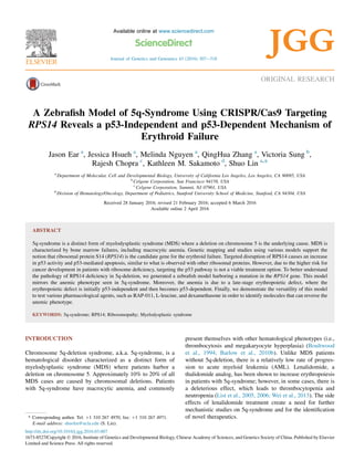
  • 1. ORIGINAL RESEARCH A Zebrafish Model of 5q-Syndrome Using CRISPR/Cas9 Targeting RPS14 Reveals a p53-Independent and p53-Dependent Mechanism of Erythroid Failure Jason Ear a , Jessica Hsueh a , Melinda Nguyen a , QingHua Zhang a , Victoria Sung b , Rajesh Chopra c , Kathleen M. Sakamoto d , Shuo Lin a,* a Department of Molecular, Cell and Developmental Biology, University of California Los Angeles, Los Angeles, CA 90095, USA b Celgene Corporation, San Francisco 94158, USA c Celgene Corporation, Summit, NJ 07901, USA d Division of Hematology/Oncology, Department of Pediatrics, Stanford University School of Medicine, Stanford, CA 94304, USA Received 28 January 2016; revised 21 February 2016; accepted 6 March 2016 Available online 2 April 2016 ABSTRACT 5q-syndrome is a distinct form of myelodysplastic syndrome (MDS) where a deletion on chromosome 5 is the underlying cause. MDS is characterized by bone marrow failures, including macrocytic anemia. Genetic mapping and studies using various models support the notion that ribosomal protein S14 (RPS14) is the candidate gene for the erythroid failure. Targeted disruption of RPS14 causes an increase in p53 activity and p53-mediated apoptosis, similar to what is observed with other ribosomal proteins. However, due to the higher risk for cancer development in patients with ribosome deficiency, targeting the p53 pathway is not a viable treatment option. To better understand the pathology of RPS14 deficiency in 5q-deletion, we generated a zebrafish model harboring a mutation in the RPS14 gene. This model mirrors the anemic phenotype seen in 5q-syndrome. Moreover, the anemia is due to a late-stage erythropoietic defect, where the erythropoietic defect is initially p53-independent and then becomes p53-dependent. Finally, we demonstrate the versatility of this model to test various pharmacological agents, such as RAP-011, L-leucine, and dexamethasone in order to identify molecules that can reverse the anemic phenotype. KEYWORDS: 5q-syndrome; RPS14; Ribosomopathy; Myelodysplastic syndrome INTRODUCTION Chromosome 5q-deletion syndrome, a.k.a. 5q-syndrome, is a hematological disorder characterized as a distinct form of myelodysplastic syndrome (MDS) where patients harbor a deletion on chromosome 5. Approximately 10% to 20% of all MDS cases are caused by chromosomal deletions. Patients with 5q-syndrome have macrocytic anemia, and commonly present themselves with other hematological phenotypes (i.e., thrombocytosis and megakaryocyte hyperplasia) (Boultwood et al., 1994; Barlow et al., 2010b). Unlike MDS patients without 5q-deletion, there is a relatively low rate of progres- sion to acute myeloid leukemia (AML). Lenalidomide, a thalidomide analog, has been shown to increase erythropoiesis in patients with 5q-syndrome; however, in some cases, there is a deleterious effect, which leads to thrombocytopenia and neutropenia (List et al., 2005, 2006; Wei et al., 2013). The side effects of lenalidomide treatment create a need for further mechanistic studies on 5q-syndrome and for the identification of novel therapeutics.* Corresponding author. Tel: þ1 310 267 4970, fax: þ1 310 267 4971. E-mail address: shuolin@ucla.edu (S. Lin). Available online at www.sciencedirect.com ScienceDirect Journal of Genetics and Genomics 43 (2016) 307e318 JGG http://dx.doi.org/10.1016/j.jgg.2016.03.007 1673-8527/Copyright Ó 2016, Institute of Genetics and Developmental Biology, Chinese Academy of Sciences, and Genetics Society of China. Published by Elsevier Limited and Science Press. All rights reserved.
  • 2. A commonly deleted region (CDR) of 5q-syndrome has been narrowed to a 1.5-Mb locus on chromosome 5 (Boultwood et al., 1994, 2002). Several genes in this CDR have been suggested to play a role in the pathogenesis of the disease, with haploinsufficiency of ribosomal protein S14 (RPS14) contributing to the anemic phenotype (Barlow et al., 2010b; Boultwood, 2011). As a consequence of this genetic aberration, expression of RPS14 is severely downregulated in patients with 5q-deletion (Boultwood et al., 2007; Wu et al., 2013). Knockdown of RPS14 using RNAi in CD34þ he- matopoietic progenitor cells severely affected erythroid dif- ferentiation, similar to what is observed in patients (Ebert et al., 2008). Furthermore, a small chromosomal deletion in a mouse model containing a deletion of RPS14 was shown to have macrocytic anemia (Barlow et al., 2010a). The anemic correlation between RPS14 deficiency and 5q-deletion led to the classification of 5q-syndrome as a ribosomopathy (a dis- order due to aberrant ribosome function or biogenesis). Haploinsufficiency of ribosomal proteins has been impli- cated in several bone marrow failure disorders including DiamondeBlackfan anemia (DBA), Dyskeratosis congenital (DKC), ShwachmaneDiamond syndrome (SDS), and Cartilage-hair hypoplasia (CHH) (Luzzatto and Karadimitris, 1998; Draptchinskaia et al., 1999; Ganapathi et al., 2007; Thiel and Rauch, 2011). A deficiency in ribosomal proteins has been shown to cause an increase in the activity of the tumor suppressor, p53. Activation of the p53-pathway leads to cell cycle arrest and apoptosis. While the exact mechanism of p53 activation remains to be fully understood, a ribosomal protein (RP)-mdm2-p53 checkpoint mechanism seems to be involved (Boultwood, 2011; Miliani de Marval and Zhang, 2011). In a mouse model of 5q-syndrome, an elevated level of p53 and cell death is observed. Furthermore, crossing the model into a p53-null background reverses the degree of cell death (Barlow et al., 2010a). While these studies may suggest targeting p53 as a potential therapeutic strategy, caution is warranted in this approach due to a risk of tumor development when targeting this pathway. Inhibiting p53 is further ill- advised when one considers the elevated risk in cancer development in patients with ribosomopathies. Recently, various studies have demonstrated that both p53-dependent and p53-independent pathways play a role in the pathology of the disease (Danilova et al., 2008; Narla et al., 2014). Targeting the p53-independent pathway, therefore, offers a more attractive therapeutic strategy. In this study, through the use of CRISPR/Cas9 gene targeting, we generated a zebrafish model of RPS14 deficiency in order to mirrorthe anemic phenotype of patients with5q-sydrome. Using this model, we demonstrate that a delay in erythroid maturation leads to anemia in embryos deficient of RPS14 through a p53-independent and a p53-dependent mechanism. This delay is partially reversed upon treatment with RAP-011 (an activin receptor type 2A ligand trap), L-leucine, as well as with dexamethasone. In all, this model will serve as a useful model to further investigate the mechanism underlying ribosome- associated disorders and to test various drug candidates in an intact animal model. RESULTS CRISPR/Cas9 targeting of RPS14 A deletion on chromosome 5, which contains the RPS14 gene, is expected to lead to haploinsufficient levels of ribosomal protein S14. The zebrafish RPS14 gene is located on chro- mosome 21 and its encoded protein shares over 99% amino acid identity to the human RPS14 (Fig. S1). To target the RPS14 gene in zebrafish, we applied CRISPR/Cas9 gene- editing technologies and generated a guide-RNA (gRNA) targeting exon 2 of the gene, near the translational start site. To facilitate validation of nuclease activity and screening, we targeted an AvrI I restriction digest site on exon 2 of the gene (Fig. 1A). Injection of the in vitro synthesized gRNA, along with Cas9 mRNA, into the zebrafish embryos successfully targeted the AvrI I restriction enzyme cut site, as indicated by the retention of an upper band after restriction digest of the PCR product with restriction endonuclease (Fig. 1B). Interestingly, a population of the injected embryos displayed a decrease in erythroid levels as revealed through the use of o- dianisidine staining to label hemoglobin positive cells throughout the entire embryo (Fig. 1C). Injected embryos were raised to adulthood and mutants were screened for transmission into germline (Fig. 1D). Progeny from positive germline carriers were genotyped, and mutations containing insertions as well as deletions were revealed, thereby confirming the success targeting of RPS14 in zebrafish (Fig. 1E). Deficiency in RPS14 leads to morphological defects and an increase in apoptosis Three mutant lines were outcrossed and maintained for further analysis (Fig. 1E and F). Two lines carried introduction of an early stop codon (RPS14D4bp and RPS14DINS) and one line (RPS14D21bp) carried an in-frame deletion (Fig. 1F). RPS14DINS was incrossed and embryos carrying two copies of the mutation displayed aplasia in the head at 24 hours post fertilization (hpf), and by 30 hpf, a decrease in pigmentation and defective yolk extension was apparent (Fig. 2A, A0 , and B). By 48 hpf, there was a slight recovery in pigmentation; however, the embryos had a smaller head, edema, and retained the defective yolk extension (Fig. 2C). Overtime, the severity of the phenotypes increased and the mutation became lethal around 6 to 7 days post fertilization (dpf) (Fig. 2DeF). These morphologically abnormal embryos were confirmed to be mutants for RPS14 through PCR vali- dation (Fig. 3A). The RPS14D4bp mutants also displayed similar morphological abnormalities to the RPS14DINS line (Figs. 2, and S2); therefore, the RPS14DINS line was used for the remainder of the work. No observable defects were present in the RPS14D21bp (Fig. S2). In fact, homozygous carriers of this mutation were able to grow to adulthood and give rise to homozygous progenies (Fig. S3). This suggests that the observed phenotype 308 J. Ear et al. / Journal of Genetics and Genomics 43 (2016) 307e318
  • 3. in the RPS14DINS and RPS14D4bp is due to a null mutation in the gene, and not through random mutation. Next, to determine if the morphological defect is accom- panied with an increase in levels of apoptosis, we performed TUNEL staining on whole embryos. At all time points analyzed, an increase in TUNEL staining positive cells was observed in mutant embryos compared to age matched sibling control (Fig. 2G). This falls in line with previous findings RPS14 Exon 1 Exon 2 Translational start codon Uninjected F0 embryos F1 embryos F0 adults F1 adults U ninjectedInjected Cas9 nuclease Guide RNA Exon 3 Exon 4 Exon 5 Avrl l cut site 48 hpf gRNA/Cas9 A B D E F C Fig. 1. Targeting RPS14 using CRISPR/Cas9. A: Illustration depicting the RPS14 target site. B: In Cas9 mRNA and RPS14 gRNA injected embryos, region flanking the AvrI I restriction digest site was PCR amplified followed by restriction digest of the PCR product with AvrI I. C: o-dianisidine staining of Cas9 mRNA and RPS14 gRNA injected embryos. Approximately 50% of embryos displayed reduction in staining. D: Screening scheme used to identify RPS14 mutants. Injected embryos were raised to adulthood and crossed for screening. Embryos (F1 embryos) were screened by restriction digest and individual clutches containing mutant carriers were raised to adulthood. F1 adults were tail cut and screened for mutations in RPS14 through restriction digest. E: Genomic sequences of mutations found in RPS14. F: Predicted protein sequence based on DNA sequence. 309J. Ear et al. / Journal of Genetics and Genomics 43 (2016) 307e318
  • 4. demonstrating that a deficiency in RPS14 lead to an increase in apoptosis. Interestingly, apoptosis was scattered throughout the embryo and not apparent in regions of blood development. To confirm that the morphologically abnormal embryos are truly deficient for RSP14, transcript measurements for RPS14 were performed using quantitative real-time PCR (qPCR). At both 24 and 48 hpf, the amount of RPS14 transcripts was significantly downregulated compared to morphologically normal sibling control embryos (Fig. 3B). This was further confirmed by whole-mount in situ hybridization (WISH) (Fig. 3C). Strong expression of RPS14 is normally observed throughout the embryo at 24 and 48 hpf; however, in our mutants we can see a severe reduction in expression levels. Additionally, there were no changes in expression levels of ribosomal protein RPL11 or RPS19 between mutants and control groups (Fig. S4), indicating that the phenotype is specifically due to loss of RPS14. Finally, we confirmed that the morphological defects in our mutants was due to a decrease in levels of RPS14 by per- forming a rescue experiment using in vitro transcribed RPS14 24 hpf 48 hpf 4 dpf 48 hpf24 hpf 30 hpf Sibling Sibling Sibling Sibling Sibling Sibling 5 dpf 3 dpf Sibling 3 dpf Sibling Mutant Mutant Mutant Mutant Mutant Mutant Mutant Mutant Mutant Sibling A C E G F D A′ B Fig. 2. RPS14 deficient embryos display physical abnormalities as well as increase in apoptosis. AeF: Morphological phenotype of RPS14 deficient embryos. At 24 hpf (A and A0 ), aplasia (arrowhead) can be seen in the head region. By 30 hpf (B), there is a delay in pigmentation. At 48 hpf (C) and 3 dpf (D), a smaller head and edema (arrowhead) are present. At 4 dpf (E), edema (arrowhead) is more apparent. At 5 dpf (F), embryos still continue to have severe edema and smaller head, and display a high degree of lethality. Most embryos die between 5 and 6 dpf. G: In situ cell death staining of morphologically abnormal embryos. Arrowheads point to the region of positive TUNEL staining. 310 J. Ear et al. / Journal of Genetics and Genomics 43 (2016) 307e318
  • 5. mRNA. Upon rescue using RPS14 mRNA, mutants showed an improvement in morphology (Fig. 3D). This suggests that a deficiency in RPS14 leads to the morphological defects seen in our mutant embryos. Erythroid failure is observed upon RPS14 deficiency Because ribosome deficiency typically leads to anemia in patients, we sought to study the effects of RPS14 deficiency on the development of red blood cells in our model. RPS14 mutants at 48 hpf and 3 dpf showed decreased levels of o-dianisidine positive cells, suggesting a defect in the production of mature erythroid cells in the embryos (Fig. 4A). This decrease in o-dianisidine staining can be restored when embryos were injected with RPS14 mRNA (Fig. S5), suggesting that the anemic phenotype is due to a deficiency in RPS14. To better assess the erythroid defect, we crossed the RPS14 mutant line into the LCR2:EGFP transgenic fish line, which specifically have all globin positive cells labeled with EGFP (Ganis et al., 2012). We observed a similar degree in the amount of EGFP positive cells between mutant and sibling control embryos at 48 hpf and a slightly lower amount of EGFP positive cells in mutant embryos at 3 dpf (Fig. 4B). Interestingly, sibling control had two populations of EGFP expressing cells (high- and low-EGFP), whereas mutants predominantly contained one population of EGFP expressing cells with an intermediate level of EGFP expression (Figs. 4C, D and S6). Analysis of the cells using flow cytometry revealed no change in the size of the cell (data not shown). The presence of EGFP positive cells and lack of o-dianisidine staining suggest that a defect in the terminal maturation of erythroid cells is impaired upon RPS14 deficiency. To further characterize the blood defect in our model, we performed WISH for various erythroid markers throughout different stages of embryonic development. gata1 is one of the earliest markers used to detect the specification of hemato- poietic cells into the erythroid lineage (Detrich et al., 1995). 1.2 0.8 0.6 0.4 0.2 0 1.0 RPS 14ΔINS WT Sibling Mutant M arker(200 bp) Relativefoldinduction 24 hpf 24 hpf Sibling Sibling Mutant Mutant Mutant Sibling 48 hpf 48 hpf 48 hpf RPS 14 Sibling + RPS14 mRNA Mutant + RPS14 mRNA * *A C D B Fig. 3. Deficiency in RPS14 is reversed by RPS14 mRNA injections. A: PCR genotype of RPS14 mutant embryos. B: qPCR of transcript levels of RPS14. *, P < 0.05. C: WISH for RPS14 in mutants and wild-type sibling at 24 hpf (left) or 48 hpf (right). D: Rescue of RPS14 deficient embryos with in vitro transcribed RPS14 mRNA. Arrowhead points to the rescue in head defect. 311J. Ear et al. / Journal of Genetics and Genomics 43 (2016) 307e318
  • 6. Other markers of erythroid cells used in zebrafish include globin transcripts such as hbbe1.1 and hbae3. There was no observable difference in expression levels of gata1, hbbe1.1, and hbae3 between mutant and sibling control embryos at 24 hpf, suggesting that RPS14 deficiency does not lead to defects in the specification of erythroid cells (Fig. 5A). Expression of gata1 was normally downregulated in the zebrafish after 24 hpf. The expression level of gata1 in the RPS14 mutants was downregulated in a similar fashion to sibling control (Fig. 5B). In addition, there was a similar expression level of global transcript at 48 hpf and only a slightly lower level of expression at 3 dpf (Fig. 5B and C). 100 80 60 40 20 0 0 102 103 104 105 Cellcount(%ofmax.) EGFP intensity Merge Sibling Mutant Mutant Mutant Mutant Mutant Mutant Mutant Mutant Mutant EGFP DAPI Sibling Sibling Sibling Sibling Sibling Sibling Sibling Sibling Sibling Mutant 48 hpf 24 hpf 3 dpf 3 dpf 48 hpf Ventral view Ventral view 3 dpfA B C D Fig. 4. RPS14 deficiency leads to anemia in mutant embryos. A: o-dianisidine staining for RPS14 between siblings or mutants at 48 hpf (left) or 3 dpf (right). Arrowheads point to the sites of weak o-dianisidine staining. B: RPS14 mutation was bred into LCR:EGFP transgenic fish line. EGFP positive cells can be seen circulating throughout mutant embryos. At 24 and 48 hpf, approximately equal number of EGFP positive cells can be seen. At 3 dpf, mutants display a slightly lower number of EGFP positive cells. C: Blood cells from 48 hpf embryos were smeared onto glass slides and immunostained for EGFP. Two populations of EGFP positive cells can be seen in sibling control (arrowhead, EGFP-high and asterisk, EGFP-low). Mutants show a more intermediate phenotype of EGFP expression. D: EGFP positive cells from 48 hpf embryos were analyzed by flow cytometry. 312 J. Ear et al. / Journal of Genetics and Genomics 43 (2016) 307e318
  • 7. When the expression pattern of mpx was analyzed, we observed a similar expression pattern between mutants and sibling control, suggesting that the specification of primitive myeloid cells is unaffected (Fig. S7). Overall, these data suggest that hematopoiesis is affected in our model of RPS14 deficiency and that the anemia is due to a late-stage terminal maturation of erythroid cells. p53-activity is upregulated in embryos deficient for RPS14 Haploinsufficiency of ribosome proteins has been shown to cause elevated levels of p53 and cell cycle arrest (Danilova et al., 2008). As expected, a mutation in RPS14 led to increased levels of p53 and p53-target genes (Fig. 6A and B). Furthermore, this increase was not restricted to hematopoietic tissues, but throughout the entire embryo (Fig. 6A). To determine if targeting the p53-pathway can alleviate the anemic phenotype in our model, we injected morpholino targeting p53 in our model (Fig. 6C). Targeting p53 led to a slight morphological rescue in our model (Fig. 6D). However, when we performed o-dianisidine staining on p53-morpholino ( p53-MO) injected embryos, no rescue was observed at 48 hpf (Fig. 6E). Interestingly, targeting p53 rescued anemic phenotype at 3 dpf (Fig. 6F). Injection of p53-MO did not show any observable changes to the erythroid levels in wild- type sibling control embryos (Fig. S8). Taken together, these findings suggest that upon RPS14 deficiency, both the p53- independent and p53-dependent pathways are involved in the anemic phenotype. RAP-011 reverses the anemia associated with RPS14 In another model of ribosome deficiency, we demonstrated that an elevated level of lft1 leads to decreased levels of mature erythroid cells (Ear et al., 2015). Furthermore, the effects of lft1 overexpression can be reversed upon treatment with the activin receptor ligand-trap, RAP-011. In RPS14 deficiency, an elevated level of lft1 is also observed (Fig. 7A), which is comparable to that seen in other models of ribosomopathies (Ear et al., 2015). Interestingly, we observe that lft1 is normally expressed in an asymmetric pattern in the embryos in presumably hatching gland cells. Upon RPS14 deficiency, the asymmetric pattern is loss and lft1 is expressed 24 hpf Sibling 24 hpf Sibling 24 hpf Sibling 48 hpf Sibling 48 hpf Sibling 3 dpf Sibling 3 dpf Sibling 48 hpf Sibling gata1 Mutant hbbe1.1 Mutant hbae3 Mutant hbae3 Mutant hbae3 Mutant A B C hbbe1.1 Mutant hbbe1.1 Mutant gata1 Mutant Fig. 5. Expression of erythroid specific transcripts is unaffected in RPS14 mutants. WISH shows that expression levels of gata1, hbbe1.1, and hbae3 in mutants are unaffected at 24 hpf (A) and 48 hpf (B). C: Slight reduction in hbbe1.1 and hbae3 expression appears at 3 dpf in mutant embryos. 313J. Ear et al. / Journal of Genetics and Genomics 43 (2016) 307e318
  • 8. in a more bilateral pattern over the yolk (Fig. 7B). Using RAP-011 in our model, we showed that it could partially reverse the anemic phenotype associated with RSP14 deficiency (Fig. 7C). Finally, we wanted to investigate if other treatments could be used in our model. We found that treatment with either L- leucine or dexamethasone could also partially rescue the anemic phenotype in our model (Fig. 7D). * * * 8 7 6 5 4 3 2 1 0 7 6 5 4 3 2 1 0 Relativefoldinduction Relativefoldinduction p53 p53 p21 p21 m dm 2 m dm 2 pum a bax 24 hpf 48 hpf 48 hpf 3 dpf 48 hpf Mutant Mutant + p53-MO p53 p21 p21p53 Sibling Sibling Sibling Sibling + p53-MO Mutant + p53-MO Mutant + p53-MO Mutant Mutant Mutant Mutant Mutant Sibling Mutant + p53-MOMutant Sibling Sibling mdm2 mdm2 A B D E F C Fig. 6. p53-independent and p53-dependent mechanisms contribute to the anemic phenotype in RPS14 deficiency. A: WISH shows that p53, p21, and mdm2 are ubiquitously expressed in RPS14 deficient embryos. B: Transcript measurement of p53 and p53-target genes ( p21, mdm2, puma, bax) was preformed by qPCR. The value is represented as fold change over sibling control. C: qPCR was used to confirm knockdown of p53 and p53-target genes in RPS14 mutants using p53-MO. *, P < 0.05. D: Slight morphological rescue (arrowhead) was observed in mutants with knockdown of p53. E and F: o-dianisidine staining of mutants with p53-MO at 48 hpf (E) and 3 dpf (F). Arrowheads in F point to the rescue in o-dianisidine staining. Observed phenotype: E, mutant ¼ 22/22, mutant þ p53-MO ¼ 19/20. F, mutant ¼ 20/20, mutant þ p53-MO ¼ 20/25. 314 J. Ear et al. / Journal of Genetics and Genomics 43 (2016) 307e318
  • 9. 2 1.5 1 0.5 0 lft1relativefoldinductiont Sibling M utant lft1 48 hpf 48 hpf 48 hpf Sibling Sibling Mutant Sibling + IgG Sibling + RAP-011 Mutant + RAP-011 Sibling + 0.2% DMSO Sibling + L-leucine Sibling + dexamethasone Mutant + dexamethasone Mutant + L-leucine Mutant + 0.2% DMSO Mutant + IgG Mutant A B C D Fig. 7. RAP-011, L-leucine, and dexamethasone recover hemoglobin levels in RPS14 deficiency. A: lft1 transcript level measured by qPCR at 48 hpf. Data are represented as fold induction over sibling control. B: WISH for lft1 in mutants or sibling control at 48 hpf. Arrowhead points to the region of lft1 expression. C: RAP-011 was injected into RPS14 mutants and o-dianisidine staining was performed to observe hemoglobin levels (arrowheads). D: L-leucine (150 mmol/L), dexamethasone (50 mmol/L), or DMSO control was used to treat RPS14 deficient embryos and hemoglobin levels (arrowheads) were analyzed by o-dianisidine staining. Observed phenotype: C, mutant þ IgG ¼ 25/25, mutant þ RAP-011 ¼ 23/26. D, mutant ¼ 20/20, mutant þ DMSO ¼ 19/19, mutant þ L-leucine ¼ 18/21, mutant þ dexamethasone ¼ 18/23. 315J. Ear et al. / Journal of Genetics and Genomics 43 (2016) 307e318
  • 10. DISCUSSION In this work, we successfully targeted RPS14 in the zebrafish using CRISPR/Cas9 gene-editing technology. Upon RPS14 deficiency, gross morphological defects can be observed and is accompanied with an elevation in p53 activity. Furthermore, the anemic phenotype that is typically seen in patients with disrupted ribosome gene function is also observed. Recent work comparing morpholino and genetic mutants has raised concern regarding the off-target effects of MO (Kok et al., 2015). Therefore, genetic models to compliment the findings using MO studies are highly suggested. We see that the phenotype in our model mirrors what is observed with MO-mediated knockdown of RPS14 in zebrafish (Narla et al., 2014). The presence of normal gata1 and globin expression but reduced levels of o-dianisidine staining, together, suggest a late-stage terminal differentiation defect. The lack of obvious apoptosis in hematopoietic tissues and normal specification of erythroid cells suggest that the anemia associated with RPS14 deficiency is due to an inability to produce sufficient levels of mature hemoglobin protein in the erythroid cells. Knockdown of p53 in our model did not restore hemoglo- bin levels at 48 hpf; however, increased levels were observed at 3 dpf. While the exact role of p53 in our model remains unclear, from our observation, we hypothesize that both p53- independent and dependent mechanisms lead to the erythroid failure and are separated temporally with the p53-independent mechanism preceding the p53-dependent mechanism. Due to the ubiquitous expression of p53 during ribosome deficiency, it is difficult to separate the cell autonomous vs. the non-cell autonomous role of p53 activity in erythroid cells; however, we believe that further analysis into the various populations of EGFP positive cells may give further insight into the mecha- nism of erythroid failure. In a previously described zebrafish model of ribosome deficiency, a defect in protein production was shown to be involved in the anemic phenotype (Zhang et al., 2014). Furthermore, RPS14 deficiency in mice revealed activation of p53 underscoring the erythroid differentiation defect (Barlow et al., 2010a). Erythroid cells require a relatively high pro- tein translational rate to meet the large globin protein requirement. It may be possible that during the early stages of erythroid development, ribosome deficiency leads to defects in protein production (and thus insufficient globin levels), which is independent of p53, and as development progresses, further deficiency in ribosome levels leads to defects that are p53- dependent. Finally, we showed that the anemia in our model is reversed upon treatment with RAP-011, L-leucine, and dexamethasone. Interestingly, both RAP-011 and L-leucine were previously demonstrated to work by p53-independent mechanisms (Narla et al., 2014; Ear et al., 2015). The fact that patients with ribosomopathies have a higher incidence for cancer development warrants caution in targeting the p53 pathway for therapeutic treatment. Therefore, our work rationalizes ongoing clinical trials investigating the use of RAP-011 and L-leucine for the treatment of patients with ribosome-associated disorders such as MDS and DBA. MATERIALS AND METHODS Zebrafish stains and husbandry Zebrafish protocol and maintenance were performed using methods approved by the University of California, Los Angeles Institutional Animal Care and Use Committee (UCLA-IACUC). Mutants were generated in wild-type (AB) strain. Cas9 mRNA and gRNA synthesis Zebrafish optimized Cas9 mRNA was in vitro transcribed from pGH-T7-zCas9 as previously described (Chang et al., 2013). 200 pg of Cas9 mRNA was injected per embryo at the one-cell stage. RPS14 gRNA was synthesized using a protocol similar to previously described methods (Chang et al., 2013). The RPS14 targeted site was flanked with a T7 promoter site at the 50 region and a scaffold sequence on the 30 region (Oligo: ATTTAGGTGACACTATAGAAGAGCAGGTCATCAGCCT GTTTTAGAGCTAGAAATAGC). This oligo was used in a PCR reaction to generate template for the gRNA synthesis. gRNA was in vitro transcribed using MAXIscript T7 Kit (Life Technologies, USA). After synthesis, DNA template was removed using TURBO DNase (Life Technologies) and pu- rified using phenol chloroform. 50 pg of gRNA (mixed with the Cas9 mRNA) was injected into the embryos at the one-cell stage. PCR validation, sequencing mutation, and genotyping of embryos To extract genomic DNA, embryos (or tail fin clips) were lysed in 50 mmol/L NaOH by boiling at 95 C for 1 h. After boiling, lysates were neutralized with 10% volume 1 mol/L Tris (pH ¼ 8.0). Crude lysates were then used in a PCR reaction. To validate the successful targeting of RPS14 and identify germline transmitted fish, the region flanking the target site was PCR amplified using RPS14 Fwd: 50 -CTGTTG ACTGTGTGTTTCAGCA-30 and RPS14 Rev: 50 -TCAAA- CATTCAATGCAACAAAAC-30 primers. PCR product was used in a restriction digest reaction using AvrI I (NEB, USA). To determine the DNA sequence, PCR products from positive carriers were used in a TOPO-TA reaction (Life Technologies) according to manufacturer direction. After generating a stable mutant line, fish lines were genotyped using primers RPS14 gel screen Fwd: 50 -ATGGCTCCTCGCAAGGGTAAGG-30 and RPS14 gel screen Rev: 50 -TGTGCACACAAAGACACTGTT-30 . The PCR product was then run on a 10% TBE-PAGE gel to differentiate between homozygous and heterozygous carriers. 316 J. Ear et al. / Journal of Genetics and Genomics 43 (2016) 307e318
  • 11. o-dianisidine staining Embryos were first fixed at room temperature for 2 h using 4% paraformaldehyde (PFA) in PBS. Next, three washes (5 min each) with PBS were performed to remove PFA. Embryos were then incubated in an o-dianisidine staining solution (40% ethanol, 0.65% H2O2, 10 mmol/L Na-Acetate, and 0.6 mg/mL o-dianisidine (Sigma, USA)) in the dark for 30 min followed by four wash times with PBS. To remove pigmentation, em- bryos were placed into a bleach solution (1% KOH, 3% H2O2) for 20 min. Finally, embryos were washed with PBS and immediately imaged. mRNA probes and in situ hybridization Plasmids for antisense probe were made using Gateway technology (Life Technologies). Genes were amplified using primers flanked with attB1 forward or attB2 reverse sequences and then used in a BP clonase II reaction with pDONR221 (Life Technologies). To generate DIG labeled antisense probes, plasmids were linearized and synthesized using T7 RNA polymerase (Promega, USA) with DIG RNA Labeling Mix (Roche, USA). For hybridization, embryos were first fixed overnight at 4 C using 4% PFA in PBS. After fixation, PFA was washed four times (5 min each) with PBS, followed by two washes (5 min each) with 100% methanol. Embryos were stored at À20 C. WISH was performed using labeled probed as previously described (Jowett, 1999). Real-time quantitative PCR To generate cDNA for real-time quantitative PCR analysis, total RNA was first isolated from whole embryos (25 embryos per group) using TRIzol Reagent (Life Technologies) following manufacture’s protocol. Next, cDNA was reverse transcribed using Oligo(dT) and SuperScript III reverse tran- scriptase (Life Technologies). All qPCR experiments were performed in triplicates. For quantitative PCR analysis, cDNA was used in a PCR reaction using FastStart SYBR Green Master (Roche). Oligos used in qPCR are as previously described (Ear et al., 2015). ACKNOWLEDGMENTS We thank Dr. Matt Veldmen, Dr. Nadia Danilova, and Dr. Anne Lindgren for their insightful discussions and input throughout the course of the work. We acknowledge Linda Dong for maintenance of the zebrafish strains used in the experiments. Also, we thank Tianna Wilson for providing invaluable technical assistance. This work was supported by Celgene (Nov022011) and National Institutes of Health (R56 DK107286) to K.S. and S.L. SUPPLEMENTARY DATA Fig. S1. Protein alignment between human and zebrafish RPS14. Fig. S2. Phenotype of RPS14 mutant alleles. Fig. S3. An inframe mutation of RPS14 does not affect development. Fig. S4. RPS14 mutants have normal expression of other ri- bosomal protein transcripts. Fig. S5. o-dianisidine staining of RPS14 mutants rescued with RPS14 mRNA. Fig. S6. Flow cytometry analysis on EGFP positive cells from 48 hpf embryos. Fig. S7. In situ hybridization of mpx transcript in RPS14 mutants at 48 hpf. Fig. S8. p53-MO injection does not affect erythroid levels under normal conditions. Supplementary data related to this article can be found at http://dx.doi.org/10.1016/j.jgg.2016.03.007. REFERENCES Barlow, J.L., Drynan, L.F., Hewett, D.R., Holmes, L.R., Lorenzo-Abalde, S., Lane, A.L., Jolin, H.E., Pannell, R., Middleton, A.J., Wong, S.H., Warren, A.J., Wainscoat, J.S., Boultwood, J., McKenzie, A.N., 2010a. A p53-dependent mechanism underlies macrocytic anemia in a mouse model of human 5q-syndrome. Nat. Med. 16, 59e66. Barlow, J.L., Drynan, L.F., Trim, N.L., Erber, W.N., Warren, A.J., McKenzie, A.N., 2010b. New insights into 5q-syndrome as a ribosomop- athy. Cell Cycle 9, 4286e4293. Boultwood, J., 2011. The role of haploinsufficiency of RPS14 and p53 acti- vation in the molecular pathogenesis of the 5q-syndrome. Pediatr. Rep. 3 (Suppl. 2), e10. Boultwood, J., Fidler, C., Lewis, S., Kelly, S., Sheridan, H., Littlewood, T.J., Buckle, V.J., Wainscoat, J.S., 1994. Molecular mapping of uncharacteris- tically small 5q deletions in two patients with the 5q-syndrome: delinea- tion of the critical region on 5q and identification of a 5q-breakpoint. Genomics 19, 425e432. Boultwood, J., Fidler, C., Strickson, A.J., Watkins, F., Gama, S., Kearney, L., Tosi, S., Kasprzyk, A., Cheng, J.F., Jaju, R.J., Wainscoat, J.S., 2002. Narrowing and genomic annotation of the commonly deleted region of the 5q-syndrome. Blood 99, 4638e4641. Boultwood, J., Pellagatti, A., Cattan, H., Lawrie, C.H., Giagounidis, A., Malcovati, L., Della Porta, M.G., Jadersten, M., Killick, S., Fidler, C., Cazzola, M., Hellstrom-Lindberg, E., Wainscoat, J.S., 2007. Gene expression profiling of CD34þ cells in patients with the 5q-syndrome. Br. J. Haematol. 139, 578e589. Chang, N., Sun, C., Gao, L., Zhu, D., Xu, X., Zhu, X., Xiong, J.W., Xi, J.J., 2013. Genome editing with RNA-guided Cas9 nuclease in zebrafish em- bryos. Cell Res. 23, 465e472. Danilova, N., Sakamoto, K.M., Lin, S., 2008. Ribosomal protein S19 defi- ciency in zebrafish leads to developmental abnormalities and defective erythropoiesis through activation of p53 protein family. Blood 112, 5228e5237. Detrich 3rd, H.W., Kieran, M.W., Chan, F.Y., Barone, L.M., Yee, K., Rundstadler, J.A., Pratt, S., Ransom, D., Zon, L.I., 1995. Intraembryonic hematopoietic cell migration during vertebrate development. Proc. Natl. Acad. Sci. USA 92, 10713e10717. Draptchinskaia, N., Gustavsson, P., Andersson, B., Pettersson, M., Willig, T.N., Dianzani, I., Ball, S., Tchernia, G., Klar, J., Matsson, H., Tentler, D., Mohandas, N., Carlsson, B., Dahl, N., 1999. The gene 317J. Ear et al. / Journal of Genetics and Genomics 43 (2016) 307e318
  • 12. encoding ribosomal protein S19 is mutated in Diamond-Blackfan anaemia. Nat. Genet. 21, 169e175. Ear, J., Huang, H., Wilson, T., Tehrani, Z., Lindgren, A., Sung, V., Laadem, A., Daniel, T.O., Chopra, R., Lin, S., 2015. RAP-011 improves erythropoiesis in zebrafish model of Diamond-Blackfan anemia through antagonizing lefty1. Blood 126, 880e890. Ebert, B.L., Pretz, J., Bosco, J., Chang, C.Y., Tamayo, P., Galili, N., Raza, A., Root, D.E., Attar, E., Ellis, S.R., Golub, T.R., 2008. Identification of RPS14 as a 5q-syndrome gene by RNA interference screen. Nature 451, 335e339. Ganapathi, K.A., Austin, K.M., Lee, C.S., Dias, A., Malsch, M.M., Reed, R., Shimamura, A., 2007. The human Shwachman-Diamond syndrome pro- tein, SBDS, associates with ribosomal RNA. Blood 110, 1458e1465. Ganis, J.J., Hsia, N., Trompouki, E., de Jong, J.L., DiBiase, A., Lambert, J.S., Jia, Z., Sabo, P.J., Weaver, M., Sandstrom, R., Stamatoyannopoulos, J.A., Zhou, Y., Zon, L.I., 2012. Zebrafish globin switching occurs in two devel- opmental stages and is controlled by the LCR. Dev. Biol. 366, 185e194. Jowett, T., 1999. Analysis of protein and gene expression. Methods Cell Biol. 59, 63e85. Kok, F.O., Shin, M., Ni, C.W., Gupta, A., Grosse, A.S., van Impel, A., Kirchmaier, B.C., Peterson-Maduro, J., Kourkoulis, G., Male, I., DeSantis, D.F., Sheppard-Tindell, S., Ebarasi, L., Betsholtz, C., Schulte- Merker, S., Wolfe, S.A., Lawson, N.D., 2015. Reverse genetic screening reveals poor correlation between morpholino-induced and mutant pheno- types in zebrafish. Dev. Cell 32, 97e108. List, A., Dewald, G., Bennett, J., Giagounidis, A., Raza, A., Feldman, E., Powell, B., Greenberg, P., Thomas, D., Stone, R., Reeder, C., Wride, K., Patin, J., Schmidt, M., Zeldis, J., Knight, R., 2006. Lenalidomide in the myelodysplastic syndrome with chromosome 5q deletion. N. Engl. J. Med. 355, 1456e1465. List, A., Kurtin, S., Roe, D.J., Buresh, A., Mahadevan, D., Fuchs, D., Rimsza, L., Heaton, R., Knight, R., Zeldis, J.B., 2005. Efficacy of lena- lidomide in myelodysplastic syndromes. N. Engl. J. Med. 352, 549e557. Luzzatto, L., Karadimitris, A., 1998. Dyskeratosis and ribosomal rebellion. Nat. Genet. 19, 6e7. Miliani de Marval, P.L., Zhang, Y., 2011. The RP-Mdm2-p53 pathway and tumorigenesis. Oncotarget 2, 234e238. Narla, A., Payne, E.M., Abayasekara, N., Hurst, S.N., Raiser, D.M., Look, A.T., Berliner, N., Ebert, B.L., Khanna-Gupta, A., 2014. L- Leucine improves the anaemia in models of Diamond Blackfan anaemia and the 5q-syndrome in a TP53-independent way. Br. J. Haematol. 167, 524e528. Thiel, C.T., Rauch, A., 2011. The molecular basis of the cartilage-hair hy- poplasia-anauxetic dysplasia spectrum. Best Pract. Res. Clin. Endocrinol. Metab. 25, 131e142. Wei, S., Chen, X., McGraw, K., Zhang, L., Komrokji, R., Clark, J., Caceres, G., Billingsley, D., Sokol, L., Lancet, J., Fortenbery, N., Zhou, J., Eksioglu, E.A., Sallman, D., Wang, H., Epling-Burnette, P.K., Djeu, J., Sekeres, M., Maciejewski, J.P., List, A., 2013. Lenalidomide promotes p53 degradation by inhibiting MDM2 auto-ubiquitination in myelodysplastic syndrome with chromosome 5q deletion. Oncogene 32, 1110e1120. Wu, L., Li, X., Xu, F., Zhang, Z., Chang, C., He, Q., 2013. Low RPS14 expression in MDS without 5q e aberration confers higher apoptosis rate of nucleated erythrocytes and predicts prolonged survival and possible response to lenalidomide in lower risk non-5q-patients. Eur. J. Haematol. 90, 486e493. Zhang, Y., Ear, J., Yang, Z., Morimoto, K., Zhang, B., Lin, S., 2014. Defects of protein production in erythroid cells revealed in a zebrafish Diamond- Blackfan anemia model for mutation in RPS19. Cell Death Dis. 5, e1352. 318 J. Ear et al. / Journal of Genetics and Genomics 43 (2016) 307e318