This document summarizes the discovery of duplicated VegfA and KDR receptor genes in zebrafish that mediate vascular development. Specifically:
- The researchers identified a duplicated zebrafish VegfA gene (VegfAb) that encodes 171- and 210-amino acid isoforms not found in the single VegfA gene.
- They also found a duplicated KDR receptor gene (Kdrb) that encodes a receptor similar to mammalian KDR.
- Knockdown experiments in zebrafish showed that both VegfAb and the duplicated KDR receptor genes play important roles in vascular development.
- Further experiments demonstrated that the VegfAb isoforms are poorly secreted compared to VegfA isoforms
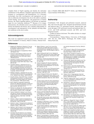
![standard tblastn algorithm, short sequence segments with homology to
mammalian VEGFA and KDR were identified using the zebrafish
genome assembly versions 2 and 3, available at the Sanger Center
website(http://www.sanger.ac.uk/Projects/D rerio/).Wefoundadupli-
cate vegfA gene, hereafter referred to as vegfAb to distinguish it from the
previously cloned vegfA gene (now termed vegfAa). This DNA se-
quence then served as the starting point for isolating the complete cDNA
from TU fish using a combination of RACE and RT-PCR. Radiation
hybrid mapping of this gene placed vegfAb on zebrafish chromosome 4,
whereas vegfAa is located on chromosome 16 (Yi Zhou, personal
written communication), November 2003).
The full-length vegfAb cDNA contained 2814 nucleotides
(nt’s) and a predicted ORF of 233 aa’s and encoded a putative
protein with stronger homology to VEGFA than VEGFB,
VEGFC, VEGFD, or PLGF (Figure 1). Therefore, the newly
identified gene appeared to be a duplicated zebrafish vegfA gene.
By RT-PCR, expression of 2 differentially spliced mRNAs was
detected from the early embryo (8 hours after fertilization [hpf])
through 4 days after fertilization (dpf) (data not shown). The
larger of 2 bands corresponded to an isoform of 210 aa’s
(VegfAb210), the smaller to a second isoform of 171 amino acids
(VegfAb171). The 2 zebrafish VegfA proteins (VegfAa and
VegfAb) were identical in size and exon structure to human
VEGFA through the first 138 aa’s (exons 1-5), suggesting
common ancestry (Figure 2A); however, insertions and dele-
tions occurred in the C-terminus, primarily in exons
6 and 7. Analysis of the genomic structure of the vegfA isoforms
by blasting against the available zebrafish genome showed 6
exons for vegfAa121, 7 for vegfAa165 and vegfAb171, and 8 for
vegfAb210; vegfAb171 and vegfAb210 share exons 1 to 5, 6, and 7
(Figure 2A). Figure 2 shows that exon use is highly conserved
between humans and zebrafish, but also that exon 6a, which
promotes mammalian VEGF binding to the basement membrane
in VEGFA145, VEGFA189, and VEGFA206, is found only in
vegfAb210. We suggest that zebrafish vegfAb210 may be the
functional equivalent of these particular VEGFA isoforms and
that vegfAb171 is orthologous to mammalian VegfA165.
By similar methods, we also obtained a 5360-nt kdrb cDNA that
encoded a novel zebrafish KDR orthologue. Alignment of critical
tyrosine residues between the zebrafish Kdrb cytoplasmic domain
and human and mouse KDR (VEGFR2) showed that the flk1-
related RTK described here is more closely related to KDR than the
previously described flk1 gene.31 In humans, tyrosine residue
Y1175 has been reported as a binding site for PLC␥1 and
Src-homology 2 protein (Shb) in -cells.42 The corresponding
tyrosine residue in the mouse, Y1173, has recently been identified
as an essential tyrosine residue for vasculogenesis and hematopoi-
esis, as knock-in mice substituting it with phenylalanine exhibited
severe vascular and hematopoietic defects, similar to KDR-null
mice.43 This critical tyrosine residue was conserved in both
zebrafish Flk1 genes (Figure 2B). However, 4 important tyrosine
residues in the C-terminal tail of KDR were conserved only in the
zebrafish RTK gene reported here (Kdrb), but not in the previously
identified RTK (Figure 2B). These residues, Y1214, Y1305,
Y1309, and Y1319, mediate binding of focal adhesion kinase
(FAK), which in turn interacts with PI3 kinase and paxillin.44-47 By
radiation hybrid mapping (Yi Zhou, personal communication), the
newly identified KDR gene was located on chromosome 20 near
the zebrafish orthologues of KIT and PDGFRA, thereby establish-
ing synteny between the zebrafish, human, and mouse KDR
chromosomal regions (Figure 1C). Based on structural conserva-
tion, synteny, and biochemical data, we suggest that this RTK gene
(Kdrb) is a duplicate of the previously cloned gene flka, which we
suggest should be called Kdra. Another duplicated zebrafish type
III RTK with homology to human FLT1 (Figure 1) is located on
zebrafish chromosome 24, near the zebrafish orthologue of IPF and
CDK8 (Yi Zhou, personal communication). Although this gene is
not the subject of this paper, we suggest that it be called flt1
henceforth. The cloning and function of this FLT1 gene will be
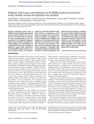
Figure 1. Evolutionary relationship between human
and zebrafish Vegfa isoforms and receptors. Protein
sequences were aligned using ClustalW (European Bioin-
formatics Institute, Cambridge, United Kingdom) with the
complete open reading frames. Species are indicated
as follows: zf, zebrafish; m, mouse; and h, human.
(A) Alignment of VegfA proteins shows a closer relation-
ship of the newly isolated gene to VEGFA than VEGFB,
VEGFC, VEGFD, and PLGF. (B) Alignment of RTK
sequences. The zebrafish gene (Kdra) formerly referred
to as flk1 shows a closer relationship to hFLT1 and mFlt1
than to hKDR (hFLK1) and mKDR (mFlk1). (C) Radiation
hybrid mapping of zebrafish kdra, flt1, and kdrb and
synteny comparison. Zebrafish kdrb is located on chromo-
some 20 in a region syntenic to human KDR, nearby the
zebrafish orthologues of kit and pdgfra, which map near
human KDR on human chromosome 4q12. Zebrafish
kdra is on chromosome 14, and flt1 near cdk8 and ipf1 in
a region on chromosome 24, which is syntenic to the
human 13q12 region where FLT1, CDK8, and IPF1 are
located.
DUPLICATED VEGF AND KDR GENES IN ZEBRAFISH 3629BLOOD, 15 NOVEMBER 2007 ⅐ VOLUME 110, NUMBER 10
For personal use only.on October 24, 2014.by guestwww.bloodjournal.orgFrom](https://image.slidesharecdn.com/63e25be3-de88-472d-9a42-666c99cca3f8-150227203716-conversion-gate01/85/Blood2007-3-320.jpg)

![separately transfected into these same cell lines with and without
vegfAa165 or vegfAb171. Both vegfAa165 and vegfAb171 were able to
activate both RTKs as demonstrated by antiphosphotyrosine anti-
body staining (Figure 6E). Therefore, the zebrafish VegfAa and
VegfAb isoforms can both bind and activate Kdra and Kdrb.
Discussion
We report here the cloning and characterization of duplicated
zebrafish genes that regulate vasculogenesis and angiogenesis. In
addition to the reported VegfA locus, now renamed vegfAa, we find
a second locus, vegfAb, which contains homologues of some of the
mammalian VegfA isoforms not encoded by vegfAa. We also
identified a second zebrafish RTK (Kdrb) with homology to
mammalian KDR.
Expression of VegfAa and VegfAb during embryonic
development
Whole-mount in situ hybridization of vegfAa and vegfAb revealed
differing expression patterns of these 2 genes (Figure 3A). Whereas
both vegfAa splice variants were broadly expressed at 12 somites,
by 24 hpf, vegfAa165 was expressed in the CNS and the somites,
whereas VegfAa121 RNA was more broadly distributed, although at
48 hpf it was expressed within/near the developing heart and
vasculature. These data differ from the reported expression of
VegfA during zebrafish embryogenesis. In that study, an unspecified
probe that detected both splice variants was similarly seen in the
prospective cardiac vasculature and somites but also in the
pronephros (seen in the present studies with vegfAb165) and was not
seen as diffusely within the CNS. The reasons for these differences
are unclear but might be related to the differing time points
analyzed and the probe chosen for use in these analyses. In the
present studies, we separately used the ORF of each isoform,
whereas the previous studies were done with an unspecified probe
that recognized both alleles, and that perhaps included 3ЈUTR
sequences as well. Our whole-mount in situ hybridization data also
correlated with our microarray data. The expression of genes
whose known embryonic whole-mount in situ pattern is within the
developing CNS and myotomes was more likely to be altered in
vegfAa morphants, whereas expression of genes in the CNS, eyes,
lens, and pharyngeal arches was altered in vegfAb morphants.
Interestingly, Hiratsuka et al (2005) examined the localization of
VegfA by immunohistochemistry during murine development and
found expression throughout the developing embryo, but especially
in the anterior part of the embryo.50 This pattern more closely, but
not completely, resembles the cumulative expression of both
zebrafish VegfA genes than any one gene or splice variant. These
expression patterns suggest that the various functions of mamma-
lian VEGFA may be encompassed within more than just these
2 zebrafish VegfA genes.
The role of zebrafish Vegf and Kdr in vascular development
In VegfAb morphants, early vascular and hematopoietic development
appeared normal at first, however as early as 2 dpf, morphants had
defects in angiogenesis. Concomitant with the vascular phenotype,
blood began to extravasate into the surrounding tissues beginning at 3
dpf (Figure 4). This extravasation of blood into the tissues surrounding
the eye and CNS in vegfAb morphants is consistent with the later
expression of vegfAb RNA in the vasculature surrounding the eye
(Figure 3A), and CNS. Similar defects in angiogenesis are seen in Kdra
morphants and the previously described zebrafish Kdra mutant.37 The
intersomitic vascular defects seen in Kdra morphants appeared to
encompass more of the embryo (Figure 5H) than those in either VegfAb
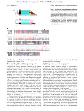
Figure 5. Kdra and Kdrb cooperate to mediate vasculogenesis during zebrafish embryogenesis. Fli-gfp transgenic embryos were injected with combinations of
morpholinos against kdra and kdrb or 5-bp mismatch control morpholinos. (A) Twenty-eight hpf embryos injected with either of 2 5-bp mismatch control morpholinos (4.5 ng
each) corresponding to separate 5ЈUTR kdrb morpholinos (m1-kdrb [n ϭ 32] and m2-kdrb [n ϭ 44]) or the previously published kdra morpholino (m-kdra [n ϭ 38]) had no
vascular abnormalities. (B) Injection of 4.5 ng kdra or kdrb morpholino has no demonstrable effect on vasculogenesis at 28 hpf (107 embryos). Vascular defects were seen at
higher concentrations of kdrb morpholinos; however, similar defects, but at a reduced penetrance, were also seen with equivalent amounts of the mismatched controls,
suggesting these effects were not directly related to Kdrb function. (C) Coinjection of either 4.5 ng m1-kdrb or m2-kdrb with 4.5ng kdra morpholino caused variable loss of
intersegmental arteries in 42 (24%) of 178 kdra/m1-kdrb or 53 (38%) of 138 kdra/m2-kdrb morpholino–injected embryos. (D) RBCs are seen in the anterior axial vasculature but
cannot circulate. (E,F) Kdra/kdrb double morphant defects are still seen at 2 dpf. (G-I) At 4 dpf, injection of 4.5 ng kdra morpholino led to defects in the angiogenesis (Š, ˜) in
34 (33%) of 101 injected embryos. Embryos coinjected with morpholinos targeting both kdra/b had severe axial vessel defects, although some subintestinal vasculature is
apparent. The results are combined from at least 4 separate experiments and the photomicrographs are representative of the visible defects.
DUPLICATED VEGF AND KDR GENES IN ZEBRAFISH 3633BLOOD, 15 NOVEMBER 2007 ⅐ VOLUME 110, NUMBER 10
For personal use only.on October 24, 2014.by guestwww.bloodjournal.orgFrom](https://image.slidesharecdn.com/63e25be3-de88-472d-9a42-666c99cca3f8-150227203716-conversion-gate01/85/Blood2007-7-320.jpg)


![Delta 32[1]](https://cdn.slidesharecdn.com/ss_thumbnails/delta321-130308152920-phpapp02-thumbnail.jpg?width=640&height=640&fit=bounds)


